
In-vitro culture of equine Achilles tendon fibroblasts (ATFs) is a feasible method to study hallmarks of tendinopathy, but ATFs should be viable and grown enough. For this purpose proper medium and supplements are required, which contain suitable growth factors for viability of ATFs. Fetal bovine serum (FBS) is used extensively in cell culture but being expensive and scarce in availability, its alternatives are needed. This study proposed autologous equine serum (AES) and adult bovine serum (ABS) to be evaluated as alternative to FBS. ATFs were cultured in DMEM-HG and supplemented with 10% FBS, 10% AES and 10% ABS. At passage 4, each group showed same morphology and appearance, there was also no significant difference between any groups, when viability was compared by using Trypan blue staining. Collagen1 was also equally expressed in all groups and there was no significant difference observed. Thus, present study concluded that AES or ABS could also be used as affordable alternatives to FBS. ABS could be preferred as it can easily be obtained from slaughter house and has almost equal potential to culture ATFs compared to FBS.
Short Communication · Volume 53, Issue 2, 28 February 2021 · Open Access
Comparing the Efficacy of Multi-Source Serum for in vitro Culture of Equine Achilles Tendon Fibroblasts
· · · ·
Affiliations & Notes ⌄
- 1Institute of Biochemistry and Biotechnology, University of Veterinary and Animal Sciences, Lahore, Pakistan 2 Institute of Molecular Biology and Biotechnology, University of Lahore, Lahore, Pakistan 3Department of Microbiology, University of Veterinary and Animal Sciences, Lahore, Pakistan , * Corresponding author: maryam.javed@uvas.edu.pk
Article Info ⌄
- Journal:
- Pakistan Journal of Zoology
- Published:
- 28 February 2021

From the article
- Viability of ATFs on staining with trypan blue showed no significant difference among FBS (93± 1.4), AES (91±0.83) and ABS (93±0.49), as this stain has ability to enter the dead cells making them to be turned out blue (Fig. 1B).
- Graphical representation also made it clear that there was no comparable significant difference among 10%FBS (41.7±0.882), 10%AES (42.0±1.00) and 10%ABS (40.0±1.73) when used as supplementation for ATFs (Fig. 2b).
- Being expensive source, FBS could be replaced with AES or ABS equally.
- However, ABS is preferable as it can be easily obtained from slaughter house, has no ethical issue and is cost-effective compared to FBS.
Selected from the article's own text.
Abstract
Materials and methods
For bovine serum preparation, blood was collected from bovine (n=06) from slaughter house in University of Veterinary and Animal Sciences, Lahore. Equine blood was collected from 6 horses by following the ethical standards. Blood was collected from the jugular vein by using 8 Gauge sterile syringe needle. Blood was allowed to clot for up to 4 h, then centrifuged at 6000 × g for 20 min. RBCs pellet was discarded and sera were collected in separate tube. All sera were kept in water bath for heat inactivation at 56°C for at least 30 min, centrifuged at 5300 × g for 10 min and the supernatant was pooled separately. Aliquots were stored at –80°C until use .
For isolation and culturing of equine tendon adult equine Achilles tendon was obtained from the horses subjected to euthanasia, within a very short time (˂ 1h). Ethical approvals were obtained from the Ethical Committee at the University of Veterinary and Animal Sciences, Lahore. Achilles tendon was washed with PBS and cut into small pieces (nearly 3mm) and allowed to digest with collagenase1 (3mg/ml) in DMEM-HG, (Sigma) for about 2 h. Cells were strained with 40 um cell strainer (Corning), cultured in DMEM containing 10% FBS (Invitrogen), 10% AES, 10% ABS plus 1% 100U penicillin/streptomycin. Conditions for incubations were general (5% CO2 and 37°C) until 70-80% confluency and then sub-cultured with trypsin-EDTA. Cells in this study were used at the stage of passage 4.
Tendon fibroblasts were stained with trypan blue stain for assessment of viable cells, by adding 100µl of this blue colored dye on each group of cells in triplicates. 6µl of this added solution was thrown on hemocytometer and counted the viable cells (colourless) under microscope.
Collagen 1 level COL1 in ATFs cultured with different sera was evaluated by staining tendon fibroblasts with anti-collagen1 primary antibody (Santa Cruz) at 1: 100 dilution. For the method, about 10,000 cells per well were plated on 24 well plate for overnight. The following day, plated cells were primarily fixed in 3.8 % paraformaldehyde for 20 min. After washing with PBS, tendon fibroblasts were incubated with the COL1 for 90 min at 37°C. After washing, incubation with FITC conjugated secondary antibody (Santa Cruz) for 60 min at 37°C was done. Following that, washing, staining with DAPI and mounting (mounting media: glycerol, PBS and ascorbic acid) was done. Fluorescence microscope (Meiji, Japan) was used for examination and images were captured with Digital Camera (Meiji, Japan).
Graph pad prism software was used for statistical analysis. One-way Analysis of Variance (ANOVA), followed by the Tukkey’s test was performed to compare means of column. References were inserted by EndNote software and images were analyzed by ImageJ software. Significance levels were predetermined at p < 0.05.
Results
Serum obtained from three different sources (FBS, AES, ABS) did not show any variation in its effect on monolayers of tendon fibroblasts. ATFs cultured in DMEM-HG with 10% of different sera exhibited the same morphological appearance at passage 4 like spindle-shaped, long and flattened (Fig. 1A). There was no detachment observed in any of the group. Viability of ATFs on staining with trypan blue showed no significant difference among FBS (93± 1.4), AES (91±0.83) and ABS (93±0.49), as this stain has ability to enter the dead cells making them to be turned out blue (Fig. 1B).
Figure 2a shows immunostaining of ATFs cultured in DMEM-HG supplemented with FBS, AES and ABS expressed same level of collagen type 1 on labelling with anti type1 collagen. Graphical representation also made it clear that there was no comparable significant difference among 10%FBS (41.7±0.882), 10%AES (42.0±1.00) and 10%ABS (40.0±1.73) when used as supplementation for ATFs (Fig. 2b).
Discussion
In-vitro model of ATFs to study hallmarks of degeneration of tendon or to develop any therapeutic agent, should be viable enough and mimic maximum characteristics comparable to in-vivo model. Thus, present study was aimed to examine viability of ATFs when supplemented with alternative sources to FBS, to give cost-effective and reliable source of supplemtation for better growth of ATFs. We supplemented cells with 10% FBS which could also serve as control, along side 10% AES and 10% ABS were also used in culturing.
Many studies reported that FBS is prone to some ethical issues as fetus has to be sacrificed for this purpose, thus it has also been suggested in other research articles that for research purpose alternative sources must be evaluated and utilized so that demand of FBS should be reduced as much as possible ; van der Valk et al., 2004; . One more drawback with FBS is its increased demand that has also made it expensive source. Sera obtained from goat, horse and porcine have been reported to be a good alternative to FBS but with limited applications ; ; . However, serum and platelet lysates obtained from human also reported reliable but limited availability makes them to be confined for therapies of human cells only . On contrary to this, serum free media was also introduced but due to cell-adaptation limitations, it has not proved to be more reliable .
A study was performed to evaluate the effect of 10% FBS and 10% horse serum (HS) on proliferation and morphology of equine bronchial fibroblasts (EBF). Results of this study were somehow related to our study as 10% FBS gave good morphological and proliferation rate. Whereas, contrary to our study 10% horse serum had limited proliferation rate and slightly different morphologic appearance with lower confluency . On the other hand supplementation with 10% AES and 10% ABS gave same morphologic and viability results as of 10 % FBS (Fig. 1). No significant difference was observed when ATFs were immunostained with primary Col type 1 antibody. Another study also reported that bovine adipocytes and sheep skeletal muscle satellite cells gave lower proliferation rate when supplemented with HS compared to FBS ; . Our study used the AES, which showed same viability, morphology and collagen 1 expression, depicts that serum obtained from same source for cells to be cultured have suitable factors for growth of cells. Some studies also supported HS for growth of equine chondrocytes and neural cells ; . Similar viability, proliferation and migration of human corneal epithelial cells (HCEC) was observed in a study, when supplemented with FBS and autologous serum (AS).
A study on the effect of ABS obtained from slaughter house on primary mouse astrocytes, primary human fibroblasts and L6 cells give similar type of results as with FBS and other sera. Another study supported ABS for increased proliferation, viability and performance of satellite cells and primary human breast cancer cells ; .
Conclusion
Both AES and ABS have equal potential to be used as supplement for ATFs. Being expensive source, FBS could be replaced with AES or ABS equally. However, ABS is preferable as it can be easily obtained from slaughter house, has no ethical issue and is cost-effective compared to FBS.
Statement of conflict of interest
The authors have declared no conflict of interest.
Figures
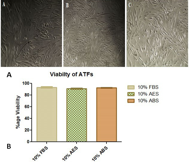
A, All cells at passage 4 showing flattened and spindle-shaped morphology, A) cultured with FBS, 100X, B) culture with AES, 100X (AES) and C) cultured with ABS, 100X (ABS). No detachment was observed under any serum group. B, Viability of ATF’s assessed by trypan blue stain method showed no significant difference and data was expressed as Mean±SEM by keeping level of significance ≤0.05.
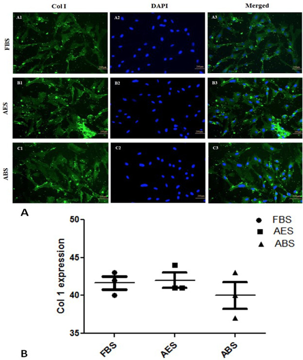
A, Expression of Collagen type1 (Col 1) on ATFs after supplementation with 10% FBS (A1-A3), 10%AES (B1-B3) and 10%ABS (C1-C3). Cells were stained with anti-type1 collagen and FITC conjugated secondary antibody whereas DAPI was used to stain the nucleus (X200). B, Expression of Collagen type1 (Col 1) in ATFs cultured with 10% FBS, AES and ABS sera. Scattered dot plot shows that there is same level of Col type1 expression all groups. Data was expressed as Mean ±SEM and level of significance was < 0.05.
References
- Ahmed, Y.A., Tatarczuch, L., Pagel, C.N., Davies, H.M., Mirams, M. and Mackie, E.J., 2007. Equine Vet. J., 39: 546-552. https://doi.org/10.2746/042516407X223699[CrossRef]
- Arigony, A.L., de Oliveira, I.M., Machado, M., Bordin, D.L., Bergter, L., Pra, D. and Henriques, J.A., 2013. Biomed. Res. Int., 2013: 597282. https://doi.org/10.1155/2013/597282[CrossRef]
- Bettger, W.J. and McKeehan, W.L., 1986. Physiol. Rev., 66: 1-35. https://doi.org/10.1152/physrev.1986.66.1.1[CrossRef]
- Brunner, D., Frank, J., Appl, H., Schoffl, H., Pfaller, W. and Gstraunthaler, G., 2010. Altern. Anim. Exp., 27: 53-62. https://doi.org/10.14573/altex.2010.1.53[CrossRef]
- Fedoroff, S. and Hall, C., 1979. In Vitro, 15: 641-648. https://doi.org/10.1007/BF02623400[CrossRef]
- Fernyhough, M.E., Hausman, G.J. and Dodson, M.V., 2008. Cells Tissues Organs, 188: 359-372. https://doi.org/10.1159/000134007[CrossRef]
- Franke, J., Abs, V., Zizzadoro, C. and Abraham, G., 2014. BMC Vet. Res., 10: 119. https://doi.org/10.1186/1746-6148-10-119[CrossRef]
- Gstraunthaler, G., Lindl, T. and van der Valk, J., 2013. A plea to reduce or replace fetal bovine serum in cell culture media. Cytotechnology, 65: 791-793. https://doi.org/10.1007/s10616-013-9633-8[CrossRef]
- Gstraunthaler, G., 2003. Altern. Anim. Exp., 20: 275-281.
- Kim, J., Kim, M., Nahm, S.S., Lee, D.M., Pokharel, S. and Choi, I., 2011. Anim. Cells Syst., 15:147-154. https://doi.org/10.1080/19768354.2011.577584[CrossRef]
- Kim, M.S., Yu, J.E., Min, K.H., Kim, J.H., Choi, I.H. and Nahm, S.S., 2012. J. Anim. Sci. Technol., 54: 59-63. https://doi.org/10.5187/JAST.2012.54.1.59[CrossRef]
- Lee, D.M., Bajracharya, P., Lee, E.J., Kim, J.E., Lee, H.J., Chun, T., Kim, J., Cho, K.H., Chang, J., Hong, S. and Choi, I., 2011. In Vitro Cell Dev. Biol. Anim., 47: 438-444. https://doi.org/10.1007/s11626-011-9427-2[CrossRef]
- Paranjape, S., 2004. Indian J. exp. Biol., 42: 26-35.
- Patterson-Kane, J.C., Becker, D.L. and Rich, T., 2012. J. comp. Pathol., 147: 227-247. https://doi.org/10.1016/j.jcpa.2012.05.010[CrossRef]
- Shanskii, Y.D., Sergeeva, N.S., Sviridova, I.K., Kirakozov, M.S., Kirsanova, V.A., Akhmedova, S.A., Antokhin, A.I. and Chissov, V.I., 2013. Bull. exp. Biol. Med., 156: 146-151. https://doi.org/10.1007/s10517-013-2298-7[CrossRef]
- van der Valk, J., Mellor, D., Brands, R., Fischer, R., Gruber, F., Gstraunthaler, G., Hellebrekers, L., Hyllner, J., Jonker, F.H., Prieto, P., Thalen, M. and Baumans, V., 2004. Toxicol. In Vitro, 18: 1-12. https://doi.org/10.1016/j.tiv.2003.08.009[CrossRef]
- Witzeneder, K., Lindenmair, A., Gabriel, C., Holler, K., Theiss, D., Redl, H. and Hennerbichler, S., 2013. Transfus. Med. Hemother., 40: 417-423. https://doi.org/10.1159/000356236[CrossRef]
- Wu, H., Ren, Y., Li, S., Wang, W., Yuan, J., Guo, X., Liu, D. and Cang, M., 2012. Cell Biol. Int., 36: 579-587. https://doi.org/10.1042/CBI20110487[CrossRef]
- Yu, J.E., Kim, M., Pokharel, S., Kim, J., Choi, I., Choe, N.H. and Nahm, S.S., 2013. Anim. Cells Syst., 17: 106-112. https://doi.org/10.1080/19768354.2013.772073[CrossRef]
- Zamansky, G.B., Arundel, C., Nagasawa, H. and Little, J.B., 1983. J. Cell Sci., 61: 289-297.
- Ziegler, A., Everett, H., Hamza, E., Garbani, M., Gerber, V., Marti, E. and Steinbach, F., 2016. BMC Vet. Res., 12: 254. https://doi.org/10.1186/s12917-016-0880-8[CrossRef]
Services
Article usage statistics
Loading…