
Toxoplasmosis is the most prevalent parasitic zoonotic disease caused by Toxoplasma gondii that infects a wide range of warm-blooded animals including humans. Congenital infection with T. gondii during pregnancy can result in severe abnormalities in infants such as hydrocephalus and mental retardation. The present study was conducted to estimate seroprevalence and potential risk factors in acquiring T. gondii infection by child-bearing age women in Dir Khyber Pakhtunkhawa, Pakistan. A cross sectional study was conducted and data regarding risk factors were recorded through questionnaire. A total of 405 women of child bearing age were serologically tested for T. gondii antibodies through immuno-chromatographic technique using strips (CTK, USA) and Indirect Enzyme Linked Immunossorbant Assay (i-ELISA). The study revealed that overall 57.28% sero-prevalence was recorded in women of child bearing age. Highest (56.46%) seroprevalence was recorded in pregnant women as compared to non-pregnant women (43.53%). Highest (57.3%) sero- prevalence was recorded in women having 21-30 years age. Notably, the highest (25%) prevalence was reported in second trimester of pregnancy. Higher (52.6%) incidence of T. gondii infection was observed in illiterate women. The study demonstrates that age, low level of education, pregnancy, contact with cat and soil are the major risk factor of T. gondii infection.
Short Communication · Volume 53, Issue 1, 30 December 2020 · Open Access
Seroepidemiology of Toxoplasma gondii Infection in Child Bearing Age Women in Dir Khyberpakhtunkhawa, Pakistan
· · · ·
Affiliations & Notes ⌄
- 1Department of Zoology, Faculty of Sciences, Shaheed Benazir Bhutto University, Sheringal, Dir Upper, Khyber Pakhtunkhwa 2Department of Animal Sciences, Shaheed Benazir Bhutto University, Sheringal, Dir Upper, Khyber Pakhtunkhwa 3Department of Biotechnology, Shaheed Benazir Bhutto University, Sheringal, Dir Upper, Khyber Pakhtunkhwa , * Corresponding author: ziaulislam@sbbu.edu.pk
Article Info ⌄
- Journal:
- Pakistan Journal of Zoology
- Published:
- 30 December 2020

From the article
- Higher seroprevalence (52.6%) was recorded in illiterate women, followed by women having primary level of education (17.2 %).
- Higher (74.60 %) seroprevalence was recorded in women living in rural areas as compared to urban areas (25.40 %).
- In present study potential risk factors to acquire T. gondii infection in child bearing age women was identified.
- The high seroprevalence of T. gondii infection was recorded in pregnant women, and those who have low level of education.
Selected from the article's own text.
Abstract
Materials and methods
A cross sectional study was conducted from May 2016 to October 2016. District Dir (L) is situated in north western part of KhyberPakhtunkhawa, Pakistan. Women population of child bearing age (14-55) was selected and blood samples were collected. Data regarding risk factors were recorded through questionnaire (age, qualification, marital status, pregnancy duration, raw meat, vegetable and milk consumption, hand washing after raw vegetable and meat consumption, residential place, house floor type, contact with soil, drinking water source, cat at home , contact with cat, livestock at home, contact with livestock, awareness about Toxoplasma). Sample size was calculated according to Thrusfield (1995). Blood samples (405) were collected and centrifuged at 3000 rpm for 10 min. The obtained serum was tested for T. gondii with the help of immuno-chromatographic technique using strips (CTK, USA). All samples were repeated through Indirect Enzyme Linked Immunossorbant Assay (i-ELISA) for T. gondii antibodies.
The data were statistical analyzed using (SPSS) and Microsoft Excel. To determine the association between seropositivity and potential risk factors Chi-square test was used.
Results and discussion
The overall seroprevalence of T. gondii infection in women of childbearing age was recorded as 57.28%, while among pregnant women seroprevalnce was recorded 56.46% and 43.53% were recorded among non-pregnant. The results of the present study are in line with the findings of earlier researchers . They reported 57.60% and 47.50 % seroprevalence among child bearing age women in Timis, Isfahan and Iran respectively. Higher seroprevalence 65.71% was recorded in women of child bearing age in Malakand agency Khyber Pakhtunkhawa, Pakistan . Seroprevalance (60-75%) was reported in northern part of Iran ; . Discrepancy in the results might be due to climatic condition, eating habits, possessing cat, life style, enrolled subjects and different sampling and analysis methods among different areas and studies.
Among positive cases of child bearing age women 7.80% were in age group 14-20 years, 57.30% were in age group 21-30 years, 26.30% were in age group of 31-40 years, 8.60% were in age group of 41-50 years. These results are in line with early findings . Prevalence in age group of 21-30 years from Khartoum State, Sudan was reported by were observed by Mohamed et al. (2013). Among positive cases of child bearing age women 7.80% were in age group of 14-20 years , 26.30% were in age group of 31-40 years, 8.60% were in age group of 41-50 years while no positive cases were recorded having age above 50 years. These results are in line with the findings Mohamed et al. (2013). Several studies have indicated an increase in seroprevalence with age ; which might be due to accumulated opportunities for contact. These differences in results may be due to target group which were in age of 14-45 years which is considered as reproductive age.
Seroprevalence of T. gondii among pregnant women during different stages of pregnancy is presented in Figure 1. Different stages of pregnancy showed different percentage of seroprevalence among women. Higher seroprevalence25.00 %was recorded at second trimester (4-6 months), 15.90 % was recorded at first trimester (1-3months), followed by (15.00 %) at third trimester (6-9 months).
The effect of different level of education on seroprevalence of T. gondii was presented in Figure 2. Education level showed a significant effect on seroprevalence of T. gondii. Those women having high education, the seroprevalence of T. gondii was minimum. Higher seroprevalence (52.6%) was recorded in illiterate women, followed by women having primary level of education (17.2 %). Similar findings were also reported by Gebremedhin et al. (2013) and Malarvizhi et al. (2012). Higher seroprevalence was recorded in illiterate women, followed by women having primary level of education. The results of the present study are an agreement with the findings of previous researchers Doni et al. (2015). Jones et al. (2001) and Daryani et al. (2014) reported that T. gondii seroprevalence reduced with the increase in education level. With higher education level knowledge about awareness, prevention and controlling of disease as a source of infection increases which decrease chance of infection.
The comparison of different risk factors for Toxoplasma seropositvity is shown in Table I. Consumption of raw vegetables and raw milk was reported in 26.70 % and 27.70 %, respectively. Among the positive cases, 84.10 % women have contact with cat. Higher (74.60 %) seroprevalence was recorded in women living in rural areas as compared to urban areas (25.40 %). Prevalence in women living in house having soil floor was 56.90 %, consuming spring water was 35.30 %, and contact with soil was 66.4%. High (65.90 %) seroprevalence was recorded in women who had contact with livestock.
The current results consonant with the early findings of Njunda et al. (2011) and Liu et al. (2009) who reported that Toxoplasma infection as associated with raw vegetable consumption. High prevalence in those individuals who consume raw vegetable may be due to poor hygienic practices. Comparatively higher seroprevalence was recorded in women who have contact with cat. Similar findings were reported by Acha and Szyfres (2003) and Negash et al. (2008). The high seroprevalence in presence of cat in the household may be due to contamination of the environment with cat shaded oocysts which become infective for a long time in water or soil . Higher prevalence was reported in rural areas as compared to urban areas. The current results are in consistent with the findings Ertug et al. (2005). The present study revealed that women living in rural areas, contact with soil and livestock, illiterate or primary education, low socioeconomic conditions are sensitive to T. gondii infection. The results of the present study are an agreement with the findings of some previous studies of Tammam et al. (2013), Senthamarai et al. (2013) and Siddiqui et al. (2014). Life style of the residents in the areas, there socioeconomic condition, contact with livestock and other related activities and favorable climatic condition may contribute for T. gondii oocysts sporulation and increase rate of infection .
Table I.- Association of risk factors and seropositivity among child bearing age women.
| Factors | Category | Number of positive samples | Prevalence (%) |
| Raw vegetable consumption | + - | 62.00 170.00 | 26.70 73.30 |
| Raw milk consumption | + - | 64.00 168.00 | 27.60 72.40 |
| Raw meat consumption | + - | 0.00 232.00 | 100.00 |
| Hand washing after handling raw meat | + - | 202.00 30.00 | 87.10 12.90 |
| Hand washing after handling raw vegetable | + - | 150.00 82.00 | 64.70 35.30 |
| Cat at home | + - | 27.00 205.00 | 11.60 88.40 |
| Contact with cat | + - | 195.00 37.00 | 84.10 15.10 |
| Exposure to soil | + - | 154.00 78.00 | 66.40 33.60 |
| Livestock at home | + - | 146.00 86.00 | 62.90 37.10 |
| Contact with livestock | + - | 153.00 79.00 | 65.90 34.10 |
| Making dong cakes | + - | 119.00 113.00 | 51.30 48.70 |
| Awareness about toxoplasmosis | + - | 48.00 184.00 | 20.70 79.30 |
+ and -” denote Yes and No, respectively.
Conclusion
In present study potential risk factors to acquire T. gondii infection in child bearing age women was identified. The high seroprevalence of T. gondii infection was recorded in pregnant women, and those who have low level of education. Women living in rural areas are at high risk. The results of the present study help to alert the government and private sector to take initiative to control the overwhelming outcome of these zoonotic diseases.
Statement of conflict of interest
The authors state that there is no conflict of interest in publishing this work.
Figures
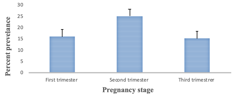
Seroprevalence of Toxoplasma gondii infection among pregnant women according to stage of pregnancy i-e first trimester (1-3 months), second trimester (4-6 months) and third trimester (7-9 months).
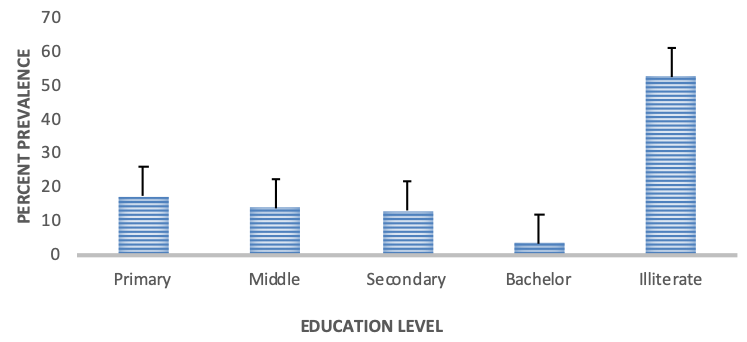
Seroprevalence of Toxoplasma gondii infection in child bearing age women having different levels of education
References
- Acha, P.N. and Szyfres, B., 2003. Zoonosis and communicable diseases common to man and animals. 3rd edition. Washington, D.C., Pan American Health Organization, pp. 76–86.
- Aleem, U., Ullah, S., Qasim, M. and Suliman, M., 2018. J. Saidu Med. Col., 8: 103-106.
- Daryani, A., Sarvi, S., Arabi, M., Mizani, A., Ahmadpour, E., Shokri, A., Rahimi, M. and Sharif, M., 2014. Acta Trop., 137: 185-194. https://doi.org/10.1016/j.actatropica.2014.05.015[CrossRef]
- Dogruman, A.I., Aslant, F.S., Alcan, S., Customer, S. and Turk, S., 2009. Int. J. Psychiat. clin. Pract., 13: 82-87. https://doi.org/10.1080/13651500802624738[CrossRef]
- Doni, N.Y.Z., Simsek, G., Gurses, F.Y., Zeyrek. and Demir, C., 2015. J. Infect. Dev. Ctries., 9: 087-093 https://doi.org/10.3855/jidc.5824.[CrossRef]
- Dubey, J.P., 2010. Toxoplasmosis of animals and humans. 2nd edition CRC Press; Boca Raton, Florida, U.S.A. pp. 1-313.
- Ertug, S.P., Okyay, M. and Yukse, H., 2005. BMC Publ. Hlth., 5: 66 https://doi.org/10.1186/1471-2458-5-66[CrossRef]
- Flegr, J., Preiss, M., Klose, J., Havlicek, J., Vitakova, M. and Kodym, P., 2003. Biol. Psychol., 63: 253-268. https://doi.org/10.1016/S0301-0511(03)00075-9[CrossRef]
- Furtado, J.M., Smith, J.R., Belfort, R., Gattey, D. and Winthrop, K.L., 2011. J. Glob. Infect. Dis., 3: 281-284. https://doi.org/10.4103/0974-777X.83536[CrossRef]
- Gebremedhin, E.Z., Abebe, A.H., Tessema, T.S., Tullu, K.D., Medhin, G. and Vitale, M., 2013. BMC Infect. Dis., 13: 101. https://doi.org/10.1186/1471-2334-13-101[CrossRef]
- Jones, J.L., Kruszon-Moran, D., Wilson, M., McQuillan, G., Navin, T. and McAuley, JB., 2001. Am. J. Epidemiol., 154: 357-365.
- Khan, M.Z., Rahman, S.U., Gul, N. and Khan, A.A., 2014. Int. J. Biosci., 5: 1-6.
- Liu, Q., Wei, F., Gao, S., Jiang, L., Lian, H., Yuan, B., Yuan, Z., Xia, Z., Liu, B., Xu, X. and Zhu, XQ., 2009. Trans. R. Soc. trop. Med. Hyg., 103: 162–166. https://doi.org/10.1016/j.trstmh.2008.07.008[CrossRef]
- Malarvizhi, A., Viswanathan, T., Lavanya, V., Malar, S.A.S. and Moorthy, K., 2012. J. Publ. Hlth. Epidemiol., 4: 170-177 https://doi.org/10.5897/JPHE12.018[CrossRef]
- Mohamed, K.A. and Elrayah, I.E., 2013. Int. J. Publ.Hlth. Epidemiol., 2: 60-66.
- Montoya, J.G. and Liesenfeld, O., 2004. Lancet, 363: 1965-1976. https://doi.org/10.1016/S0140-6736(04)16412-X[CrossRef]
- Montoya, J.G. and Rosso, F., 2005. Clin. Perinatol., 32: 705–726. https://doi.org/10.1016/j.clp.2005.04.011[CrossRef]
- Mostafavi, N.B., Ataei, Z.N., Monfared, L.J., Yaran, M., Ataie, M. and Babak, A., 2012. Adv. biomed. Res., 1: 60. https://doi.org/10.4103/2277-9175.100181[CrossRef]
- Negash, T.G. and Medhin, G., 2008. East Afri. J. Publ.Hlth., 5: 211–214.
- Njunda, A.L., Assob, J.C.N., Nsagha, D.S., Kamga, H.L., Nde, P.F. and Yugah, V.C., 2011. J. Publ. Hlth., 2: 240-251. https://doi.org/10.4081/jphia.2011.e16[CrossRef]
- Petersen, E., Vesco, G., Villari, S. and Buffolano, W., 2010. Zoonoses Publ. Hlth., 57: 8–17. https://doi.org/10.1111/j.1863-2378.2009.01278.x[CrossRef]
- Senthamarai, S., Sivasankari, S., Apurba, S.S., Sandhya, B.K., Kumudavathi, M.S., Anitha, C. and Amshavathani, S.K., 2013. Disease, 3: 29-32.
- Sharif, M.A., Ajami, A., Daryani, H. and Khalilian, A., 2006. Int. J. Mol. Med. Adv. Sci., 2: 134-7.
- Siddiqui, N.F., Shujatullah., H.M., Rabbani, T. and Khan, P.A., 2014. J. Immunol. Vac. Technol., 1: 101-103.
- Sroka, S., Bartelheimer, N., Winter, A., Heukelbach, J., Ariza, L., Ribeiro, H., Oliveira, F.A., Queiroz, A.J.N., Alencar, A.J.N.J. and Liesenfeld, O., 2010. Am. J. trop. Med. Hyg., 83:528–533. https://doi.org/10.4269/ajtmh.2010.10-0082[CrossRef]
- Tammam, A.E., Haridy, M.A., Abdellah, A.H., Ahmed, S.R., Fayed, H.M. and Sammani, M.A., 2013. Egypt. J. clin. Diagn. Res., 7: 2870-2873.
- Techalew, S., Mekashaw, T., Endale, T., Belete, T. and Ashenafi, T., 2009. BMC Res. Notes., 2: 213.
- Tenter, A.M., Heckeroth, A.R. and Weiss, L.M., 2000. Int. J. Parasital., 30: 1217-1258. https://doi.org/10.1016/S0020-7519(00)00124-7[CrossRef]
- Thrusfield, M., 1995. Veterinary epidemiology. 2ed edition. Back well scientific Ltd. UK, pp. 182-198.
- Torgerson, P.R. and Macpherson, C.N.L., 2011. Vet. Parasitol., 182: 79– 95
- Youssefi, M.R., Sefidgar, A.A., Mostafazadeh, A. and Omran, S.M., 2007. Pak. J. biol. Sci., 10:1550-1552. https://doi.org/10.3923/pjbs.2007.1550.1552[CrossRef]
Services
Article usage statistics
Loading…