Short Communication · Volume 13, Issue 10, 11 October 2025 · Open Access
The Effect of Insulin Like Growth Factor-1 (IGF-1) Supplementation in Maturation Media on Transforming Growth Factor-Beta (TGF-β) and Growth Differentiation Factor-9 (GDF-9) Expression in Kacang Goat Oocytes
· · · · · ·
Affiliations & Notes ⌄
1Reproductive Biology, Universitas Airlangga, Surabaya
2Division of Veterinary Anatomy, Faculty of Veterinary Medicine, Universitas Airlangga, Surabaya, Indonesia
3Division of Veterinary Reproduction, Faculty of Veterinary Medicine, Universitas Airlangga, Surabaya, Indonesia
4Division of Animal Husbandry, Faculty of Veterinary Medicine, Universitas Airlangga, Surabaya, Indonesia
5Faculty of Veterinary Medicine, Brawijaya University, Malang, Indonesia. *Correspondence | Widjiati Widjiati, Division of Veterinary Anatomy, Faculty of Veterinary Medicine, Universitas Airlangga, Surabaya, Indonesia
The correlation significance results are presented in Table 4, where the data were tested using two-tailed Kendall’s tau and Spearman’s rho statistical methods to assess the relationship between the expressions of TGF-β and GDF-9 during oocyte maturation with IGF-1 supplementation.
This study recommends considering IGF-1 supplementation at a dosage of 100 ng/ml in the in vitro maturation medium for Kacang goat oocytes, as it significantly increases the expression of TGF-β and GDF-9, essential for oocyte development in Kacang goats.
The findings underscore the potential of IGF-1 supplementation to improve oocyte maturation in Kacang goats, hence enhancing the genetic quality of this significant indigenous breed.
Selected from the article's own text.
Abstract
Kacang goats, native to Indonesia, play a vital role in enhancing food security and supporting rural economies. Nonetheless, their population and genetic quality are still not at an optimal level. In vitro maturation (IVM) plays a crucial role in genetic conservation, by impacting oocyte competence and embryonic development. This research assessed the impact of insulin-like growth factor-1 (IGF-1) supplementation in the maturation medium on the expression of TGF-β and GDF-9 in Kacang goat oocytes. Oocytes were harvested from goat ovaries and subjected to in vitro maturation with IGF-1 at concentrations of 0 (control), 50, 100, and 150 ng/mL. Immunocytochemistry evaluated the expression of TGF-β and GDF-9. Oocytes exposed to 100 ng/mL IGF-1 exhibited a marked increase in the expression of TGF-β and GDF-9 compared to other groups (p < 0.05). A significant positive correlation exists between TGF-β and GDF-9 (r = 0.499; τ = 0.424; p < 0.05). The results indicate that 100 ng/mL IGF-1 effectively promotes oocyte maturation through the activation of signaling pathways. In summary, administering 100 ng/mL IGF-1 is advised to enhance oocyte quality in Kacang goats, which may lead to improved IVF outcomes.
The Kacang goat is an indigenous Indonesian ruminant widely reared by rural communities, serving as an important source of animal protein and supporting food security . Economically, it provides additional income and improves dietary protein intake. However, its population growth and genetic quality remain suboptimal . The genetic quality of this native breed is threatened by frequent crossbreeding, which reduces its purity and highlights the urgent need for conservation efforts . To address this issue, reproductive technologies have been developed. Technologies such as artificial insemination, embryo transfer, in vitro fertilization (IVF), cryopreservation, and genetic engineering can improve genetic quality in Kacang goats . IVF, in particular, offers potential to increase productivity , but its success is often limited by low oocyte quality and availability .
In vitro maturation (IVM) is a critical stage in the IVF process, largely determined by the maturation medium composition . IVM outcomes are influenced by medium constituents, hormonal stimulation, species-specific traits, exposure time, and intrinsic oocyte quality, particularly morphology ; ; . Environmental factors such as contamination, temperature, pH, and osmolality also affect maturation . Supplementation with growth factors, hormones, or antioxidants has been shown to enhance maturation efficiency and oocyte quality .
Insulin-like growth factor-1 (IGF-1), produced by almost all tissues, acts via IGF1R receptors to activate MAPK and PI3K pathways, promoting cumulus expansion and oocyte maturation . During maturation, oocytes and cumulus cells secrete Transforming growth factor-β (TGF-β) and growth differentiation factor-9 (GDF-9), both detected in Kacang goat oocytes , 2011). TGF-β regulates follicular cell proliferation, meiosis, apoptosis, and cumulus expansion ; ; , while GDF-9 supports follicular growth, oocyte maturation, and embryo quality ; .
Given this background, further research is warranted to investigate the effects of IGF-1 supplementation on TGF-β and GDF-9 expression during Kacang goat oocyte maturation, contributing to improved reproductive performance and genetic preservation of this native breed.
MATERIALS AND METHODS
Ethical approval
The Animal Care and Use Committee of the Faculty of Veterinary Medicine, Universitas Airlangga, Surabaya, authorized the experimental procedure. The study was performed in compliance with established protocols and adhered to animal welfare and medical ethical standards. The ethical clearance number is 1.KEH.77.05.2025.
Materials
The materials used in this study included physiological NaCl (0.95%), Phosphate-Buffered Saline (PBS), Gentamicin (PT. Indofarma), mineral oil (Vitrolife, Sweden), G-MOPS (Vitrolife®), G-IVF (Vitrolife®), Recombinant Goat IGF-1 (MedikBio®, Catalogue number: MDB-EP01108600), Poly-L lysine-coated slides, vaseline, 3% hydrogen peroxide, Growth Differentiation Factor-9 (GDF-9) Polyclonal Antibody (Bioss), TGF-β Antibody (TGF-β 1 [3C11]: sc-130348, Santa Cruz Biotechnology), 70% alcohol, methanol, 1% aceto-orcein acid, 0.025% trypsin, Biotinylated Link (Yellow) drops, Streptavidin (Red) drops, 3,3-Diaminobenzidine tetrahydrochloride (DAB) chromogen, and methylene green.
Ovarium collection
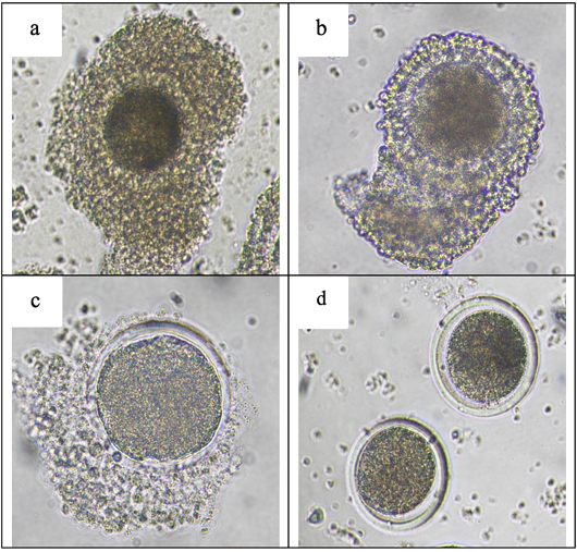
The ovaries were rinsed with 0.9% NaCl with 100 µL gentamicin. Preantral follicles measuring 2–6 mm were aspirated with a 10 mL syringe fitted with an 18G needle containing medium, followed by three washes in G-MOPS and two washes in G-IVF maturation media. Oocytes were classified morphologically as follows: Grade 1, characterized by homogeneous cytoplasm with three or more cumulus layers; Grade 2, exhibiting one to three layers and homogeneous cytoplasm; Grade 3, presenting few or no cumulus layers and heterogeneous cytoplasm; Grade 4, displaying abnormal morphology with detached cumulus and heterogeneous cytoplasm, indicative of apoptosis , 2024). Figure 1 illustrates oocyte grading.
In-vitro maturation
Maturation process of the oocytes was conducted using G-IVF medium as the maturation substrate. The medium was supplemented with IGF-1 at concentrations of 0 (control), 50, 100, and 150 ng/mL, following the protocols of Javvaji et al. (2020) and Jimenez et al. (2016). For each experimental group, Petri dishes were prepared with four aliquots of the maturation medium (50 µg/mL), each covered with mineral oil to prevent evaporation and maintain osmolarity. The oocytes were then incubated under controlled conditions of 5% CO₂, incubation temperature at 38.5 °C, with approximate relative humidity by 95%, and for a duration of 20–22 hours ; .
Identification of TGF-β and GDF-9 expression by immunocytochemistry
Matured oocytes were placed on Poly-L-lysine-coated slides, loosely covered with a cover glass sealed with vaseline, and fixed for 24 hours. They were then treated with 3% hydrogen peroxide for 5–10 minutes, followed by two PBS washes (pH 7.4) for 5 minutes each. Oocytes were incubated with 0.025% trypsin at 37°C for 15 minutes and rewashed with PBS.Ultra V block was applied for 5 minutes, followed by incubation with either anti-TGF-β or anti-GDF-9 antibodies for 60 minutes at room temperature. Biotinylated Link (Yellow) was added for 30 minutes, then washed with PBS, followed by Streptavidin (Red) for 30 minutes. DAB chromogen was applied for 6–10 minutes, followed by PBS and aquadest washes. The final steps included rinsing with distilled water (aquadest) for 5 minutes, staining with methylene green for 5–10 minutes, and gently blotting excess stain to reduce moisture . The oocytes then observed under an Olympus® CX-41 microscope at 400× magnification, Expression assessment was performed using the Remmele scale, known as the Immunoreactive Score (IRS). The outcome were obtained by the multiplication of the positive cells percentage with the staining intensity score. The scoring system for the percentage are shown in Table 1 .
Table 1: The index remmele scale.
Percentage of positive cells (A)
Staining intensity (B)
Score 0: No positive cells
Score 0: No color reaction
Score 1: Positive cells < 10%
Score 1: Weak color intensity
Score 2: Positive cells 11%–50%
Score 2: Moderate color intensity
Score 3: Positive cells 51%–80%
Score 3: Strong color intensity
Score 4: Positive cells > 80%
Data analysis
The data were analyzed using the Kruskal–Wallis test, and when a significant difference was detected (p < 0.05), pairwise comparisons were carried out with the Mann–Whitney U test. Correlation analyses were conducted using two-tailed Kendall’s tau and Spearman’s rho coefficients. The analyses assumed that the population under study was normally distributed, homogeneous, and composed of independent samples. All statistical procedures were performed using the Statistical Package for the Social Sciences (SPSS).
RESULTS
Immunocytochemical staining results of TGF-β and GDF-9
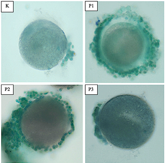
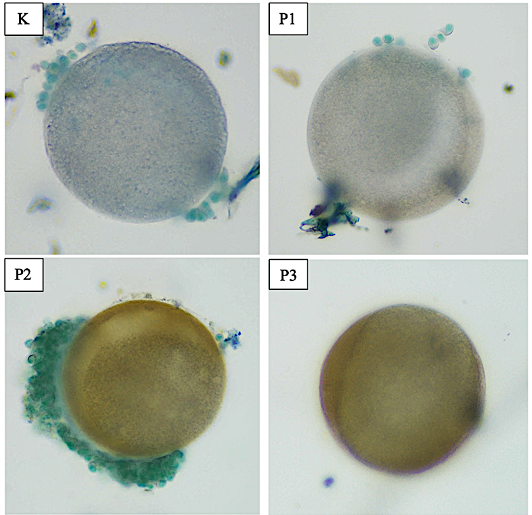
The expressions of TGF-β and GDF-9 were identified using the immunohistochemical staining method. All slides were examined microscopically and assessed semi-quantitatively using the Remmele method. The immunocytochemical staining results are shown in Figures 2 and 3. The expressions of both markers are indicated by brown coloration in the oocytes. In contrast, oocytes that did not express both markers appeared green, reflecting the methylene green counterstain. The brown coloration signifies the presence of antigen–antibody–chromogen binding, indicating positive expression. Conversely, green coloration in oocytes or cumulus cells indicates the absence of this binding and, therefore, no expression by the two protein. These staining differences served as the basis for determining the expression scores.
Expression analysis
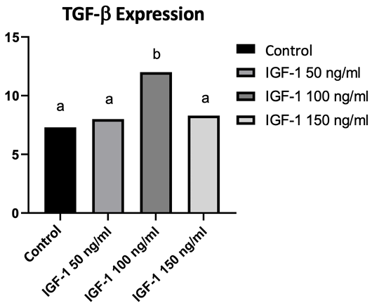
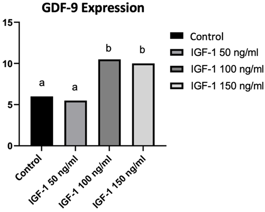
TGF-β expression was significantly highest in the 100 ng/mL IGF-1 treatment group, with a mean rank of 20.50 as shown in Table 2. This group exhibited significantly higher TGF-β expression (p < 0.05) compared to the control group (6.67), the 50 ng/mL IGF-1 group (9.92), and the 150 ng/mL IGF-1 group (12.92). These findings are illustrated in Figure 4. Similarly, GDF-9 expression was also significantly highest in the 100 ng/mL IGF-1 treatment group, with a mean rank of 18.83 as shown in Table 3. The control group had a mean rank of 6.50, which was significantly lower than both the 100 ng/mL (18.83) and 150 ng/mL (17.50) groups, but not significantly different from the 50 ng/mL group. Both the 100 ng/mL (18.83) and 150 ng/mL (17.50) IGF-1 groups showed significantly higher GDF-9 expression compared to the control (6.50) and 50 ng/mL (7.17) groups (p < 0.05). The results are also depicted in Figure 5.
Table 2: Mean rank of TGF-β expression.
Group
Mean rank of TGF-β expression
(K) Control
6.67a
(P1) IGF-1 50 ng/ml
9.92a
(P2) IGF-1 100 ng/ml
20.50b
(P3) IGF-1 150 ng/ml
12.92a
Table 3: Mean rank of GDF-9 expression.
Group
Mean rank of GDF-9 expression
(K) Control
6.67a
(K) Control
6.50a
(P1) IGF-1 50 ng/ml
7.17a
(P2) IGF-1 100 ng/ml
18.83b
Correlation analysis of immunohistochemical staining results
The correlation significance results are presented in Table 4, where the data were tested using two-tailed Kendall’s tau and Spearman’s rho statistical methods to assess the relationship between the expressions of TGF-β and GDF-9 during oocyte maturation with IGF-1 supplementation. The analysis showed a statistically significant correlation (p<0.05) between the two variables, with a p-value of 0.013. Table 5 displays the correlation coefficients, with Kendall’s tau (τ) and Spearman’s rho (r) values of 0.424 and 0.499, respectively. The positive values in both tests indicate a positive relationship between the two variables. This suggests that an increase in TGF-β expression is likely accompanied by an increase in GDF-9 expression, indicating a potential significant positive association between these two proteins in supporting the in vitro maturation of Kacang goat oocytes.
Table 4: Significance analysis of the correlation between TGF-β and GDF-9 expression.
TGF-β
GDF-9
TGF-β
0.013*
GDF-9
0.013*
Table 5: Correlation coefficient analysis between TGF-β and GDF-9 expression.
TGF-β
GDF-9
Kendall’s tau (τ)
TGF-β
1
0.424
Spearman’s rho (r)
GDF-9
0.499
1
DISCUSSION
IGF-1 is a growth factor produced by almost all tissues, playing an essential role in cell survival, proliferation, and differentiation, including oocytes ; . By binding to the IGF-1 receptor (IGF1R), it activates intracellular signaling cascades that support oocyte maturation, notably the PI3K/Akt, ERK/MAPK, and phospholipase C pathways . In the present study, supplementation with 100 ng/mL IGF-1 in the maturation medium resulted in the highest expression of TGF-β and GDF-9, with significantly higher levels (p < 0.05) than the control and other treatment groups. IGF-1 appears to enhance TGF-β via the PI3K/Akt pathway, involving Akt phosphorylation and NF-κB activation ; , which in turn upregulates EGF-like growth factors—AREG, EREG, and BTC that promote oocyte maturation . The ERK/MAPK pathway further supports maturation by phosphorylating Connexin 43 (CX43), diminishing gap junction communication and facilitating meiotic resumption ; .
IRS, Grb2, Sos, and Raf, which activates USF-1, a transcription factor binding E-box motifs within the GDF-9 promoter ; ; . GDF-9 enhances cumulus expansion via Smad2/3 activation upon binding ALK5 and BMPR2, leading to transcription of HAS2, GREM1, and PTGS2 ; .
Both TGF-β and GDF-9 contribute to in vitro maturation by lowering cAMP levels, activating ERK/MAPK in follicular cells, and closing gap junctions (Cx43 and Cx37), thereby restricting cGMP transfer and permitting meiotic progression , 2011, 2012; ; . Meiotic arrest is maintained by elevated cAMP and PKA activity, supported by GPR3 and cGMP-mediated PDE3 inhibition . ERK-mediated gap junction closure permits PDE3 activation, reducing cAMP and PKA, thereby activating MPF (CDK1–Cyclin B), inducing GVBD and meiotic resumption ; .
IGF-1 circulates as a ternary complex with IGFBP and ALS, with IGFBP regulating its bioavailability ; . Notably, the 150 ng/mL IGF-1 dose was less effective than 100 ng/mL, possibly due to receptor saturation or negative feedback mechanisms that impair maturation or trigger apoptosis .
Both TGF-β and GDF-9 belong to the TGF-β superfamily, and their expression has been shown to be enhanced by IGF-1. The upregulation of these markers may reflect a synergistic relationship in regulating oocyte maturation, as evidenced by the significant positive correlation between TGF-β and GDF-9 expression observed in this study (p < 0.05; Kendall’s tau = 0.424; Spearman’s rho = 0.499). Although TGF-β was originally thought to be expressed exclusively by follicular cells, it is also produced by oocytes, acting via the Smad2/3 pathway to modulate gene expression within the follicular environment, whereas GDF-9, secreted by oocytes, regulates cumulus cell function .
Before the identification of GDF-9 and BMP-15, recombinant TGF-β1 and TGF-β2 were commonly applied to improve in vitro maturation, with these factors exhibiting positive feedback through the Smad2/3 pathway ; . Notably, GDF-9 has also been shown to enhance TGF-β expression by activating the TGF signaling pathway through binding to the BMPR2–TGFBR1 complex, which initiates a Smad2/3-mediated phosphorylation cascade leading to increased TGF-β transcription ; . In contrast to TGF-β, however, GDF-9 does not appear to regulate its own expression, relying instead on USF-1-mediated transcription , and primarily acts in a paracrine or autocrine manner on somatic follicular cells, with no evidence of autoregulation within oocytes ; .
CONCLUSIONS
This study recommends considering IGF-1 supplementation at a dosage of 100 ng/ml in the in vitro maturation medium for Kacang goat oocytes, as it significantly increases the expression of TGF-β and GDF-9, essential for oocyte development in Kacang goats. The findings underscore the potential of IGF-1 supplementation to improve oocyte maturation in Kacang goats, hence enhancing the genetic quality of this significant indigenous breed. Additional research is required to examine the pathways connecting biomarker levels to their influence on enhancing oocyte quality during in vitro maturation.
Acknowledgements
The authors extend their heartfelt appreciation to the Faculty of Veterinary Medicine, Universitas Airlangga, for affording the essential facilities and support for the completion of this research. The authors aspire that the findings in this publication will enhance understanding and facilitate future research in in vitro maturation (IVM) and in vitro embryo production (IVEP).
Novelty Statement
This study is unique in recommending IGF-1 supplementation in the in vitro maturation medium for Kacang goat oocytes to increase the expression of TGF-β and GDF-9, essential for oocyte development in Kacang goats. The findings underscore the potential of IGF-1 supplementation to improve oocyte maturation in Kacang goats.
Author’s Contribution
Mohamad Raviansyah Jawindra: Responsible for conceptualization, provision of resources, and drafting the original manuscript. Widjiati Widjiati: Responsible for data verification, data evaluation, supervision, critical revision of the manuscript, and securing funding. Rimayanti Rimayanti: Contributed to data verification, data interpretation, supervision, and critical review and editing of the manuscript. Suherni Susilowati: Contributed to data verification, data assessment, and overall supervision of the study. Epy Muhammad Luqman: Contributed to data verification, data examination, and supervision of the research process. Mirni Lamid: involved in data verification, data review, and supervisory roles. Viski Fitri Hendrawan: Contributed to writing, critical review, and editing of the manuscript.
Generative AI and AI-assisted technology statement
The authors declare that no Genrative AI was used in the creation of this manuscript.
Conflicts of interest
The authors hereby declare that no actual or potential conflicts of interest exist with respect to the research, authorship, and/or publication of this article.
Figures
Figure 1:.
Morphological appearance of Grade 1 oocyte (a), Grade 2 (b), Grade 3 (c), and Grade 4 (d) based on Widjiati et al. (2024) (Personal documentation).
Figure 2:.
Differences in TGF-β expression in goat oocytes after in vitro maturation with IGF-1 supplementation. (K) represents the control group without treatment, (P1) with IGF-1 at a dose of 50 ng/ml, (P2) with a dose of 100 ng/ml, and (P3) with a dose of 150 ng/ml. The highest expression was observed in P2 (100 ng/ml), indicated by strong positive brown staining. The images were taken at 400× magnification using Olympus CX-41 microscope.
Figure 3:.
Differences in GDF-9 expression in goat oocytes after in vitro maturation with IGF-1 supplementation. (K) represents the control group without treatment, (P1) with IGF-1 at a dose of 50 ng/ml, (P2) with a dose of 100 ng/ml, and (P3) with a dose of 150 ng/ml. The highest expression was observed in P2 (100 ng/ml), indicated by strong positive brown staining. The images were taken at 400× magnification using Olympus CX-41 microscope.
Figure 4:.
Diagram of TGF-β expression during oocyte maturation of Kacang goats supplemented with various doses of IGF-1.
Figure 5:.
Diagram of GDF-9 expression during oocyte maturation of Kacang goats supplemented with various doses of IGF-1.
References
Bai D, Ueno L, Vogt PK (2009). Akt-mediated regulation of NFκB and the essentialness of NFκB for the oncogenicity of PI3K and Akt. Int. J. Cancer, 125(12): 2863-2870. https://doi.org/10.1002/ijc.24748[CrossRef]
Coticchio G, Dal CM, Mignini RM, Guglielmo MC, Brambillasca F, Turchi D, Novara PV, Fadini R (2015). Oocyte maturation: Gamete-somatic cells interactions, meiotic resumption, cytoskeletal dynamics and cytoplasmic reorganization. Hum. Reprod. Update, 21(4): 427-454. https://doi.org/10.1093/humupd/dmv011[CrossRef]
Daldello EM, Luong XG, Yang CR, Kuhn J, Conti M (2019). Cyclin B2 is required for progression through meiosis in mouse oocytes. Development, 146(8): 172734. https://doi.org/10.1242/dev.172734[CrossRef]
Das D, Arur S (2022). Regulation of oocyte maturation: Role of conserved ERK signaling. Mol. Dev. Dev., 89: 353-374. https://doi.org/10.1002/mrd.23637[CrossRef]
Datta TK, Rajput SK, Wee G, Lee KB, Folger JK, Smith GW (2015). Requirement of the transcription factor USF1 in bovine oocyte and early embryonic development. Reproduction, 149(2): 203-212. https://doi.org/10.1530/REP-14-0445[CrossRef]
del Llano E, Iyyappan R, Aleshkina D, Masek T, Dvoran M, Jiang Z, Kubelka M, Susor A (2022). SGK1 is essential for meiotic resumption in mammalian oocytes. Eur. J. Cell Biol., 101(2): 151210. https://doi.org/10.1016/j.ejcb.2022.151210[CrossRef]
Faizah Z, Aswin RH (2021). Expression of transforming growth factor β and growth differentiation factor 9 in sheep oocytes vitrified after and before in vitro maturation. J. Vet., 22(1): 109-115. https://doi.org/10.19087/jveteriner.2021.22.1.109[CrossRef]
Faizal MA, Nuraini H, Prastowo S, Rachmawati R (2024). Expression of HAS2, GREM1, and PTGS2 genes as markers of oocyte and embryo quality in in vitro fertilization technology: A systematic review. Reprod. Biol. J., 15(1): 25-36. https://doi.org/10.3389/fendo.2024.1274376[CrossRef]
Fang L, Chang HM, Cheng JC, Leung PC, Sun YP (2014). TGF beta1 induces COX 2 expression and PGE₂ production in human granulosa cells through Smad signaling pathways. J. Clin. Endocrinol. Metab., 99(7): E1217-E1226. https://doi.org/10.1210/jc.2013-4100[CrossRef]
Fang L, Sun YP, Cheng JC (2023). The role of amphiregulin in ovarian function and disease. Cell. Mol. Life Sci., 80: 60. https://doi.org/10.1007/s00018-023-04709-8[CrossRef]
Fountas S, Petinaki E, Bolaris S, Kargakou M, Dafopoulos S, Zikopoulos A, Moustakli E, Sotiriou S, Dafopoulos K (2024). The roles of GDF-9, BMP-15, BMP-4 and EMMPRIN in Folliculogenesis and in vitro fertilization. J. Clin. Med., 13(13): 3775. https://doi.org/10.3390/jcm13133775[CrossRef]
Gilchrist RB, Lane M, Thompson JG (2008). Oocyte-secreted factors: Regulators of cumulus cell function and oocyte quality. Hum. Reprod. Update, 14(2): 159–177. https://doi.org/10.1093/humupd/dmm040[CrossRef]
Gode F, Gulekli B, Dogan E, Korhan P, Dogan S, Bige O, Cimrin D, Atabey N (2011). Influence of follicular fluid GDF9 and BMP15 on embrio quality. Fertil. Steril., 95(22): 74-78. https://doi.org/10.1016/j.fertnstert.2011.03.045[CrossRef]
Hakuno F, Takahashi S (2018). IGF1 receptor signaling pathways. J. Mol. Endocrinol., 61: 169-186. https://doi.org/10.1530/JME-17-0311[CrossRef]
Hao X, Yuan F, Cui Y, Zhang M (2022). Oocyte-secreted factor TGFB2 enables mouse cumulus cell expansion in vitro. Mol. Reprod. Dev., 89(11): 554–562. https://doi.org/10.1002/mrd.23646[CrossRef]
Herta AC, Lolicato F, Smitz JEJ (2018). In vitro follicle culture in the context of IVF. Reproduction, 156(1): 59-73. https://doi.org/10.1530/REP-18-0173[CrossRef]
Hough C, Radu M, Doré JJE (2012). TGF-beta induced Erk phosphorylation of smad linker region regulates smad signaling. PLoS One, 7(8): e42513. https://doi.org/10.1371/journal.pone.0042513[CrossRef]
Huang C, Du Z, Shi Y, Sun N, Zhu Z, Yu X, Li A (2024). Growth differentiation factor 9 activates the TGF-β pathway in follicle atresia of Muscovy ducks. Poult. Sci., 103(12): 104278. https://doi.org/10.1016/j.psj.2024.104278[CrossRef]
Ilmi LUNL, Lestari TD, Widjiati, Safitri E, Restiadi TI, Mafruchati M, Amrullah MF, Khairullah AR, Kurniawan SC, Abani S, Hasib A, Samodra MEE, Silaen OSM (2023). Supplementation of alpha lipoic acid on Kacang goat (Capra hircus) oocyte to the growth of GDF-9 and BMP-15. J. Adv. Vet. Res., 14(1): 135-138.
Javvaji PK, Dhali A, Francis JR, Kolte AP, Roy SC, Selvaraju S, Mech A, Sejian V (2021). IGF-1 treatment during in vitro maturation improves developmental potential of ovine oocytes through the regulation of PI3K/Akt and apoptosis signaling. Anim. Biotechnol., 32: 795-805. https://doi.org/10.1080/10495398.2020.1752703[CrossRef]
Jiang W, Cai F, Xu H, Lu Y, Chen J, Liu J, Cao N, Zhang X, Chen X, Huang Q, Zhuang H, Hua ZC (2020). Extracellular signal regulated kinase 5 promotes cell migration, invasion and lung metastasis in a FAK-dependent manner. Protein Cell, 11(11): 825-845. https://doi.org/10.1007/s13238-020-00701-1[CrossRef]
Jimenez CR, de Azevedo JL, Silveira RG, Penitente-Filho J, Carrascal-Triana EL, Zolini AM, Araujo VR, Torres CAA, Goncalves WG (2016). Effects of IGF-1 on in vitro culture of bovine preantral follicles are dose-dependent. Reprod. Domest. Anim., 51: 435–444. https://doi.org/10.1111/rda.12698[CrossRef]
Juengel JL, Bibby AH, Reader KL, Lun S, Quirke LD, Haydon LJ, McNatty KP (2004). The role of transforming growth factor-beta (TGF-beta) during ovarian follicular development in sheep. Reprod. Biol. Endocrinol., 2: 78. https://doi.org/10.1186/1477-7827-2-78[CrossRef]
Kalous J, Tetkova A, Kubelka M, Susor A (2018). Importance of ERK1/2 in regulation of protein translation during oocyte meiosis. Int. J. Mol. Sci., 19(3): 698. https://doi.org/10.3390/ijms19030698[CrossRef]
Kasman AAMN, Santoso B, Widjiati (2020). The effect of vitrification after warming on the expressions of p38, CDK1, and cyclin B in immature goat oocytes followed by in vitro maturation. Vet. World, 13(10): 2126-2132. https://doi.org/10.14202/vetworld.2020.2126-2132[CrossRef]
Kusindarta DL (2009). Effect of maturation duration and fertilization incubation time on the fertility rate of Ongole crossbred cattle oocytes in vitro. J. Kedokt. Hewan, 3(1): 185–193. https://doi.org/10.21157/j.ked.hewan.v3i1.3071[CrossRef]
Lavoie H, Therrien M (2015). Regulation of RAF protein kinases in ERK signalling. Nat. Rev. Mol. Cell Biol., 16: 281-298. https://doi.org/10.1038/nrm3979[CrossRef]
Li Y, Chang HM, Zhu H, Sun YP, Leung PCK (2024). EGF-like growth factors upregulate Pentraxin 3 expression in human granulosa-lutein cells. J. Ovarian Res., 17: 97. https://doi.org/10.1186/s13048-024-01404-5[CrossRef]
Miescher I, Rieber J, Calcagni M, Buschmann J (2023). In vitro and in vivo effects of IGF-1 delivery strategies on Tendon Healing: A Review. Int. J. Mol. Sci., 24(3): 2370. https://doi.org/10.3390/ijms24032370[CrossRef]
Mustofa I, Susilowati S, Suprayogi TW, Akitunde AO, Oktanella Y, Purwanto DA (2023). Epigallocatechin-3-gallate chitosan nanoparticles in an extender improve the antioxidant capacity and post-thawed quality of Kacang goat semen. F1000 Res., 12: 32. https://doi.org/10.12688/f1000research.127744.1[CrossRef]
Najiati AY, Restiadi TI, Madyawati SP, Mustofa I, Mafruchati M, Widjiati, Hendrawan VF, Luqman EM (2022). Calcium ionophore supplementation in in vitro fertilization medium towards fertilization rates of Kacang goats (Capra hircus). Ecol. Environ. Conserv., pp. 635-641. https://doi.org/10.53550/EEC.2022.v28i02.010[CrossRef]
Nursadida M, Widjiati W, Mustofa I, Safitri E, Susilowati S, Suprayogi TW (2024). The effects of duration of the in vitro maturation process on the maturation level and apoptosis of Kacang goat. Vet. Stanica, 55(1): 79–85. https://doi.org/10.46419/vs.55.1.8[CrossRef]
Rameshwar P, Narayanan R, Qian J, Denny TN, Colon C, Gascon P (2000). NF-B as a central mediator in the induction of TGF-in monocytes from patients with idiopathic myelofibrosis: An inflammatory response beyond the realm of homeostasis. J. Immunol., pp. 165. https://doi.org/10.4049/jimmunol.165.4.2271[CrossRef]
Ramirez G, Palomino J, Aspee K, De Los Reyes M (2020). GDF-9 and BMP-15 mRNA levels in canine cumulus cells related to cumulus expansion and the maturation process. Animals, 10(3): 462. https://doi.org/10.3390/ani10030462[CrossRef]
Silva JRV, Figueiredo JR, van den Hurk R (2009). Involvement of growth hormone (GH) and insulin-like growth factor (IGF) system in ovarian folliculogenesis. Theriogenology, 71: 1193-1208. https://doi.org/10.1016/j.theriogenology.2008.12.015[CrossRef]
Soto-Heras S, Menéndez-Blanco I, Catalá MG, Izquierdo D, Thompson JG, Paramio MT (2019). Biphasic in vitro maturation with C-type natriuretic peptide enhances the developmental competence of juvenile-goat oocytes. PLoS One, 14(8): e0221663. https://doi.org/10.1371/journal.pone.0221663[CrossRef]
Suprayogi TW, Susilowati S, Hernawati T, Hafidha FG, Wening CA, Purnawan AB (2022). Improved quality of Kacang goat sexing frozen semen with the addition of green tea extract. J. Adv. Vet. Anim. Res., 9(3): 412-418. https://doi.org/10.5455/javar.2022.i609[CrossRef]
Susilowati S, Mustofa I, Wurlina W, Triana IN, Utama S, Utomo B (2021). A 150 kDa protein derived from bull seminal plasma extended the survival time of Kacang goat sperm stored at 5 oC. Vet. Med. Int., 1470209. https://doi.org/10.1155/2021/1470209[CrossRef]
Vitt UA, Mazerbourg S, Klein C, Hsueh AJ (2002). Bone morphogenetic protein receptor type II is a receptor for growth differentiation factor-9. Biol. Reprod., 67: 473–480. https://doi.org/10.1095/biolreprod67.2.473[CrossRef]
Widjiati W, Faizah Z, Darsini N, Hendrawan VF, Karima HN, Chotimah C, Sumitro SB, Luqman EM (2020). Indentification of caspase 3 intensity in Kacang goat oocyte post vitrification using confocal laser scanning microscopy. Adv. Anim. Vet. Sci., 8(12): 1362-1366. https://doi.org/10.17582/journal.aavs/2020/8.12.1362.1366[Full text][CrossRef]
Widjiati W, Faizah Z, Darsini N, Hendrawan VF, Karima HN, Chotimah C, Sumitro SB, Yustinasari LR, Kasman AAMN, Ntoruru JM, Luqman EM (2022). Calcium (Ca2+) expression and intensity in cumulus-oocyte complex (COCs) in Kacang goat after vitrification. Pol. J. Vet. Sci., 25(1): 19-26. https://doi.org/10.24425/pjvs.2022.140836[CrossRef]
Widjiati W, Hestianah EP, Luqman EM, Taqwa SF, Caesar J, Shabira Z, Mega RL (2024). Effect of heat shock through MDA and 8-OHdG levels of post-thawing goat oocytes. Int. J. Vet. Sci., 13(5): 611-616.
Widjiati W, Wulansari D, Wurlina W, Widjaja N (2011). Identification of growth differentiation factor-9 (GDF-9) in in vitro matured bovine oocytes using immunocytochemistry. J. Kedokt. Hewan, 5: 59–62. https://doi.org/10.21157/j.ked.hewan.v5i2.356[CrossRef]
Widjiati, Boediono A, Sumitro SB, Hinting A, Aulani’am, Susilowati T (2012). Isolation and identification of transforming growth factor β from in vitro matured cumulus oocyte complexes. Hayati J. Biosci., 19(1): 6-10. https://doi.org/10.4308/hjb.19.1.6[CrossRef]
Widjiati, Rimayanti, Arief, Agus (2010). In vitro oocyte maturation induced by oocyte-derived transforming growth factor β in cumulus-oocyte complexes. Dissertation. Universitas Brawijaya, Malang.
Wungo WAL, Sudarma IMA, Kaka A (2023). Effect of mung bean extract supplementation in tris-egg yolk extender on the semen quality of Kacang goats. Proc. Sustain. Agric. Technol. Innov. (SATI), 1(1): 265–271.
Yan X, Xiong X, Chen YG (2018). Feedback regulation of TGF-β signaling. Acta Biochim. Biophys. Sin., 50(1): 37–50. https://doi.org/10.1093/abbs/gmx129[CrossRef]
Yang J, Zhang Y, Xu X, Wang H, Luan H, Wang L, Qi H (2019). Transforming growth factor-β is involved in maintaining oocyte meiotic arrest by promoting natriuretic peptide type C expression in mouse granulosa cells. Cell Death Dis., 10(8): 558. https://doi.org/10.1038/s41419-019-1797-5[CrossRef]