Medicinal plants and potential herbs are still very widely exposed for improvement to be used in the field of human, animal, and poultry health, especially those that have bioactive functions such as antimicrobial, antioxidant, anti-diabetics, antiparasitic, anti-cancerous, and other functions (Haniarti et al., 2019; Jamil et al., 2022). There is a general increasing demand to reduce applying antibiotics in poultry farms and replace them with new natural alternatives (Raza et al., 2022). Curcumin extracted from Curcuma longa herbal plant is widely investigated in this respect as it has anti-inflammatory, antimicrobial, antiproliferative, neuroprotective and wound healing properties for humans and animals (Willenbacher et al., 2019).
It was recorded that curcumin has anti-protozoal and antioxidant activity, effectively reducing coccidia and other poultry protozoal infections (Abbas et al., 2010). Curcumin possesses many medical benefits but must be used at high concentrations (10 g/L) to induce considerable sporulation inhibition to the immature Eimeria spp. oocysts (El-Khtam et al., 2014; Yadav et al., 2020). Moreover, crude curcumin is unstable in the working solution and degrades during long-timed sample preparation (Ajay et al., 2012).
For these causes, the direction towards developing and manufacturing nano-curcumin was performed to gain the benefits of curcumin after using a low dose level and small-sized particles with higher stability. In this respect, the present study was initiated to evaluate the anti-coccidial effects of native original newly formulated nano-curcumin molecules after loading at particles of olive oil.
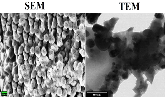
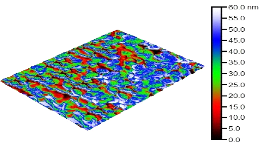
C-Oo.Nc was produced in Departement of Pharmacology Faculty of Veterinary Medicin Cairo University, Giza, Egypt. Characterization of the produced C-Oo.Nc using TEM, SEM, and AFM revealed that the obtained particles are spherical structures with maximum thickness less than 50 nm. Moreover, it was noticed that C-Oo.Nc particles do not tend to form agglomeration in certain areas. Zeta size and potential values demonstrated that the produced molecules are 63 nm sized with good stability in the aqueous media. The obtained particle size was within the range previously described for the suitable size of nanoparticles. These characteristics are identical to the findings of (Kamel et al., 2019) who characterized and described the successful nanoparticles characters.
In this study, anti-coccidial effect of the produced C-Oo.Nc was evaluated in Department of Parasitology, Faculty of Veterinary Medicine, Cairo University, Egypt. The current study evaluated the inhibitory effects of C-Oo.Nc on un-sporulated and sporulated oocysts of five Eimeria spp.; aiming to determine C-Oo.Nc LC50 and LC90 to be used in the second part of the study versus infected chicken in vivo. Efficacy of the product was evaluated by measuring its sporulation inhibitory % of unsporulated oocysts, while the sporulated oocysts exposed to the calculated LC50 & LC90 were evaluated after inoculation to groups of broiler chickens. In addition, DNA damage of the exposed and control oocysts was also evaluated using the comet assay.
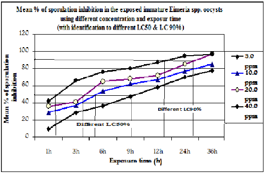
The current study proved that C-Oo.Nc inhibited the sporulation ability of the exposed immature Eimeria spp. oocysts. This increased with the increase in C-Oo.Nc concentration and exposure time. This was in agreement with (El-Banna et al., 2005; Ramadan et al., 1997; El-Khtam et al., 2014). The mean rate of inhibition was increased from 9.82 ±0.88 % after exposure of the oocysts to 5 ppm/1h to reach 77.50±0.75 % after 36 h exposure time. Also, the % of sporulation inhibition in oocysts exposed to 10ppm was increased from 28.66 ±0.97% to 85.24±1.25 % with an increasing the exposure time from 1h to 36h Exposure to 20 ppm/1h revealed 36.0 ± 1.5 % sporulation inhibition rate increased to 97,33±1.7 % after 36 h. The maximum inhibitory effects were recorded after exposure to 40 ppm/1h as it reached 42.0± 1.2% increased to 95 then to 97.66±0.95 % after 48h & 36 h respectively. Increasing the inhibitory rate of sporulation in exposed Eimeria spp. oocysts In vitro agreed with that previously described by Felici et al., (2021).
From the previous screening, the calculated LC50 was 5.0 ppm/9 h, 10 ppm/5 h, 20 ppm/4 h, or 40 ppm/90 minutes of exposure time. The LC90 was recorded after exposure to 20 ppm and 40 ppm only after 28 and 15 hours of exposure time, respectively. These results agreed with Felici et al. (2021). The recorded high efficacy of the tested C-Oo.Nc at the lower concentrations has come in agreement with Gogoi et al. (2019) who reported that nano-curcumin has a great anti-coccidial effect on the experimentally infected broiler chicks at low doses. Moreover, Tsai et al. (2011) reported that the nano form of curcumin is easily dissolvable in water and has a significantly higher penetration and distribution rate. In addition, Partovi et al. (2019) conducted that dietary supplementation with nanocurcumin at low doses is safe and effective on carcass characteristics and physical and chemical properties of broiler chicken breast meat infected with Eimeria acervulina, Eimeria maxima, and Eimeria tenella.
However, these results were contrary to that mentioned by El-Khtam et al. (2014), as they obtained 60% inhibition in sporulation of the exposed oocysts at a very high dose (0.8 to 10 g/L during 6-18 hours of exposure with no significant difference between the different concentrations and exposure time. This may be due to variations in the level of the solubility of the type of curcumin they evaluated. Moreover, Curcumin’s biological action is dependent on its bioavailability. Curcumin bioavailability was affected by the amount and concentration at which it is absorbed, occurs in the plasma, and reaches its target area (Paulraj et al., 2019).
The recorded efficacy of newly formed C-Oo.Nc on the level of this study gives this formulation special superiority related to induction of good target effect at low concentrations. On the contrary of this, nano-chemicals such as nano-silver materials proved potent effects at low doses than the other similar nano-plant extracts such as nano-curcumin, but the nano chemicals usually have a narrow safety margin than that of plant one (Elnabarawy et al., 2020; Madbouly et al., 2022).
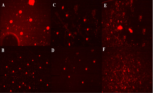
The damage in DNA of the exposed and control Eimeria spp oocysts was evaluated using the comet assay on immature oocysts exposed to the tested four concentrations of C-Oo.NC after 24 h exposure time, for sporulated oocysts exposed to LC90 (20 ppm/26 h & 40 ppm/15 h) as well as in control non-exposed oocysts. Comet assay revealed a direct relationship between the increase in the dose of C-Oo.NC and the degree of the overall DNA genotoxic damage which is represented by variations in the tail length (μm), % of DNA in the tail segment, and tail moment (μm). Investigating the migration of DNA fragments by agarose gel electrophoresis revealed no significant difference (p ≤ .05) in the mean values of destruction in DNA parameters in unsporulated oocysts exposed to 40 ppm and sporulated ones exposed to LC90 as well as that exposed to the control reference drugs. Moreover, significant very low destruction (2.30±0.31) in DNA and the other estimated parameters were recorded in the control non-exposed oocysts. The ability of nano-curcumin to induce DNA damage in the exposed agents was previously described by Kumar et al. (2015) and Attaullah et al. (2020) as they mentioned that changes in the tail length investigated by comet assay in comparison with the control non-exposed stages was reflected marked DNA damage and this damage was increased by the increase in the concentration of tested curcumin extract.
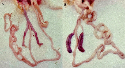
To investigate the effect of the C-Oo.Nc on the exposed sporulated oocysts. The sporulated oocysts exposed to the previously estimated LC50 & LC90 were inoculated to separate groups of 14th days old broiler chickens. The results revealed a marked decrease in the mean total number of oocysts shed from each bird during the whole period (5th to 10th dpi) (2200 ± 50 and 1580 ± 50 ) in the group inoculated by oocysts exposed to both calculated LC90 (20 ppm/28 h and 40 ppm/15 h) as well as a significant decrease (p ≤ 0.05) in the mean total number of shed oocysts in groups exposed to LC50 (11520 ± 120 to 10080 ± 100) in comparison with that shed from control birds (33680 ± 150) inoculated by un-exposed sporulated oocysts. This decrease in pathogenicity of the exposed sporulated oocysts was clearer at PM inspection performed at the end of the experiment (12th dpi) as very slight or un-apparent reddens without thickening in the wall of ceci. This was closely related to the normal un-infected control was recorded in the group inoculated by oocysts exposed to LC90. This was in contrast to the group inoculated by sporulated oocysts exposed to LC50. The direct relation between infectivity inhibition and the degree of the produced lesion score after exposure to different concentrations of the tested drugs was in agreement with that recorded in previous works of Ramadan et al. (1997) and El-Banna et al. (2005) on other anti-coccidial drugs. The ability of the used medical plants to inhibit the infectivity of the exposed oocysts was previously mentioned by several authors, for example, Tsai et al. (2011) and El-Khtam et al. (2014), as a clear sign of the success of treatment and control of the disease. It is worth mentioning that the marked severe effect of PM was observed in control birds in ceca due to increasing the % of E.tenella oocysts in the original inoculum (Ramadan et al. 1997; El-Banna et al. (2005).