
Salvadora persica is one of the commonly used plants in Islamic countries.The root extracts of S. persica has been tested for its anticoccidial activity against Eimeria paillata induced infection in mice jejunum. Three different doses, 300, 600 and 900 mg/kg were used after infection of mice with sporulated oocysts. A dose of 300 mg/kg reduced the number of oocyst output in mice faeces by about 56.8 % and also improved the body weight. In addition, the root extract decreased the number of parasitic stages and the number of goblet cells in the jejunal villi in the infected mice. Moreover, the expression of the goblet cell mucin gene, muc2 was upregulated after treatment with the root extract. Based on our results, it could be concluded that, S. persica root extracts acts as anticoccidial agent.
Research Article · Volume 49, Issue 1, 2 October 2016 · Open Access
In vivo Anticoccidial Activity of Salvadora persica Root Extracts
· ·
Affiliations & Notes ⌄
- 1Department of Zoology, College of Science, King Saud University, Saudi Arabia 2Department of Zoology and Entomology, Faculty of Science, Helwan University, Egypt Email: mohameddkhil@yahoo.com
Article Info ⌄
- Journal:
- Pakistan Journal of Zoology
- Published:
- 2 October 2016

From the article
- S. persica root extracts was significantly able to suppress the oocyst output by 56.8, 22.8 and 32.2 %, respectively (Table I).
- After treatment with 300 mg⁄kg S. persica root extracts, the loss of the original weight was reduced to -1.89 % (Fig. 1).
- Examination of Alcian blue stained sections showed that the infection induced a significant decrease in the number of goblet cells inside the jejuna villi (Table II).Treatment of mice with S. persica root extracts (Table II) could significantly increase the number of goblet cells compared to the infected group.
- S. persica root extract could significantly downregulate the MUC2 expression due to infection however, this mechanism of downregulation has not been studied before.
Selected from the article's own text.
Abstract
Introduction
Intestinal eimeriosis caused by the parasite, Eimeria spp. are considered to be one of the major threats affecting animal health and production . Such parasite has a great economic importance as it causes severe reduction in meat and milk production and death of domestic agriculture animals . There are many anticoccidial agents used against eimeriosis. However these agents have many side effects ; . Researchers efforts are now directed towards finding natural agents with reduced or with no side effects on the host infected with Eimeria. Extracts of pomegranate, neem, palm date, garlic and others were found to be good agents against E. papillata induced infection in mice jejunum .
In this study, we used Salvadora persica, commonly known as miswak, and used most extensively against microbes . The propagation and use of miswaks is due to the spread of Islamic culture in many countries .
It was reported that, S. persica, has antimicrobial activity against numerous oral pathogens . Recently, Abdel-Baki et al. (2016) investigated the antiparasitic activity of S. persica against against Echinococcus granulosus.
S. persica extracts contain important phytochemicals like vitamin C, salvadorine, salvadourea, alkaloids, trimethylamine, cyanogenic glycosides, tannins, saponins and salts mostly as chlorides .
Miswak was harvested from the root of S. persica and methanolic extract was prepared for studying its potential against E. papillata infection in mice.
Materials and Methods
Collection and extraction of Salvadora persica roots
Fresh roots of Salvadora persica were harvested from Jizan region, Kingdom of Saudi Arabia. A botanist, Department of Botany, College of Science, King Saud University, Saudi Arabia authenticated the samples. S. persica root methanolic extracts were prepared according to Amer et al. (2013). Briefly, the roots were dried, grinded by an electronic blender into a powder that extracted by 70% methanol. The used collected residue was dissolved in water for the inoculation in mice.
Infection and experimental design
Adult male C57BL/6 mice were obtained from the Department of Comparative Medicine, King Faisal Specialist Hospital and Research Center, Riyadh, Saudi Arabia. The mice were bred under specified pathogen-free conditions and fed a standard diet and water ad libitum. The experiments were approved by the state authorities and followed Saudi Arabian rules on animal protection.
To prepare Eimeria papillata oocysts for infection, the stored sporulated E. papillata were inoculated into mice using an epigastric tube. Every mouse received 100 µl saline contained 1000 sporulated oocysts.
Animals were divided into 5 groups of 5 mice per group. The first group was the non-infected control group. Mice of this group were daily inoculated with 100 µl saline for 5 days. The second, third, fourth and fifth groups group were infected with 1000 sporulated E. papillata oocysts. One hour later, the third, fourth and fifth groups were daily treated with 300, 600 and 900 mg/kg, respectively.
The faeces were daily collected from each mouse, weighed and prepared for counting using McMaster chamber . To investigate the change in weight, all mice were weighed at the beginning and at the end of the experiment.
Samples collection and counting of parasitic stages of E. papillata
Jejuna of mice were collected after 5 days postinfection with E. papillata. Each jejunum was washed and then fixed in 10% formalin. Tissue sections were prepared for histological examination . To calculate the number of parasitic stages, the sections were stained with hematoxylin and eosin.
Goblet cell response
To calculate the number of goblet cells in non-infected, infected and infected-treated mice, jejunum tissue sections were stained with Alcian blue and the number of goblet cells was scored in 10 well orinted jejuna villi .
Goblet cell mucin gene
To determine the expression of MUC2-mRNA, we used a quantitative real-time PCR. In brief, the total RNA was isolated from mice jejuna using Trizol (Invitrogen). RNA samples were treated with DNase (Applied Biosystems, Darmstadt, Germany) and then converted into cDNA using the reverse transcription kit (Qiagen, Hilden, Germany). Quantitative real-time PCR (qRT-PCR) was performed using the ABI Prism 7500HT sequence detection system (Applied Biosystems, Darmstadt, Germany) with SYBR green PCR master mix from Qiagen (Hilden, Germany). MUC2 primer was obtained from Qiagen . mRNA levels were normalized to 18S rRNA. The fold induction of mRNA expression upon infection with E. papillata was determined using the 2−ΔΔCT method .
Statistical analysis
Statistical analysis was carried out using one-way ANOVA, and Duncan’s test was used for comparison between groups.
Results
On day 5 p.i. the expelled oocysts of E. papillata were at its maximum level being about 6242.7 ± 731.5 oocysts / g faeces in infected group. S. persica root extracts was significantly able to suppress the oocyst output by 56.8, 22.8 and 32.2 %, respectively (Table I).
Table 1.- S. persica induced reduction in oocyst output.
| Group | Oocyst output/g faeces | Suppression of oocyst shedding (%) |
| Infected | 6242.7 ± 731.5 | 0% |
| Infected (300 mg/kg) | 2696.7 ± 441* | 56.8% |
| Infected (600 mg/kg) | 4820.7 ± 2650 | 22.8% |
| Infected (900 mg/kg) | 4230.6 ± 2531 | 32.2% |
Values are means ± SD.
*Significance at P≤0.001.
It is clear now that the dose 300 mg/kg is the best dose for suppression of oocyst output. So, we used only 300 mg/kg root extracts to investigate the histological changes as well as the goblet cell response to infection and/or treatment.
Control non-infected mice grew normally resulting in an average weight gain of about 5.8 %. Whereas experimentally infected mice with E. papillata had lost their original weight by about 2.8 %. After treatment with 300 mg⁄kg S. persica root extracts, the loss of the original weight was reduced to -1.89 % (Fig. 1).
Table II shows the change in the number of parasitic stages on day 5 p.i. due to treatment of mice with 300 mg/kg S. persica root extracts. The developmental stages of E. papillata clearly appeared inside the intestinal epithelial of the villi and we found no parasites inside goblet cells.
Examination of Alcian blue stained sections showed that the infection induced a significant decrease in the number of goblet cells inside the jejuna villi (Table II).Treatment of mice with S. persica root extracts (Table II) could significantly increase the number of goblet cells compared to the infected group.
Table II.- S. persica induced reduction in the number of parasitic stages, goblet cells and weight change of mice infected with Eimeria papillata.
| Group | Uninfected | Infected | Infected with extract (300 mg/kg) |
| Number of parasitic stages | - | 116.75±3.5 | 75.25 ± 13.8 |
| Number of goblet cells/10 VCU | 73.25±8.5 | 35.75±5.3 | 53± 5.6* |
| Weight change (%) | 5.8±1.10 | −2.8±1.2 | - 1.8±3.08* |
Values are means ± SD.
*Significance at P≤0.001.
The expression of the jejunal goblet cell gene, MUC2 was downregulated due to infection of mice with E. papillata (Fig. 2). S. persica root extracts could significantly upregulate MUC2 in the infected jejuna (Fig. 2).
Discussion
Recently, several studies used plant extracts as antiparasitic agents ; ; . The results of the current study indicated that, S. persica root extracts exhibit good anticoccidial properties. This effect may be due to the content of the extract. Khan et al. (2010) found that the root extracts contain flavonoids, alkaloids, carbohydrates, glycosides, saponins, steroids and tannins.
S. persica was able to improve the weight loss due to infection with E. papillata. This weight loss was previously studied by others (Anwar et al.,2008; ; and explained this loss due to decrease in water and food intake (Anwar et al., 2008). Also, Metwaly et al. (2013) related the weight loss due to the consumption of the parasitic stages to the carbohydrate content inside the intestinal villi and also led to structural alterations of the villi. The most structural change is the disruption of the host epithelium due to the discharge of the developed oocysts to the intestinal lumen.
Through the examination of the infected jejunum tissue sections, we observed that, different stages of E. papillata develop in the crypt region. Also, Cheng (1974) reported that, crypt region contains the stem cells producing goblet cells. These cells were invaded by the parasite leading to the decrease in the number of produced goblet cells in the jejunal villi. These observations were also documented by Dkhil (2013) during the investigation of the anticoccidial action of neem extract and its ability to increase the reduced goblet cell number due to E. papillata infection.
Dkhil et al. (2013) reported that, MUC2 gene was downregulated in the mice jejuna due to the infection with E. papillata. This gene is responsible for the immune response against pathogen-induced injury. S. persica root extract could significantly downregulate the MUC2 expression due to infection however, this mechanism of downregulation has not been studied before.
The high potential effect of S. persica is due to the antioxidant and anti-inflammatory activities of the plant extract constituents. The dose 300 mg/kg was also proved to have a significant anti-inflammatory activity on rat induced paw oedema (Ahmed et al., 2011) but until now there was no anti-coccidial study about this and our study was the first one dealing with the anti-eimerial properties of S. persica extract.
Conclusion
Based on these findings, we conclude that S. persica root extract exhibits antieimerial activities and could be used with the normal food of animals as an additive.
Acknowledgment
This research project was supported by a grant from the “Research Center of the Center for Female Scientific and Medical Colleges”, Deanship of Scientific Research, King Saud University.
Conflict of interest statement
We declare that we have no conflict of interest.
Figures
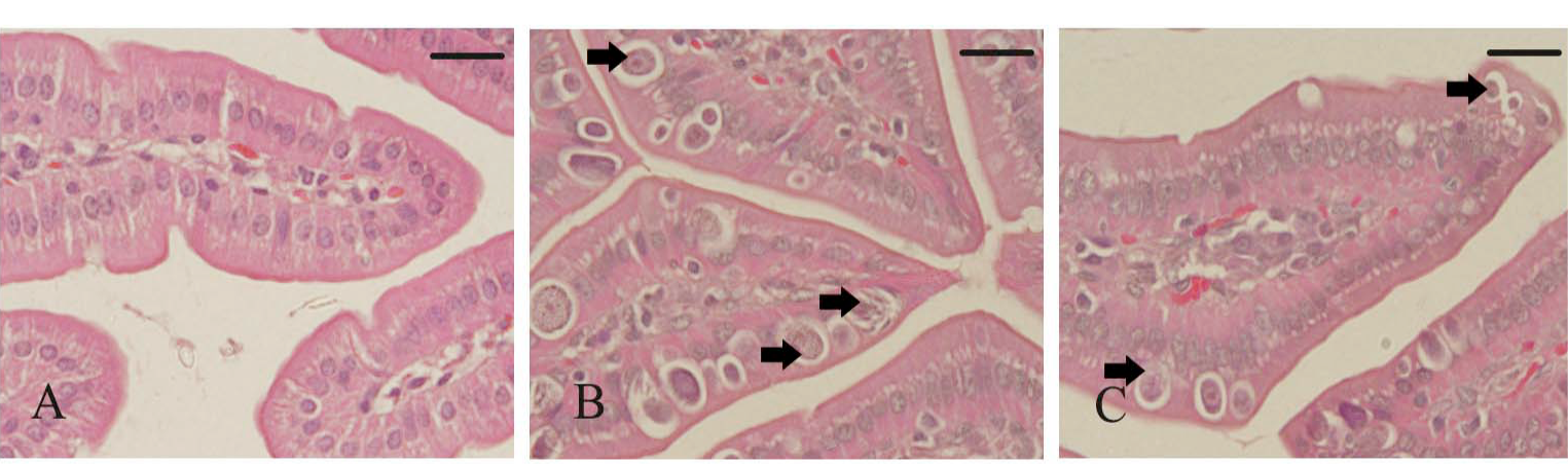
S. persica reduced change in E. papillata parasitic stages. A, uninfected control jejunum; B, Iinfected jejunum containing developmental stages (arrows); C, Infected treated mouse jejunum (300 mg/Kg) with reduced numbers of developmental stages. Sections were stained with hematoxylin and eosin. Bar=20 um.
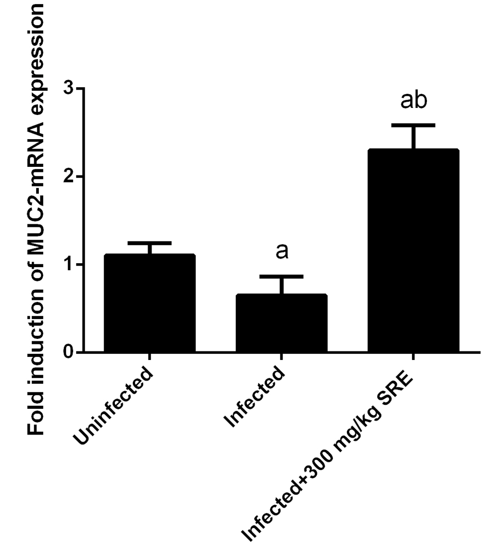
MUC2 gene expression in the jejunum of mice. Expression was normalized to 18S rRNA signals, and relative expression is given as fold increase compared to the non-infected control mice. Values are means±SD. *Significant change at P < 0.01 with respect to infected mice.
References
- Abdel-Baki, A.A., Almalki, E., Mansour, L. and Al-Quarishy, S., 2016. In vitro scolicidal effects of Salvadora persica root extract against protoscolices of Echinococcus granulosus. Korean J. Parasitol., 54:61-66. http://dx.doi.org/10.3347/kjp.2016.54.1.61[CrossRef]
- Adam, H. and Caihak, G., 1964. Grosses zoologisches parktikum tell. Arbeitsmethoden der makroskopischen und mikroskopischen anatomie mit Abbildungen. Gustav Fischer Verlag, Stuttgart.
- Ahmad, M., Imran, H., Yaqeen, Z., Rehman, Z., Rahman, A., Fatima, N. and Sohail, T., 2011. Pharma-cological profile of Salvadora persica. Pak. J. Pharm. Sci., 24:323-30.
- Alali, F., Hudaib, M., Aburjai, T., Khairallah, K., and Al-Hadidi, N., 2005. GC-MS analysis and antimicrobial activity of the essential oil from the stem of the Jordanian toothbrush tree Salvadora persica. Inform. Hlthcare., 42:577-580.
- Allen, A., Hutton, D.A., Leonard, A.J., Pearson, J.P. and Sellers, L.A., 1986. The role of mucus in the protection of the gastroduodenal mucosa. Scand. J. Gastroenterol., 125:71–78. http://dx.doi.org/10.3109/00365528609093820[CrossRef]
- Alzahrani, F., Al-Shaebi, E.M., Dkhil, M.A. and Al-Quraishy, S., 2016. In vivo anti-Eimeria and in vitro anthelmintic activity of Ziziphus spinachristi leaves extract. Pakistan J. Zool., 48:409-413.
- Amer, S.O., Dkhil, M.A. and Al-Quraishy. S. 2013. Antischistosomal and hepatoprotective activity of Morus alba leaves extract. Pakistan J. Zool., 45:387-393.
- Anwar, M.I., Akhtar, M., Hussain, I., Haq, A.U., Muhammad, F., Abdul Hafeez, M., Mahmood, M.S.
- and Bashir, S., 2008. Field evaluation of Eimeria
- tenella (local isolates) gametocytes vaccine and its comparative efficacy with imported live vaccine, LivaCox®. Parasitol. Res., 104:135–143. http://dx.doi.org/10.1007/s00436-008-1171-5[CrossRef]
- Bos, G., 1993. The miswak, an aspect of dental care in Islam. Med. Hist., 37:68–79. http://dx.doi.org/10.1017/S0025727300057690[CrossRef]
- Cheng, H., 1974. Origin, differentiation and renewal of the four main epithelial cell types in the mouse small intestine. II. Mucous cells. Am. J. Anat., 141:481–501. http://dx.doi.org/10.1002/aja.1001410405[CrossRef]
- Dkhil, M.A., 2013. Anti-coccidial, anthelmintic and antioxidant activities of pomegranate (Punica granatum) peel extract. Parasitol. Res., 112:2639-2646. http://dx.doi.org/10.1007/s00436-013-3430-3[CrossRef]
- Dkhil, M. A., Delic, D, and Al-Quraishy S., 2013. Goblet cells and mucin related gene expression in mice infected with Eimeria papillata. Scient. World J., 2013:1-6. http://dx.doi.org/10.1155/2013/439865[CrossRef]
- Dkhil, M.A., Abdel-Baki, A.S., Wunderlich, F., Sies, H. and Al-Quraishy, S., 2011. Anticoccidial and antiinflammatory activity of garlic in murine Eimeria papillata infections. Vet. Parasitol., 175:66–72. http://dx.doi.org/10.1016/j.vetpar.2010.09.009[CrossRef]
- Dkhil, M.A., Abdel-Baki, A.S., Wunderlich, F., Sies, H. and Al-Quraishy, S., 2014. Dietary selenium affects intestinal development of Eimeria papillata in mice. Parasitol. Res., 113:267–274. http://dx.doi.org/10.1007/s00436-013-3653-3[CrossRef]
- Elvin-Lewis, M., 1982. The therapeutic potential of plants used in dental folk medicine. Odontostomatol. Trop. 5:107–117.
- Ezmirly, S.T. and Cheng. J.C., 1979. Wilson SR. Saudi Arabian medicinal plant: Salvadora persica. Planta Med., 35:191–192. http://dx.doi.org/10.1055/s-0028-1097205[CrossRef]
- Khan, W., Mujum, A., Shaikh, T., Katekar, S.M. Tambe, R. and Rub, R.A., 2010. Pharmacognostic and preliminary phytochemical investigation of Salvadora persica Linn (Salvadoraceae). Res. J. Pharmacog. Phytochem., 2:319-23.
- Khatak, M., Khatak, S., Siddqui, A.A., Vasudeva, N., Aggarwal, A. and Aggarwal, P., 2010. Salvadora persica. Pharmacogn. Rev., 4:209–214. http://dx.doi.org/10.4103/0973-7847.70920[CrossRef]
- Klimpel, S., Abdel-Ghaffar, F., Al Rasheid, K.A.S., Aksu, G., Fischer, K., Strassen, B. and Mehlhorn, H., 2011. The effects of different plant extracts on nematodes. Parasitol. Res., 108:1047–1054. http://dx.doi.org/10.1007/s00436-010-2168-4[CrossRef]
- Livak, K.J, and Schmittgen, T.D., 2001. Analysis of relative gene expression data using real-time quantitative PCR and the 2 – ∆∆ C T method. Methods, 25: 402-408. http://dx.doi.org/10.1006/meth.2001.1262[CrossRef]
- Mehlhorn, H. (ed), 2014. Encyclopedic reference of parasitology, 4th edn. Springer, Berlin.
- Metwaly, M.S., Dkhil, M.A., Al-Quraishy, S, and Al Omar, S.Y., 2015. Protective effects of palm pollen aqueous extract against Eimeria papillata induced intestinal damage in mice. Pakistan J. Zool., 47:971-979.
- Metwaly, S., Dkhil, M.A., Gewik, M.M., AL-Ghamdy, A. and Al-Quraishy, S., 2013. Induced metabolic disturbance and growth depression in rabbits infected with Eimeria coecicola. Parasitol. Res., 112:3109-3114. http://dx.doi.org/10.1007/s00436-013-3485-1[CrossRef]
- Mohamed, S.A., and Khan, J.A., 2013. Antioxidant capacity of chewing stick miswak Salvadora persica. BMC Complem. Altern. Med., 13:40. http://dx.doi.org/10.1186/1472-6882-13-40[CrossRef]
- Poureslami, H., Makarem A. and Mojab F., 2007. Paraclinical effects of miswak extract on dental plaque. Dent. Res. J., 4:106–110.
- Schito, M.L. and Barta, J.R., 1997. Nonspecific immune responses and mecha- nisms of resistance to Eimeria papillata infections in mice. Infect. Immun., 65:3165–3170.
- Wunderlich, F., Al-Quraishy, S., Steinbrenner, H., Sies, H. and Dkhil, M.A., 2014. Towards identifying novel anti-Eimeria agents: trace elements, vitamins, and plant-based natural products. Parasitol. Res. 113:3547– 3556. http://dx.doi.org/10.1007/s00436-014-4101-8[CrossRef]
Services
Article usage statistics
Loading…