Research Article · Volume 12, Issue 5, 20 March 2024 · Open Access
Study The Pathological, Immunological Effects After Repeated Exposure to Tetrodotoxin in Liver of Albino Male Rats
·
Affiliations & Notes ⌄
1Department of Pathology and Poultry Diseases, College of Veterinary Medicine, Baghdad University, Baghdad, Iraq. *Correspondence | Taghreed Jabbar Humadai, Department of Pathology and Poultry Diseases, College of Veterinary Medicine, Baghdad University, Baghdad, Iraq
The current study aimed to examine the immunological and histopathological effects after intraperitoneal injection of tetrodotoxin (TTX) on liver of male albino rats. At the age of 8–9 weeks and weighing 190–200 gm, 60 male albino rats were separated into 3 groups: First group of 20 rats received distilled water and regular pellets. Second group of 20 rats received weekly injections of 0.5 µg/kg b.w. by I/P route, which was equivalent to 1/20 LD50 of tetrodotoxin. Third group of 20 rats received weekly injections of 1 µg/kg b.w. by I/P route, which was equivalent to 1/10 LD50 of tetrodotoxin. All animals were sacrificed at the conclusion of the experiment (90 days) in order to conduct biochemical, histopathological, and Immunohistochemical analyses. The oxidative stress result showed elevation in Malon Di-Aldehyde concentration in liver tissue treated with tetrodotoxin. The pathological lesion in liver of animals exposed to toxic dose of tetrodotoxin was characterized by dilation and congestion of portal vein, Focal aggregation of mononuclear cells formation granulomatous like lesion, Various form of nuclear necrosis with number apoptotic hepatocyte, nuclear pyknosis with evidence of councilman body in liver tissue, The Immunohistochemistry study revealed an increase in the scoring of IL1 expression in liver that showed score (3) compared to control group, TNF alpha expression showed score (1) in liver tissue when compared to untreated control group . According to the findings of the current study, tetrodotoxin has adverse toxic effects on liver tissue of male albino rats.
The most well-known kind of fish intoxication is puffer fish intoxication, which has been initially identified by Dr. Yoshizumi Tahara from globefish ovaries in 1909 . The powerful neurotoxin tetrodotoxin (TTX) is present in the muscle, skin, liver and gonads of many species of marine puffer fish . One naturally occurring toxin that has caused human intoxications and deaths is called tetrodotoxin (TTX) . TTX was thought to be restricted to South East Asia, but more recent studies have revealed that it has spread to other parts of the world such as Australia, Bangladesh, New Zealand, Spain, Taiwan and United States of America (California, Hawaii) . However, puffer fish have long been known to be toxic to people. Many marine organisms contain TTX, a highly potent neurotoxin ; Luo et al., 2012). In a few terrestrial ones as well . The fact that TTX has no known counteragent and is more than a thousand times more toxic to humans than cyanide is frequently using to highlight how harmful it is ; . It is a blocker of channels containing sodium. The toxin attaches itself to the sodium channels in the victim’s excitable tissues, such as nerves and muscle; this effectively immobilizes the tissues by blocking the flow of sodium ions through the channels . The onset and intensity of TTX poisoning symptoms in humans are dose-dependent . Ataxia and muscle weakness may develop from the initial symptoms, which include tingling (paresthesia) of the lips and tongue, headache, and vomiting, either simultaneously or sequentially. In extreme situations, heart failure and/or respiratory failure may result in death .
Vinagre et al. (2003) and Pinho et al. (2005), Al-Sabaawy and Al-Kaisie (2021) found that some toxins produce oxidative stress and disrupt crustacean osmotic and ionic processes such as oral administration of microcystin to the estuarine crab Chasmagnathus granulatus (Decapoda, Brachyura) that causes elevate antioxidant enzyme level (superoxide dismutase, catalase-(CAT), Glutathione-S-transferase- (GST)) also this toxin (microcystin) increase the oxygen consumption. Saoudi et al. (2011) investigated that tetrodotoxin produce from lagocephalus lagocephalus are well documented to have a range of effects on organism cells. Saoudi et al. (2011) examined Lagocephalus lagocephalus meat and liver extracts’ hepato- and nephrotoxic effects on rats. Using raw or cooked Lagocephsalus lagocephalus extracts can lower serum level of transaminases (AST, ALT), ALP, LDH, and γ-GT enzyme and increase these enzymes in liver . Hepatocyte damage may be connected to these biochemical alterations in the liver profile . Long-term toxin delivery causes the mitochondrial and monooxygenase systems to malfunction, which can lead to changes in these systems including an increase in reactive oxygen species (ROS) production . Oxidative stress and cell damage may result from increased ROS generation that exceed antioxidant defense and repair capabilities ; . Reactions that result in toxicity can be caused by or result from lipid peroxidation . Recent research has demonstrated that certain marine toxins cause lipid peroxides, ROS, DNA damage, and antioxidant enzyme activity in hepatocytes through the action of multispecific bile acid transporters .
According to Elshaer (2016), Male rats injected with TTX derived from the muscles of the porcupine fish Diodon hystrix exhibited moderate histopathological effects on the liver tissue after two and four hours. This was indicated by a mild infiltrate of polymorphic leukocytes, congestion in the central vein, pyknotic nuclei in some hepatocytes, and likely necrosis or vacuolation. By injection, the liver’s trabecular structure was obscured, with larger cells, more compact nuclear chromatin, and less obvious, somewhat smaller nucleoli, when TTX was isolated from Diodon hystrix ovaries was used. Histocytes underwent necrotization; pyknotic nuclei containing condensed chromatin and very acidophilic cytoplasm were seen. Therefore, the current study concentrated on the immunological effect and histopathological changes found in the liver of a male rat that had received an intraperitoneal injection of tetrodotoxin.
Materials and Methods
Chemicals
Tetrodotoxin (Purity: >=98%, Formula: C11H17N3O8, Molecular Weight: 319.27) obtained from Wuhan ChemFaces Biochemical CO., Ltd. Company, china, Store in a well closed container. Protected from air and light, refrigerate or freeze (2-8°C). Distilled water was used to dissolve and dilute tetrodotoxin to the needed amounts.
Experimental animals
Sixty mature white male albino rats weighing between 190 and 200 grams and (8-9) weeks age were used in this investigation. They were procured for adaptation from the veterinary and Medical College’s animal facility at Baghdad University. The animals were housed in plastic cages within a room that was equipped with air conditioning and maintained at a temperature of 25±2 degrees Celsius at all times. The bedding in the plastic cages was made of hard-wood chips, and it was changed on a regular basis to maintain a clean environment. Ad libitum water and food pellets were provided to the rats. In compliance with ethical norms, the College of Veterinary Medicine at the University of Baghdad’s local committee for animal care and use awarded its ethical permission (Number 2284/P. G at 18/10/2023).
Experimental design
Sixty mature white male albino rats used in this experiment were separated into three groups of twenty each, as follows: 1st group: control group contains (20) rats were given distilled water and normal rat pellet for 90 days, 2nd group: contains (20) rats were given (0.5 µg/kg b.w.) weekly I/P injection by Disposable sterile syringes for 90 days,3rd group: conations (20) rats were given (1 µg/kg b.w. ) weekly I/P injection by Disposable sterile syringes for 90 days. After 90 days, animals from each group were sacrificed for study the oxidative stress induction, histopathological investigations and immunohistochemistry study.
Parameters of experiment
Biochemical analysis (determination of tissue malon di aldehyde assay (MDA) by elisa technique
ELISA Kit purchased from China’s ELK Biotechnology used to measure the amount of malon Di-aldehyde in male rats liver tissue as pictograms per milliliter (pg/mL), and this test was carried out in accordance with the manufacturer’s procedure.
Pathological examination
Animals were chloroform-anesthetized and then scarified at the conclusion of the trial, which lasted 12 weeks. The specimen (liver) was collected and preserved for fixation in a 10% formaldehyde solution. The histopathological protocol was used as specified by .
Immunohistochemistry assays
The immunohistochemistry was performed using Dako EnVision detection immunohistochemistry kit (Envision FLEX, Dako, K8000, Denmark), Anti-IL-1A primary antibody (E-AB-40407, Elabscience, China) and anti-TNF alph primary antibody (E-AB-40015, Elabscience, China) were used for detection IL-1A and TNF alpha in liver tissue in current study and this assay was carried out in accordance with the manufacturer’s procedure.
Scoring
In each region, a minimum of 10 randomly selected high-power fields were utilized to determine the quantity of positive cells present in the tissue samples dyed with dye. Additionally, the quantity of positive cells in the fields was determined. The immune-stained sections were scrutinized by two observers using a light microscope equipped with a 400X objective lens. To assess the number of immune cells in the liver specimens, a count of one thousand cells was performed on each section. The scores listed below were utilized to calculate the quantitative evaluation of compound expression, according to Zenclussen et al. (2003): zero (one to ten), one (fifteen to thirty), two (thirty-one to fifty), three (fifty-one to one hundred) and four (one hundred twenty to two hundred) and more positive cells).
Statistical analysis
Statistical analysis was done using SAS 9.1 (SAS, 2010). A one-way analysis of variance (ANOVA) and a post hoc test using the least significant differences (LSD) were used to determine the significance of mean differences; statistical significance is reached when P < 0.05.
RESULTS AND DISCUSSION
Tissue malon di-aldehyde (MDA) assay
The results of Malon Di-Aldehyde in the liver tissue treated with tetrodotoxin for 90 day showed significant increase (P<0.05) in their concentration in liver (1116.88±0.21) at dose 0.5 µg/kg b.w., while at dose 1 µg/kg b.w. reached to (1150.27±0.22) compared with control group (1075.51±0.23) the result was shown in Table 1.
Table 1: The effect of Tetrodotoxin on (Malon Di-Aldehyde assay) MDA (pg/ ml) in liver tissue of rat for chronic toxicity.
Organ/group
Liver
Control
1075.51±0.23c
0.5 µg/kg b.w.
1116.88±0.21b
1 µg/kg b.w.
1150.27±0.22a
LSD
0.66
*Means with a different letter in the same column are significantly different (P<0.05).
Histopathological study
Second group (0.2 µg/kg b.w. (1/20 of LD50))
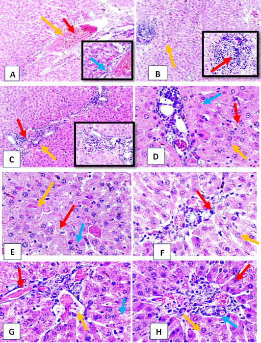
The main pathological changes in liver tissue were dilation and congestion of central vein, adjacent hepatic sinusoid and portal vein, Focal aggregation of mononuclear cells formation granulomatous like lesion with slight presence of edematous substance in adjacent hepatic parenchyma. Various form of nuclear necrosis with number apoptotic hepatocyte, Moderate nuclear pyknosis of hepatocyte with slight sinusoid congestion with evidence of councilman body Figure 2.
Third group (1/10 of LD5O) (1 µg/kg b.w)
The histopathological section of rat liver showed perivascular and periductal mononuclear cells infiltration in liver parenchyma mainly in portal regain with evidence of nuclear pykinosis of adjacent cells, moderate necrotic finding of surrounding hepatocyte Also hepatocyte vessels dilation and congestion with new neutrophil in lumen and binucleted hepatocyte, Moderate perivascular mononuclear cells with scattered apoptotic cells Figure 1.
Immunohistochemistry study
IL1 expression
The results of IL1 expression in the liver tissue treated with tetrodotoxin for 90 day showed significant elevation (P<0.05) in score of liver (42.67±0.33) at dose 0.5 µg/kg b.w, while at dose 1 µg/kg b.w reached to (70.67±0.67) compared with control group (0.00±0.00), the result was shown in Table 2.
Table 2: The effect of tetrodotoxin on scoring for IL1 expression in liver tissue of rat for chronic toxicity.
Organ/ group
Liver
Score
1st group (Control)
0.00±0.00c
0
2nd group (0.5 µg/kg b.w) (1/20)
42.67±0.33b
2
3rd group (1µg/kg b.w) (1/10)
70.67±0.67a
3
LSD
1.48
*Means with a different letter in the same column are significantly different (P<0.05).
Imunohistochamical sections for IL1 detection
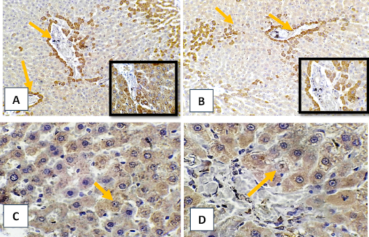
The result of Immunohistochemistry section of liver tissue in treated group showed moderate immune positive cells for IL1 expression mainly around portal regain and central vein and in liver parenchyma mostly in 3rd group Figure 2 when compared to control group which show negative result.
TNF alpha expression
The results of TNF alpha expression inliver tissue treated with tetrodotoxin for 90 day showed significant elevation (P< 0.05) in score of liver (15.67±0.33) at dose 0.5 µg/kg b.w, while at dose 1 µg/kg b.w reached to (21.67±0.33) compared with control group (0.00±0.00), the result was shown in Table 3.
Table 3: The effect of tetrodotoxin on scoring for TNF alpha expression in liver tissue of rat for chronic toxicity.
Organ/ group
Liver
Score
1st group (Control)
0.00±0.00c
0
2nd group (0.5 µg/kg b.w) 1/20)
15.67±0.33b
1
3rd group (1 µg/kg b.w) (1/10)
21.67±0.33a
1
LSD
0.94
*Means with a different letter in the same column are significantly different (P<0.05).
Imunohistochamical sections for tnf alpha detection
The result of immunohistochemistry section of liver tissue showed scattered immune positive cells for TNFα antibody in hepatocyte Figure 2. compared to control untreated group that showed negative result.
Lipid peroxidation of polyunsaturated fatty acids produces malon Di-aldehyde. The amount of malon Di-aldehyde in tissues can be used to evaluate the degree of lipid peroxidation ; . Malon Di-aldehyde is produced when polyunsaturated lipids are broken down by reactive oxygen species. The synthesis of this molecule, a reactive aldehyde, is employed as a biomarker to estimate an organism’s level of oxidative stress because it is one of the several reactive electrophile species that induce toxic stress in cells ; . The finding of the chronic toxicity exposure of malon Di-aldehyde in animal tissues were not consistent, with liver tissue exhibiting significantly higher concentrations (P < 0.05) throughout along experimentation period and for both doses. This might be because tetrodotoxin incite the production of lipid peroxides, ROS, DNA damage, and antioxidant enzyme activity in hepatocytes through the action of multispecific bile acid transporters . Moreover, as indicated by the increased levels of thiobarbituric acid reactive substances (TBARS), this raises the body’s oxidative stress. This is also associated with decreased levels of the scavenging enzymes in the liver, kidney, and spleen, including catalase, glutathione peroxidase activity (GPx), and superoxide dismutase (SOD) ; Neama and Yousif, 2022). The present investigation looks at whether the liver of the treated group is more susceptible to oxidative damage. The liver is a highly functioning organ that is especially sensitive to toxins, which helps to explain this discovery. Because of their important involvement in the biotransformation of xenobiotic, high cytochrome P450 concentration, and metabolic activity, hepatocytes are particularly susceptible to oxidative stress in vivo, the multiple liver diseases that have been connected to oxidative stress serve as evidence for this . This result was in line with that of , who showed that Tunisian pufferfish meat (muscle and skin) was hazardous and resulted in oxidative stress and hepatotoxicity.
The microscopic finding of chronic toxicity showed severe pathological changes in many organs and the severity of the lesions was dose dependent. Numerous reports have confirmed that the major target organs are the liver, kidney and central nervous system exposure to tetrodotoxin, blocking of sodium channels by tetrodotoxin causes gastrointestinal, neurologic and cardiac failure and in many cases respiratory paralysis is the primary cause of death . The kidney and liver are often threatened by toxic effects of xenobiotic because of its ability to accumulate these substances by active transport ; . the present study, there are many lesions reported in liver varying from congestion of central vein and portal area with dilation of sinusoid which causes compression on adjacent hepatocyte, also necrosis with mononuclear cells aggregation and pyknosis nuclei with evidence of binuclated hepatocyte was seen in many sections which indicated that some toxic metabolites may be transported from intestine to liver, resulting in these changes. The presence of distinct necrosis suggested that the toxic metabolites could cause oxidative stress in hepatocytes, as evidenced by changes in the mitochondria and involvement of the monooxygenase system; these changes could be followed by an increase in the production of reactive oxygen species (ROS), which would lead to cell death ; . This finding was agree with Elshaer (2016) who investigated that liver of rat treated by tetrodotoxin extracted from gonads and muscles of porcupine fish species causes congestion of central vein, dilation of sinusoid, necrosis with mononuclear cells aggregation and pyknotic nuclei. Councilman body is the final common pathological pathway of liver damage arising from a wide variety of liver diseases. Councilman body also known as a Councilman hyaline body or apoptotic body, is an eosinophilic globule of apoptotic hepatocyte cells fragments. Ultimately, the fragments are taken up by macrophages or adjacent parenchymal cells that it occurs due to liver injury or inflammation in most form of liver disease .
The current results of Immunohistochemical study showed increased in IL1 expression in the different rat organs that treated chronically with tetrodotoxin, the score of liver (3) specially at 3rd group. These results are due to the tetrodotoxin toxicity that causes injury in these organs so many proinflammatory cytokines will be released to the circulation like Interleukin 1. IL1 is a potent proinflammatory cytokine capable of triggering multiple physiological processes such as activation of lymphocytes, induction of acute-phase hepatic proteins, infiltration of leukocytes at sites of infection, fever and anorexia , 2009; , clearly indicating that this cytokine is critical for innate immune response . IL1 mainly produced by macrophages during defensive reactions . The term IL-1 refers to two cytokines, IL-1α and IL-1α . IL-1α released from necrotic cells is considered a major inducer of sterile inflammation in several ischemic conditions, by inducing neutrophil and macrophage recruitment and IL-1 β expression and maturation ; . Furthermore, the liver’s production of IL-1 β, together with pro-inflammatory IL-6 and TNF alpha, has a role in activating native immune cells and attracting additional leucocytes to the injured liver, ultimately resulting in chronic inflammation .
The liver (1) score in the current study’s immunohistochemistry results. This is because the toxicity of the tetrodotoxin causes injury and inflammation in the treated organs, leading to the release of various pro-inflammatory cytokines into the bloodstream, including tumor necrosis factor alpha (TNF alpha). TNF alpha is a cytokine that affects different kinds of cells in pleiotropic ways. It is known to play a role in the etiology of some inflammatory and autoimmune disorders and has been identified as a key regulator of inflammatory responses ; . It is primarily produced by T lymphocytes, natural killer cells, activated macrophages (including alveolar macrophages, microglia, Langerhans cells, Kupffer cells, and astrocytes) and T lymphocytes . It is functionally known to initiate a cascade of other inflammatory chemicals, such as chemokines and additional cytokines. On the other hand, excessive or improper TNF alpha signaling activation is linked to chronic inflammation and may eventually cause the emergence of clinical consequences including autoimmune disorders . TNF-α is a crucial cytokine that can cause inflammation and apoptosis, and it is essential in the disturbance of macrovascular and microvascular circulation both in vivo and in vitro ; and is a significant cytokine that has the ability to cause inflammation and apoptosis . Increased synthesis of TNF-α occurs when ROS are present, and TNF-α signaling exacerbates oxidative stress . According to Zelová and Hošek (2013), there is a correlation between altered coagulation propensity and TNF-α overexpression. TNF-α, in brief, contributes to leukocyte adherence to the epithelium via expressing adhesion molecules causing vasodilatation and edema development. Moreover, it controls blood coagulation, increases oxidative stress at inflammatory sites, and indirectly raises body temperature . TNF alpha contributes to liver inflammation in the liver, and persistent liver inflammation causes liver fibrosis. According to Yang and Seki (2015), TNF alpha induces apoptosis, proliferation, and inflammation.
CONCLUSION AND RECOMMENDATION
Based on the experimental findings from the current investigation, we demonstrated that tetrodotoxin has adverse toxic effect on liver tissue of male albino rats. Additional efforts are required to raise awareness that eating fish species that contain tetrodotoxin should be avoided year-round dufe to the potentially fatal effects of TTX. Guidelines for handling these intoxications appropriately.
ACKNOWLEDGEMENTS
The authors are thankful to the head College of Veterinary Medicine, University of Baghdad for their assistance during work.
NOVELTY STATEMENT
The novelty of the study is focus on the histopathological effects of tetrodotoxin as well as the immunohistochemistry investigation in reproductive system of male rats.
AUTHORS CONTRIBUTION
These authors each contributed equally.
Conflict of interest
The authors have declared no conflict of interest.
Figures
Figure 1:.
Histopathological section of liver tissue from tetrodotoxin treated group (A, B, C, D, E) (0.5 µg/kg b.w.) of male albino rats (A) obvious dilation and congestion of portal vein (red arrow) with mild cellular infiltration (yellow arrow) with evidence of scattered apoptotic hepatic cells (blue arrow). (B) moderate vascular congestion and dilation of hepatic vessels (red arrow) with focal aggregation of mononuclear cells formation granulomatous like lesion (yellow arrow). (C) moderate periductal mononuclear cells aggregation (red arrow) with slight presence of edematous substance in adjacent hepatic parenchyma (yellow arrow). (D) various form of nuclear necrosis (red arrow) with number apoptotic hepatocyte (yellow arrow) with periductal mononuclear cells infiltration (blue arrow). (E) moderate nuclear pyknosis of hepatocyte (red arrow) with slight sinusoid congestion (yellow arrow) with evidence of councilman body (blue arrow). (F, G, H) (1 µg/kg b.w) (F) perivascular and periductal mononuclear cells infiltration (red arrow) with moderate necrotic finding of surrounding hepatocyte (yellow arrow) (G) hepatocyte vessels dilation and congestion (red arrow) with new neutrophil in lumen (yellow arrow) with binucleted hepatocyte (blue arrow) (H) moderate perivascular mononuclear cells (res arrow) with numerous binucleated hepatocyte (yellow arrow) with scattered apoptotic cells (blue arrow) (H&E stain, X40).
Figure 2:.
Immunohistochemistry section of liver tissue from tetrodotoxin treated group (A) (0.5 µg/kg b.w), (B) (1µg/kg b.w) of male albino rats. (A) moderate immune positive cells for IL1 antibody (arrow) mainly around portal regain and central vein (B) moderate immune positive cells for IL1 antibody (arrow) mainly around portal regain and liver parenchyma. (C) (0.5 µg/kg b.w), (D) (1 µg/kg b.w) (C) scattered immune positive cells for TNFα antibody (arrow) in hepatocyte. (D) weak reactive immune positive cells for TNFα antibody (arrow) in hepatocyte of portal area. (DAB- chromogen, X40).
References
A’maal AS, Al-Shawi NN, Jwaied AH, Mahmood DM, Hussain SA (2006). Protective effect of melatonin against chlorpromazine-induced liver disease in rats. Saudi Med. J., 27(10): 1477-1482.
Abd ST, Gathwan KH (2022). Protective effect of garlic on pesticide (Cypermethrin) induced hepatotoxicity on liver of mice. J. Anim. Vet. Sci., 1(1): 1-3.
Abu-Amra E, El-Shater AR, Khalifa MH, Abd-Elsatter M (2002). Effect of the tetrodotoxins extracted from the ovary and liver of puffer fish Arothron hispidus on kidney function of male albino rat. J. Egypt. Ger. Soc. Zool., 37(A): 353-367.
Al-Ghurabi BH (2013). Impact of smoking on the IL-1 [Beta], IL-8, IL-10, IL-17 and TNF-[alpha] production in chronic periodontitis patients. J. Asian Sci. Res., 3(5): 462.
Al-Okaily BN, Ali EH (2019). Effect of pomegranate seed oil against hepatotoxicity- induced by sodium fluoride in adult female rats (Part II). Iraqi J. Vet. Med., 43(1): 102–112. https://doi.org/10.30539/iraqijvm.v43i1.480[CrossRef]
Al-Sabaawy HB, Al-Kaisie BI (2021). Effects of sub lethal concentrations of sodium fluoride on sperm activity and on the level of sex hormones of adult male albino rats. Iraqi J. Vet. Med., 44(2): 92–98. https://doi.org/10.30539/ijvm.v44i2.980[CrossRef]
Al-Samarraae IAA, Al-Maadhidi RNA (2018). Evaluation of interleukins (2, 6 and 8) in immunized white rats by Salmonella enterica subspecies typhimurium and Cryptococcus neoformans antigens. Iraqi J. Vet. Med., 41(2): 72–76. https://doi.org/10.30539/iraqijvm.v41i2.51[CrossRef]
Assumaidaee AAM, Ali NM, Fadhil AA (2019). Effect of Vitamin E as α-Tocopherol Acetate on Mercuric Chloride-Induced Chronic Oxidoreductive Stress and Nephrotoxicity in Rats. Iraqi J. Vet. Med., 43(2): 98-108.
Bane V, Lehane M, Dikshit M, O’Riordan A, Furey A (2014). Tetrodotoxin: Chemistry, toxicity, source, distribution and detection. Toxins, 6: 693-755. https://doi.org/10.3390/toxins6020693[CrossRef]
Benjamin N, Kushwah A, Sharma RK, Katiyar AK (2006). Histopathological changes in liver, kidney and muscles of pesticides exposed malnourished and diabetic rats. Indian J. Exp. Biol., 44: 228-232.
Bertheloot D, Latz E (2017). HMGB1, IL-1a, IL-33 and S100 Proteins: Dual function alarmins. Cell Mol. Immunol., 14: 43–64. https://doi.org/10.1038/cmi.2016.34[CrossRef]
Boraschi D (2022). What is IL-1 for? The functions of interleukin-1 across evolution. Front. Immunol., 13: 872155. https://doi.org/10.3389/fimmu.2022.872155[CrossRef]
Bradley JR (2008). TNF-mediated inflammatory disease. J. Pathol., 214: 149–160. https://doi.org/10.1002/path.2287[CrossRef]
Chulanetra M, Sookrung N, Srimanote P, Indrawattana N, Thanongsaksrikul J, Sakolvaree Y, Chongsa-Nguan M, Kurazono H, Chaicumpa W (2011). Toxic marine puffer fish in Thailand Seas and tetrodotoxin they contained. Toxins, 18(3): 1249-1262. https://doi.org/10.3390/toxins3101249[CrossRef]
Damjanov MD (2012). PhD: Pathology for health professions, 4th ed, St. Louis, Saunders Elsevier, pp. 19. 1-26.
Dinarello CA (2006). Interleukin 1 and interleukin 18 as mediators of inflammation and the aging process. Am. J. Clin. Nutr., 83: 447S–455S. https://doi.org/10.1093/ajcn/83.2.447S[CrossRef]
Dinarello CA (2009). Immunological and inflammatory functions of the interleukin-1 family. Annu. Rev. Immunol., 27: 519–550. https://doi.org/10.1146/annurev.immunol.021908.132612[CrossRef]
Dinarello CA, Simon A, van der Meer JWM (2012). Treating inflammation by blocking interleukin-1 in a broad spectrum of diseases. Nat. Rev. Drug Discovery, 11: 633–652. https://doi.org/10.1038/nrd3800[CrossRef]
Ding WX, Shen HM, Zhu HG, Ong CN (1998). Studies on oxidative damage induced by cyanobacteria extract in primary cultured rat hepatocytes. Environ. Res., 78(1): 12-18. https://doi.org/10.1006/enrs.1998.3843[CrossRef]
Elshaer FM (2016). Comparative histopathological studies on kidney and liver of rat treated by tetrodotoxin (TTX) extracted from gonads and muscles of porcupine fish species. Int. J. Fish. Aquat. Stud., 4(2): 355-360.
Fabisiak JP, Borisenko GG, Liu SX, Tyurin VA, Pitt BR, Kagan VE (2002). Redox sensor function of metallothioneins. J. Methods Enzymol., 353: 268–281. https://doi.org/10.1016/S0076-6879(02)53055-X[CrossRef]
Gehringer MM, Shephard EG, Downing TG, Wiegand C, Neilan BA (2004). An investigation into the detoxification of microcystin-LR by the glutathione pathway in Balb/c mice. Int. J. Biochem. Cell Biol., 36(5): 931-941. https://doi.org/10.1016/j.biocel.2003.10.012[CrossRef]
Halliwell B, Gutteridge JM (1986). Oxygen free radicals and iron in relation to biology and medicine: Some problems and concepts. Arch. Biochem. Biophys., 246(2): 501-504. https://doi.org/10.1016/0003-9861(86)90305-X[CrossRef]
Horiuchi T, Mitom H, Harashima S, Tsukamoto H, Shimoda T (2010). Transmembrane TNF-alpha: Structure, function and interaction with anti-TNF agents. Rheumatology, 49: 1215–1228. https://doi.org/10.1093/rheumatology/keq031[CrossRef]
How CK, Chern CH, Huang YC, Wang LM, Lee CH (2003). Tetrodotoxin poisoning. Am. J. Emerg. Med., 21: 51-54. https://doi.org/10.1053/ajem.2003.50008[CrossRef]
Islam QT, Razzak MA, Islam MA, Bari MI, Basher A, Chowdhury FR, Sayeduzzaman AB, Ahasan HA, Faiz MA, Arakawa O, Yotsu-Yamashita M (2011). Puffer fish poisoning in Bangladesh: Clinical and toxicological results from large outbreaks in 2008. Trans. R. Soc. Trop. Med. Hyg., 105(2): 74-80. https://doi.org/10.1016/j.trstmh.2010.10.002[CrossRef]
Ito E, Satake M, Ofuji K, Kurita N, McMahon T, James K, Yasumoto T (2000). Multiple organ damage caused by a new toxin azaspiracid, isolated from mussels produced in Ireland. Toxicon, 38(7): 917-930. https://doi.org/10.1016/S0041-0101(99)00203-2[CrossRef]
Jang DI, Lee AH, Shin HY, Song HR, Park JH, Kang TB, Lee SR, Yang SH (2021). The role of tumor necrosis factor alpha (TNF-) in autoimmune disease and current TNF- inhibitors in therapeutics. Int. J. Mol. Sci., 22: 2719. https://doi.org/10.3390/ijms22052719[CrossRef]
Kaneko N, Kurata M, Yamamoto T, Morikawa S, Masumoto J (2019). The role of interleukin-1 in general pathology. Inflamm. Regen., 39: 12. https://doi.org/10.1186/s41232-019-0101-5[CrossRef]
Kang YJ (2006). Metallothionein redox cycle and function. Exp. Biol. Med., (Maywood), 231: 1459–1467. https://doi.org/10.1177/153537020623100903[CrossRef]
Katikou P, Gokbulut C, Kosker A R, Campàs M, Ozogul F (2022). An updated review of tetrodotoxin and its peculiarities. Mar. Drugs, 20(1): 47. https://doi.org/10.3390/md20010047[CrossRef]
Laura J, Niedernhofer J, Scott D, Carol A, Rouzer RE, Greene, Lawrence JM (2003). Malondialdehyde, a product of lipid peroxidation, is mutagenic in human cells. J. Biol. Chem., 278(33): 31426–31433. https://doi.org/10.1074/jbc.M212549200[CrossRef]
Luo X, Yu RC, Wang XJ, Zhou MJ (2012). Toxin composition and toxicity dynamics of marine gastropod Nassarius spp. collected from Lianyungang, China. Food Addit. Contam., 29: 117-127. https://doi.org/10.1080/19440049.2011.615069[CrossRef]
Malhi H, Gores GJ (2020). Hepatocyte apoptosis: Mechanisms and relevance in liver diseases. The liver: Biology and Pathobiology, Sixth Edition. https://doi.org/10.1002/9781119436812.ch17[CrossRef]
Marcil J, Walezak JS, Guindon J, Ngoe AH, Lu S, Beaulieu P (2006). Antinociceptive effects of tetrodotoxin (TTX) in rodents. Br. J. Anaesth., 96: 761-768. https://doi.org/10.1093/bja/ael096[CrossRef]
Mebs D, Yotsu-Yamashita M (2012). Tetrodotoxin in North- American newts. Toxicon, 2(60): 120. https://doi.org/10.1016/j.toxicon.2012.04.051[CrossRef]
Muhammad Assumaidaee AA, Ali NM, Fadhil AA (2022). Effect of vitamin E as α-tocopherol acetate on mercuric chloride-induced chronic oxidoreductive stress and nephrotoxicity in rats. J. Vet. Med., 43(2): 98–108. https://doi.org/10.30539/iraqijvm.v43i2.538[CrossRef]
Neamah GAK, Yousif EH (2022). Determination of depleted uranium concentration and histopathological changes in local Iraqi fish and chickens. Iraqi J. Vet. Med., 43(2): 86–97. https://doi.org/10.30539/iraqijvm.v43i2.537[CrossRef]
Noguchi T, Ebesu JS (2001). Puffer poisoning: Epidemiology and treatment. J. Toxicol. Toxin Rev., 20(1): 1-10. https://doi.org/10.1081/TXR-100103080[CrossRef]
Pinho GL, da Rosa CM, Maciel FE, Bianchini A, Yunes JS, Proenca LA, Monserrat JM (2005). Antioxidant responses and oxidative stress after microcystin exposure in the hepatopancreas of an estuarine crab species. Ecotoxicol. Environ. Saf., 61(3): 353-360. https://doi.org/10.1016/j.ecoenv.2004.11.014[CrossRef]
Racine R, Gueguen Y, Gourmelon P, Veyssiere G, Souidi M (2009). Modifications of the expression of genes involved in cerebral cholesterol metabolism in the rat following chronic ingestion of depleted uranium. J. Mol. Neurosci., 38: 159–165. https://doi.org/10.1007/s12031-008-9145-8[CrossRef]
Saha P, Singh D, Venu S, Ram BS (2015). Effect of tetrodotoxin of puffer fish Arothron immaculatus on Oreochromis mossambica from South Andaman, Indian EEZ. Int. J. Environ. Sci. Toxic. Res., 3(6): 85-91.
Saoudi M, Abdelmouleh A, El-Feki A (2010). Tetrodotoxin: A potent marine toxin. Toxin Rev., 29: 60-70. https://doi.org/10.3109/15569543.2010.487631[CrossRef]
Saoudi M, Abdelmouleh A, Ellouze F, Jamoussi K, El-Feki A (2009). Oxidative stress and hepatotoxicity in rats induced by poisonous pufferfish (Lagocephalus lagocephalus) meat. J. Venom Anim. Toxins Incl. Trop. Dis., 15(3): 424-443. https://doi.org/10.1590/S1678-91992009000300006[CrossRef]
Saoudi M, Messarah M, Boumendjel A, Abdelmouleh A, Kammoun W, Jamoussi K, El-Feki A (2011). Extracted tetrodotoxin from puffer fish Lagocephalus lagocephalus induced hepatotoxicity and nephrotoxicity to Wistar rats. Anim. J. Biol., 10(41): 8140-8145. https://doi.org/10.5897/AJB10.2531[CrossRef]
SAS (2010). SAS/STAT users guide for personal computer. Release 9.13.SAS Institute, Inc., Cary, N.C., USA.
Suehiro M (1994). Historical review on chemical and medical studies of globefish toxin before World War II. Jpn. Soc. Hist. Pharm., 29: 428-434.
Suzuki M, Bartlett JD (2014). Sirtuin 1 and autophagy protect cells from fluoride-induced cell stress. Biochim. Biophs. Acta (BBA)-Mol. Basis Dis., 1842(2): 242-255. https://doi.org/10.1016/j.bbadis.2013.11.023[CrossRef]
Tilg H, Moschen AR, Szabo G (2016). Interleukin-1 and inflammasomes in alcoholic liver disease/acute alcoholic hepatitis and nonalcoholic fatty liver disease/nonalcoholic steatohepatitis. Hepatology, 64: 955–965. https://doi.org/10.1002/hep.28456[CrossRef]
Vinagre TM, Alciati JC, Regoli F, Bocchetti R, Yunes JS, Bianchini A, Monserrat JM (2003). Effect of microcystin on ion regulation and antioxidant system in gills of the estuarine crab Chasmagnathus granulatus (Decapoda, Grapsidae). Comp. Biochem. Physiol. C Toxicol. Pharmacol., 135(1): 67-75. https://doi.org/10.1016/S1532-0456(03)00076-0[CrossRef]
Yahya LT, Hassan SL, Hasan HF (2021). Toxicopathological changes in internal organs of albino mice after treatment with tartrazine. Biochem. Cell. Arch., 21(1): 411-416.
Yamagishi SI, Adachi H, Matsui T, Nakamura K, Enomoto M, Fukami A, Otsuka M, Kumagae SI, Nanjo Y, Imaizumi T (2009). Decreased High-Density Lipoprotein Cholesterol Level is an Independent Correlate of Circulating Tumor Necrosis Factor-α in a General Population. Clin. Cardiol., 32(9): E29-E32. https://doi.org/10.1002/clc.20517[CrossRef]
Yang G, Shao G F (2016). Elevated serum IL-11, TNF alpha, and VEGF expressions contribute to the pathophysiology of hypertensive intracerebral hemorrhage (HICH). Neurol. Sci., 37: 1253–1259. https://doi.org/10.1007/s10072-016-2576-z[CrossRef]
Yang YM, Seki S (2015). TNFa in liver fibrosis. Curr. Pathobiol. Rep., 3: 253–261. https://doi.org/10.1007/s40139-015-0093-z[CrossRef]
Yar HS, Ismail DK, Alhmed MN (2012). Hepatoprotective effect of Carthamus tinctorius L. against carbon tetrachloride induced hepatotoxicity in rats. Pharma. Glob., 3(9): 1.
Zegura B, Sedmak B, Filipic M (2003). Microcystin-LR induces oxidative DNA damage in human hepatoma cell line HepG2. Toxicon, 41(1): 41-48. https://doi.org/10.1016/S0041-0101(02)00207-6[CrossRef]
Zelová H, Hošek J (2013). TNF-alpha signalling and inflammation: Interactions between old acquaintances. Inflamm. Res., 62: 641–651. https://doi.org/10.1007/s00011-013-0633-0[CrossRef]
Zenclussen AC, Blosis S, Stumpo R (2003). Murine absorption is associated with enhanced interleukin -6 level at the feto-materal interfeace cytokine. Radiat. Res., 24(4): 50-60. https://doi.org/10.1016/j.cyto.2003.08.002[CrossRef]
Zhang H, Park Y, Wu J, Chen XP, Lee S, Yang J, Dellsperger KC, Zhang C (2009). Role of TNF-α in vascular dysfunction. Clin. Sci., 116(3): 219-230. https://doi.org/10.1042/CS20080196[CrossRef]