Research Article · Volume 12, Issue 4, 25 February 2024 · Open Access
Potential Pathological Effects After Repeated Exposure to Tetrodotoxin in Reproductive System of Albino Male Rats
·
Affiliations & Notes ⌄
1Department of Pathology and Poultry Diseases, College of Veterinary Medicine, University of Baghdad, Baghdad, Iraq. *Correspondence | Taghreed Jabbar Humadai, Department of Pathology and Poultry Diseases, College of Veterinary Medicine, University of Baghdad, Baghdad, Iraq
Lipid peroxidation of polyunsaturated fatty acids produces malon Di-aldehyde.
Malon Di-aldehyde is produce when polyunsaturated lipids are broken down by reactive oxygen species.
The synthesis of this molecule, a reactive aldehyde, employed as a biomarker to estimate an organism’s level of oxidative stress because it is one of the several reactive electrophile species that induce toxic stress in.
Therefore, it is best to avoid eating fish species that contain tetrodotoxin in order to reduce the risks to one’s health.
Selected from the article's own text.
Abstract
The high light of this study was tetrodotoxin has a harmful toxic effect on rats that reflected on oxidative stress and increase IL1 and TNF alpha expression also induce hypospermatogenisis in testes and sperm stasis in epididymis. So, the objective of the present study was to evaluate the toxic effect of tetrodotoxin on the histological structure of male reproductive system and oxidative stress induction in male albino rats. The methods included sixty male albino rats at age 8-9 weeks and weighted 190-200 gm were divided into three groups: The first group: Consisted of 20 rats control were given distilled water and normal rat pellet, the second group: Consisted of 20 rats injected weekly 0.5µg /kg. b.w. by I/P route which represented 1/20 LD50 of tetrodotoxin, the third group: It also consisted of 20 rats injected weekly 1 µg /kg. b.w. by I/P route which represented 1/10 LD50 of tetrodotoxin. At the end of experiment (90 days) all animals were scarified for study the biochemical, histopathological investigations and Immunohistochemistry study. The results of this study showed oxidative stress elevation in Malon Di-Aldehyde concentration in testes, epididymis tissue treated with tetrodotoxin. The pathological lesion in testes of animals exposed to toxic dose of tetrodotoxin was characterized by obvious disruption of seminiferous tubules with absence of leydig cells and congestion, in epididymis tissue the most histopathological section showed widening of intertubular distance with evidence of sperm stasis, focal hyperplasia of epididymal lining with blood vessels congestion and sever mononuclear cells infiltration in the vascular connective tissue, fibrovascular tissue with newly form capillaries between epididymal tubules. The Immunohistochemistry study revealed an increase in the scoring of interleukin1 (IL1) expression in testes and epididymisthat showed the highest score (3), whereas the tumor necrosis factor alpha (TNF α) expression showed highest score (4) in the testes and epididymis tissue. The conclusion of the recent investigation found that tetrodotoxin has a harmful toxic effect on rats that indicated the effects of oxidative stress and increase interleukin1 (IL 1) and tumor necrosis factor alpha (TNF α) expression also induce hypospermatogenisis in testes and sperm stasis in epididymis.
A wide range of creatures, including pufferfish, blue-ringed octopuses, several crabs, and numerous amphibians, contain tetrodotoxin (TTX). A strong neurotoxin known as TTX was initially discovered in a few Tetraodontidae pufferfish species. It is unbound by proteins, low molecular weight, tasteless, odorless, and heat-stable . Fast voltage-gated sodium channels are blocked by tetrodotoxin, which results in toxicity and clinical symptoms related to the heart, brain, and gastrointestinal systems . Time and dose depend on when tetrodotoxin toxicity first manifests and how severe it gets. Clinical signs and symptoms might vary from uncertain, mild complaints to respiratory failure, paralysis of the muscles, and even death . At much lower dosages, tetrodotoxin exhibits therapeutic benefits; it is presently primarily used to treat visceral, neuropathic, and/or cancer-related pain. .
Vinagre et al. (2003) and Pinho et al. (2005), Al-Sabaawy and Al-Kaisie (2021) found that some toxins produce oxidative stress and disrupt crustacean osmotic and ionic processes. The toxins of lagocephalus lagocephalus (tetrodotoxin) are well documented to have a range of effects on organism cells. Saoudi et al. (2011) examined lagocephalus lagocephalus meat and liver extracts’ hepato- and nephrotoxic effects on rats. Using raw or cooked lagocephalus lagocephalus extracts can lower serum level of transaminases (Aspartate Aminotransferase, Alanine transaminase) Alkaline Phosphatase, Lactic Dehydrogenase, and Gamma Glutamyl Transferase enzyme and increase of these enzyme in liver . Long-term toxin delivery causes the mitochondrial and monooxygenase systems to malfunction, which can lead to changes in these systems including an increase in reactive oxygen species (ROS) production . Oxidative stress and cell damage may result from increased reactive oxygen species (ROS) generation that exceeds antioxidant defense and repair capabilities , . According to Özogul and Hamed (2018), anatoxin and tetrodotoxin are both Marian toxins with similar clinical symptoms, which means that the histopathological alterations caused by anatoxin are the same as those caused by tetrodotoxin. In the testes of mice, anatoxin caused dose-dependent histopathological changes that included seminiferous tubule degenerations, spermatogenetic cell line intercellular disassociation, germ cell sloughing into the tubular lumen, vacuolization in Sertoli cells, and germ cell loss . Additionally, in all treatment groups, there was a significant decrease in the epithelial thickness of seminiferous tubules in a dose-dependent manner. According to He et al.’s investigation from 2022, exogenous tetrodotoxin (TTX) administration mostly caused feminization of the testis gene expression pattern, upregulation of additional metabolic pathways, and an increase in apoptosis in Takifugu rubripes. More evidence supporting the negative effects of high tetrodotoxin on testicular development. According to Yavasoglu et al. (2008), anatoxin (marange toxin) significantly reduces cauda epididymis weights in treated groups compared to controls. All treatment groups showed dose-dependent decreases in cauda epididymis sperm relative to controls. Therefore, the current work’s emphasis was on histopathological, immunohistochemical changes and biochemical effects recorded in testes and epididymis of male rats injected intraperitoneally with tetrodotoxin.
MATERIALS AND METHODS
Chemicals
Tetrodotoxin (Purity: >=98%, Formula: C11H17N3O8, Molecular Weight: 319.27) obtained from Wuhan ChemFaces Biochemical CO., Ltd. Company, china, Store in a well closed container. Protected from air and light, refrigerate or freeze. (2-8°C). Distilled water was used to dissolve and dilute tetrodotoxin to the needed amounts.
Experimental Animals
Sixty mature white male albino rats weight between 190 and 200 grams and (8-9) weeks age were used in this investigation. They were procured for adaptation from the veterinary and Medical College’s animal facility at Baghdad University. The animals were housed in plastic cages within a room that was equipped with air conditioning and maintained at a temperature of 25±2 degrees Celsius at all times. The bedding in the plastic cages was made of hard-wood chips, and it was changed on a regular basis to maintain a clean environment. Ad libitum water and food pellets were provided to the rats. The University of Baghdad’s College of Veterinary Medicine’s local committee for animal care and use has approved the use of animals (number 2284/P. G at 18/10/2023) in compliance with ethical norms.
Experimental Design
Sixty mature white male albino rats used in this experiment were separated into three groups according to under press) each group consisted of twenty animals as follows:1st group: control group contains (20) rats were given distilled water and normal pellet for 90 days, 2nd group: contains (20) rats were given (0.5 µg/kg.bw) weekly I/P injection by Disposable sterile syringes (1 ml) for 90 days,3rd group: conations (20) rats were given (1µg/kg.bw) weekly I/P injection by Disposable sterile syringes (1 ml) for 90 days. After 90 days, animals from each group were sacrificed for study the biochemical, histopathological investigations and immunohistochemistry study.
Parameters Of Experiment
Biochemical Analysis (Determination Of Tissue Malon Di-Aldehyde Assay (Mda) By Elisa Technique
ELISA Kit purchased from China’s ELK Biotechnology used to measure the amount of malon Di-aldehyde in male rats’ testicles and epididymis tissue as pictograms per milliliter (Pg/mL), and this test was carried out in accordance with the manufacturer’s procedure.
Pathological Analysis
Animals were chloroform-anesthetized and then scarified at the conclusion of the trial, which lasted 90 days. The specimens (tests, epididymis) were collected and preserved for fixation in a 10% neutral buffered formalin and processed them normally using a histokinete after that Paraffin blocks with implanted tissue slices were sectioned by microtome and staining by Hematoxylin and eosin stain then investigated in light microscope .
Immunohistochemistry Assays
The immunohistochemistry was performed using Dako EnVision detection immunohistochemistry kit (Envision FLEX, Dako, K8000, Denmark), Anti-IL-1primary antibody (E-AB-40407, Elabscience, China) and anti-TNF alph primary antibody (E-AB-40015, Elabscience, China) were used for detection interleukin1 (IL-1) and tumor necrosis factor alpha (TNF alpha) in testes and epididymis tissue in current study and this assay was carried out in accordance with the manufacturer’s procedure.
Scoring
In each region, a minimum of 10 randomly selected high-power fields were utilized to determine the quantity of positive cells present in the tissue samples dyed with dye. Additionally, the quantity of positive cells in the fields was determined. The immune-stained sections were scrutinized by two observers using a light microscope equipped with a 400X objective lens. To assess the number of immune cells in the liver, brain, testis, and epididymis specimens, a count of one thousand cells was performed on each section. The scores listed below were utilized to calculate the quantitative evaluation of compound expression, according to Zenclussen et al. (2003): zero (one to ten), one (fifteen to thirty), two (thirty-one to fifty), three (fifty-one to one hundred) and four (one hundred twenty to two hundred) and more positive cells).
Statistical Analysis
Statistical analysis was done using SAS 9.1 (SAS, 2010). A one-way analysis of variance (ANOVA) and a post hoc test using the least significant differences (LSD) were used to determine the significance of mean differences; statistical significance is reached when P < 0.05.
RESULTS AND DISCUSSION
Tissue Malon Di-Aldehyde (Mda) Assay
The results of Malon Di-Aldehyde in the tissue treated with tetrodotoxin for 90 day showed significant increase (P< 0.05) in their concentration in testis and epididymis (40.74±0.27, 31.30±0.18) respectively at dose 0.5µg/kg.bw, while at dose 1µg/kg.bw reached to (52.30±0.27, 46.69±0.19), respectively, compared with control group (1075.51±0.23, 51.07±0.21, 31.25±0.19, 16.06±0.18), the result was shown in Table (1).
Table 1: The effect of Tetrodotoxin on (Malon Di aldehyde assay) MDA (pg/ ml) in testes and epididymis tissue of rat for chronic toxicity.
Organ/Group
Testis
Epididymis
Control
31.25±0.19c
16.06±0.18c
0.5µg/kg.bw
40.74±0.27b
31.30±0.18b
1µg/kg.bw
52.30±0.27a
46.69±0.19a
LSD
0.74
0.55
*Means with a different letter in the same column are significantly different (P<0.05)
Clinical Signs And Symptoms:
In experimental animals, the effects that are observed upon acute exposure include skeletal muscle fasciculation, apathy, lethargy, ataxia, ascending progressive paralysis and death.
Histopathological Study
First Group (Control)
No changes observed in the histopathological section of this croup
Second Group (0.5 Μg /Kg. B.w.)
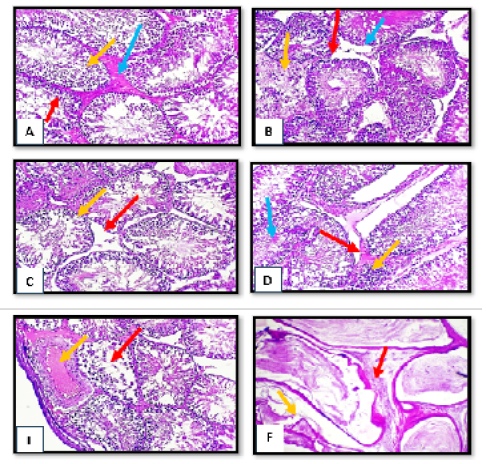
The main histopathological changes of rat testes showed sever degenerated changes of spermatogonia cells with irregular appearance of spermatid and presence of edematous substance between degenerated seminiferous tubules. In other section obvious disruption of seminiferous tubules with necrotic finding of some tubules with absence of leydig cells and evidence of hypospematogensis. Also intertubler edema with mild mononuclear cells infiltration and necrotic of immature spermatid with obvious subcapsulear vascular congestion and dilation. The histopathological section of rat epididymis showed irregularity of epididymal tubules with obvious atrophy of some tubules (Fig. 1).
Third Group (1 Μg /Kg. B.w.)
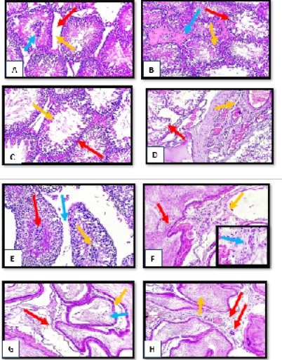
The main histopathological section showed obvious disruption of seminiferous tubules with absence of leydig cells with central clamping of necrotic spermatogonia. Also there is sever degenerative changes of spermatogonia cells with irregular appearance of spermatid and presence of edematous substance between degenerated seminiferous tubules. Marked reduction of spermatogonia cells lining with sperm irregularity. In other section there is disorganized irregular seminiferous tubules with subscapular dilation and congestion. Also clumping of degenerated spermatocyte with necrotic sperms and obvious widening of interlobular septa. The histopathological section of rat epididymis showed Focal hyperplasia of epididymal lining with blood vessels congestion and sever mononuclear cells infiltration in the vascular connective tissue. In other section there is moderate widening of some epididymal tubules with giant tubules appearance and slight degeneration of mature sperms. Fibrovascular tissue and newly form capillaries between epididymal tubules with evidence of sperms stasis. (Fig. 2)
Immunohistochemistry Study
Interleukin1 (Il1) Expression
The results of IL1 expression in the tissue treated with tetrodotoxin for 90 day showed significant elevation (P<0.05) in their score in testes and epididymis (42.67±0.33, 47.33±0.33) respectively at dose 0.5µg/kg.bw, while at dose 1µg/kg.bw reached to (99.00±0.57, 97.33±0.33) respectively compared with control group (0.00±0.00), the result was shown in Table 2.
Table 2: The effect of tetrodotoxin on scoring for interleukin1 (IL1) expression in rat tissues for chronic toxicity.
Group/Organ
Testis
Score
Epididymis
Score
1st group
(Control)
0.00±
0.00c
0
0.00±
0.00c
0
2nd group
(0.5µg/kg.bw)(1/20)
42.67±
0.33b
2
47.33±
0.33b
2
3rd group
(1µg/kg.bw)(1/10)
99.00±
0.57a
3
97.33±
0.33a
3
LSD
1.33
0.94
*Means with a different letter in the same column are significantly different(P<0.05)
Imunohistochamical Sections For Interleukin1 (IL 1) Detection
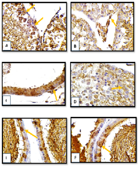
The result of Immunohistochemistry section of testis tissue for treated group showed moderate immune positive cells for interleukin 1 (IL1) antibody mainly in sertoli cells and spermatogonia and in interstitial leydig cells mostly in 3rd group. result of Immunohistochemistry section of epididymis tissue in treated group showed numerous immune positive cells for interleukin 1 (IL1) antibody in the epididymal epithelial cells lining mostly in 3rd group (Fig. 3).
The results of tumor necrosis factor alpha (TNF α) expression in the tissue treated with tetrodotoxin for 90 day showed significant elevation (P< 0.05) in their score in testes and epididymis (75.67±0.33, 78.67±0.33) respectively at dose 0.5µg/kg.bw, while at dose 1µg/kg.bw reached to (113.67±0.33, 119.67±0.33), respectively, compared with control group (0.00±0.00), the result was shown in Table 3.
Table 3: The effect of tetrodotoxin on scoring for tumor necrosis factor alpha (TNF alpha) expression in rat tissues for chronic toxicity.
Group/Organ
Testis
Score
Epididymis
Score
1st group (Control)
0.00±
0.00c
0
0.00±
0.00c
0
2nd group
(0.2µg/kg.bw)(1/20)
75.67±
0.33b
3
78.67±
0.33b
3
3rd group
(1µg/kg.bw)(1/0)
113.67±
0.33a
4
119.67±
0.33a
4
LSD
0.94
0.94
*Means with a different letter in the same column are significantly different (P<0.05).
Imunohistochamical Sections For Tumor Necrosis Factor Alpha (TNFα ) Detection
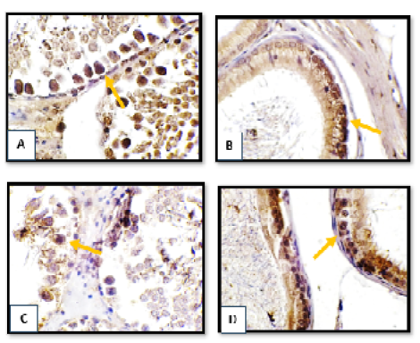
The result of Immunohistochemistry section of testes tissue showed numerous immune positive cells for TNFα antibody in sertoli cells and spermatogonia. The result of Immunohistochemistry section of epididymis tissue showed numerous and moderate immune positive cells for TNFα antibody in the epididymal epithelial cells lining (Fig. 4).
DISCUSSION
Lipid peroxidation of polyunsaturated fatty acids produces malon Di-aldehyde. The amount of malon Di-aldehyde in tissues can be used to evaluate the degree of lipid peroxidation . Malon Di-aldehyde is produce when polyunsaturated lipids are broken down by reactive oxygen species. The synthesis of this molecule, a reactive aldehyde, employed as a biomarker to estimate an organism’s level of oxidative stress because it is one of the several reactive electrophile species that induce toxic stress in
cells and . The findings of the chronic toxicity exposure of malon Di-aldehyde in animal tissues were not consistent, with the testes and epididymis exhibiting significantly higher concentrations (P<0.05) throughout every experimentation period and for both doses. This might be because tetrodotoxin incite the production of lipid peroxides, reactive oxygen species (ROS), DNA damage, and antioxidant enzyme activity in hepatocytes through the action of multispecific bile acid transporters . Moreover, as indicated by the increased levels of thiobarbituric acid reactive substances (TBARS), this raises the body’s oxidative stress. This is also associated with decreased levels of the scavenging enzymes in the liver, kidney, and spleen, including catalase, glutathione peroxidase activity (GPx), and superoxide dismutases (SOD) , Neama and Yousif, 2022). Furthermore, due to the testicular tissue’s extremely rapid rate of cell division, mitochondrial oxygen consumption, and relatively greater quantities of unsaturated fatty acids than in other tissues, oxidative stress plays a significant role in the development of male infertility . Oxidative stress caused by free radicals has a major role in sperm production and growth, sperm count reduction, sperm DNA fragmentation, and aberrant sperm production. Infertility is the result of these changes in sperm DNA alterations . Lipid peroxidation is inducing in spermatozoa by increased reactive oxygen species (ROS) generation, and this has two significant effects: 1) Diminishing the sperm-oocyte combination; 2) rising the spermatozoa’s capacity to adhere to the transparent region (zona placida) . Additionally, oxidative stress damaged sperm DNA, which is why it’s critical to preserve the accuracy and integrity of the DNA in the sperm nucleus in order to transfer genetic material entirely from one generation to the next. This is because genetic material disorders result in faulty genetic information being transmitted to the embryo. DNA deterioration increases under oxidative stress , . According to Wu et al. (2020), oxidative stress induced long-term alterations in the growing spermatozoa and epididymis that degrade the quality of sperm. In humans and other mammals, epididymal maturation is an essential stage in the development of viable and healthy spermatozoa . Following spermatogenesis, immature, immotile, completely formed spermatozoa enter the epididymis. As they exit, they develop the ability to move and physical characteristics that maximize their capacity for fertilization . Along with sperm maturation, the epididymis supplies spermatozoa with vital proteins through epididymosomes to preserve their biological functions and shield them from possible harm including oxidative stress-dependent injuries .
The microscopic finding of chronic toxicity in the current study showed severe pathological changes in many organs and the severity of the lesions was dose dependent. The supporting guide derived from our histopathological changes of testes and epididymis in both tetrodotoxin concentrated groups showed different pathological lesions due to tetrodotoxin toxicity but these changes was more intensive in 3rd groups. Testes and epididymis in both group showed degenerated changes of spermatogonia cells, irregular appearance of spermatid, presence of edematous substance between degenerated seminiferous tubules, necrosis of some tubules, absence of leydig cells, central clamping of necrotic spermatogonia ,mononuclear cells infiltration, reduction of spermatogonia cells lining with sperm irregularity, evidence of hypospermatogensis, irregularity and atrophy of some epididymal tubules, sperm stasis, hyperplasia of epididymal lining, sever mononuclear cells infiltration. These result indicate that tetrodotoxin responsible for increasing oxidative stress which induce alteration in plasma membrane by increasing lipid peroxidation and causes inhibition of sperm viability due to testicular and epididymal dysfunction . As well, the sperm is produced by the testes required furthermore processing in the epididymis to mature and acquired the ability to fertilize . The epididymis is an important organ for maturing of sperm and gain their capability to fertilize . These findings are consistent with those of Yavasoglu et al. (2008), who found that anatoxin-a (Marian toxin)-treated mice had lower sperm counts in their cauda epididymis across all treatment groups as compared to control animals. Tetrodotoxin also reduced sperm motility and density. In mouse testes, anatoxin-a induces histopathological alterations that include degenerations of seminiferous tubules, spermatogenetic cell line intercellular disassociation, sloughing of germ cells into tubular lumen, vacuolization in Sertoli cells, and loss of germ cells. The epithelial thickness of seminiferous tubules significantly decreased in all treatment groups.
The current results of immunohistochemically study showed increased in interleukin 1 (IL1) expression in the different rat organs that treated chronically with tetrodotoxin, the highest score was in the testes and epididymis (3) specially at 3rd group. This results are due to the tetrodotoxin toxicity that causes injury in these organs so many proinflammatory cytokines will be release to the circulation like Interleukin 1. (IL1) is a potent proinflammatory cytokine capable of triggering multiple physiological processes such as activation of lymphocytes, induction of acute-phase hepatic proteins, infiltration of leukocytes at sites of infection, fever and anorexia (Dinarello, 2006, , , clearly indicating that this cytokine is critical for innate immune response (AL-Samarraae and AL –Maadhidi, 2018). Interleukin 1 (IL1) mainly produced by macrophages during defensive reactions . The term interleukin 1(IL-1) refers to two cytokines, interleukin 1 alpha (IL-1α) and interleukin 1 bata (IL-1β) . interleukin 1 alpha (IL-1α) released from necrotic cells is considered a major inducer of sterile inflammation in several ischemic conditions, by inducing neutrophil and macrophage recruitment and interleukin 1 bata (IL-1 β) expression and maturation ; . Additionally, it has been observed that testicular macrophages, postmeiotic germ cells, and Sertoli cells all produce interleukin 1 alpha (IL-1 α). Testicular macrophages, Sertoli cells, Leydig cells, and germ cells have all been found to carry interleukin 1 IL-1 receptors, indicating both autocrine and paracrine activities. Although this proinflammatory cytokine plays a significant role in maintaining normal testicular homeostasis, increased expression of it can cause dysfunctions in the testicles by tipping the scales in favor of inflammatory and immune responses. Once immune cells such as neutrophils and lymphocytes are either drawn to the testis or activated there such as by macrophages, germ cell apoptosis is seen, and spermatogenesis is interrupted .
The results of immunohistochemistry in the current study showed increase in tumor necrosis factor alpha (TNF α) expression in organ of rat treated with tetrodotoxin chronically the highest score was in the testes and epididymis (4) this result is due to the toxicity of tetrodotoxin that causes injury and inflammation in these treated organ which lead to various proinflammatory cytokines released to the bloodstreams such as tumor necrosis factor alpha (TNF alpha). tumor necrosis factor alpha (TNF α) is a cytokine that affects different kinds of cells in pleiotropic ways. It is known to play a role in the etiology of some inflammatory and autoimmune disorders and has been identified as a key regulator of inflammatory responses , . It is primarily produced by T lymphocytes, natural killer cells, activated macrophages (including alveolar macrophages, microglia, Langerhans cells, Kupffer cells, and astrocytes) and T lymphocytes . It is functionally known to initiate a cascade of other inflammatory chemicals, such as chemokines and additional cytokines. On the other hand, excessive or improper tumor necrosis factor alpha (TNF α) signaling activation is linked to chronic inflammation and may eventually cause the emergence of clinical consequences including autoimmune disorders . TNF-α is a crucial cytokine that can cause inflammation and apoptosis, and it is essential in the disturbance of macrovascular and microvascular circulation both in vivo and in vitro ; . . Increased synthesis of TNF-α occurs when ROS are present, and tumor necrosis factor alpha (TNF-α) signaling exacerbates oxidative stress . According to Zelová and Hošek (2013), there is a correlation between altered coagulation propensity and tumor necrosis factor alpha overexpression. tumor necrosis factor alpha, in brief, contributes to leukocyte adherence to the epithelium via expressing adhesion molecules causing vasodilatation and edema development. Moreover, it controls blood coagulation, increases oxidative stress at inflammatory sites, and indirectly raises body temperature . According to Yang and Seki (2015), tumor necrosis factor alpha (TNF α) induces apoptosis, proliferation, and inflammation. Testicular macrophages, pachytene spermatocytes, and round spermatids all release tumor necrosis factor alpha (TNF alpha) in the testes. Numerous studies indicate that tumor necrosis factor alpha (TNF α) in the normal testis functions through a paracrine pathway, and tumor necrosis factor receptors have been identified on Sertoli and Leydig cells. Under normal physiological settings, the testis produces tumor necrosis factor alpha (TNFα), which is crucial for preserving testicular function. Nonetheless, in some pathological situations, such as testicular torsion, testicular inflammation, and immunological response including the stimulation of immune cells (lymphocytes, neutrophils, macrophages) to testes, there is a rise in the expression of this cytokine. Testes with spermatogenesis disruption also exhibit apoptosis .
CONCLUSION AND RECOMMENDATION
On the basis of the current study’s experimental results, we showed that tetrodotoxin has a harmful toxic effect on rats that indicated the effects of oxidative stress and increase interleukin 1 (IL1) and tumor necrosis factor alpha (TNFα) expression also induce pathological lesion in the testes characterized by absence of leydig cells and necrotic of immature spermatid with central clamping of necrotic spermatogonia, disorganized irregular seminiferous tubules with subscapular dilation and congestion, in epididymis tissue the most histopathological section showed widening of intertubular distance with evidence of sperm stasis and moderate cellular infiltration in peritubular tissue, focal hyperplasia of epididymal lining with blood vessels congestion and sever mononuclear cells infiltration in the vascular connective tissue, Fibrovascular tissue with newly form capillaries between epididymal tubules. Therefore, it is best to avoid eating fish species that contain tetrodotoxin in order to reduce the risks to one’s health.
ACKNOWLEDGEMENTS
The authors are thankful to the head College of Veterinary Medicine, University of Baghdad for their assistance during work.
NOVELTY STATEMENT
insufficient knowledge on tetrodotoxin from current veterinary studies, hence the current study attempted to identify potential pathological effects in albino male rats repeatedly exposed to tetrodotoxin
AUTHORS CONTRIBUTION
these authors each contributed equally
CONFLICT OF INTEREST
The authors have declared no conflict of interest.
Figures
Figure 1:.
Histopathological section of testes and epididymis tissue from tetrodotoxin treated group (0.5 µg/kg.bw) of male albino rats (A) sever degenerated changes of spermatogonia cells (red arrow) with irregular appearance of spermatid (yellow arrow) with presence of edematous substance between degenerated seminiferous tubules. (B) obvious disruption of seminiferous tubules (red arrow) with necrotic finding of some tubules (yellow arrow) with absence of leydig cells (blue arrow). (C) complete loss of leydig cells (red arrow) with evidence of hypospematogensis (yellow arrow). (D) intertubler edema (red arrow) with mild mononuclear cells infiltration (yellow arrow) with necrotic of immature spermatid (blue arrow). complete necrosis of spermatogonial cells (red arrow) with obvious subcapsulear vascular congestion and dilation (yellow arrow). (E) complete necrosis of spermatogonial cells (red arrow) with obvious subcapsulear vascular congestion and dilation (yellow arrow). H&E, x10. (F) marked irregularity of epididymal tubules (red arrow) with obvious atrophy of some tubules (yellow arrow). H&E, x40
Figure 2:.
Histopathological section of testes and epididymis tissue from tetrodotoxin treated group (1µg/kg.bw) of male albino rats (A) disruption of seminiferous tubules (red arrow) with absence of leydig cells (yellow arrow) with central clamping of necrotic spermatogonia (blue arrow).(B) sever degenerative changes of spermatogonia cells (red arrow) with irregular appearance of spermatid (yellow arrow) with presence of edematous substance between degenerated seminiferous tubules (blue arrow).(C) marked reduction of spermatogonia cells lining (red arrow) with sperm irregularity (yellow arrow).(D) disorganized irregular seminiferous tubules (red arrow) with subcapsular dilation and congestion (yellow arrow).(E) clumping of degenerated spermatocyte (red arrow) with necrotic sperms (yellow arrow) with obvious widening of interlobular septa (blue arrow).(F) focal hyperplasia of epididymal lining (red arrow) with blood vessels congestion (yellow arrow) with sever mononuclear cells infiltration in the vascular connective tissue (blue arrow) (H&E stain, X10, inset X40).(G) moderate widening of some epididymal tubules (red arrow) with giant tubules appearance (yellow arrow) with slight degeneration of mature sperms (blue arrow) (H&E stain, X10, inset X40).(I) fibrovascular tissue with newly form capillaries between epididymal tubules (red arrow) with sperms stasis (yellow arrow). (H&E stain, X10).
Figure 3:.
Immunohistochemistry section of testes and epididymis tissue from tetrodotoxin treated group (A,B,C) (0.5µg/kg.bw),(D,E,F) (1µg/kg.bw) of male albino rats (A),(B) moderate immune positive cells for IL 1 antibody (arrow)mainly in sertoli cells and spermatogonia.(C) numerous immune positive cells for IL 1 antibody (arrow)in the epididymal epithelial cells lining.(D) numerous immune positive cells for IL 1 antibody (arrow) in spermatocyte. (E) moderate immune positive cells for IL1 antibody (arrow) in epididymal epithelial cells lining.(F) numerous immune positive cells for IL 1 antibody (arrow) in epididymal epithelial cells lining. (DAB- chromogen, X40).
Figure 4:.
Immunohistochemistry section of testes and epididymis tissue from tetrodotoxin treated group (A, B) (0.5µg/kg.bw), (C, D) (1µg/kg.bw) of male albino rats (A) numerous immune positive cells for TNFα antibody (arrow) in sertoli cells and spermatogonia. (B) moderate immune positive cells for TNFα antibody (arrow) epididymal epithelial cells lining. (C) mild immune positive cells for TNFα antibody (arrow) mainly in spermatogonia. (D) numerous immune positive cells for TNFα antibody (arrow) in epididymal epithelial cells lining. (DAB- chromogen, X40).
References
Abd ST, Gathwan, KH (2022). Protective Effect of Garlic on Pesticide (Cypermethrin) Induced Hepatotoxicity on Liver of Mice. ACE J. Anim. Vet. Sci., 1(1): 1-3.
Ahmed HA, Ali HA, Mutar, TF (2021). Protective effects of olive leaf extract against reproductive toxicity of the lead acetate in rats. Environ. Sci. Pollut. Res., 28(44): 63102-63110. http://doi.org/10.1007/s11356-021-15240-3[CrossRef]
Al-Ghurabi, BH (2013). Impact of smoking on the IL-1 [Beta], IL-8, IL-10, IL-17 and TNF-[alpha] production in chronic periodontitis patients. J. Asian Scient. Res., 3(5): 462-470.
Alhatali B, Al Lawatia S, Khamis, F, Kantur S, Al-Abri S, KapilV, Thomas J (2022). A cluster of tetrodotoxin poisoning in Oman. Clin. Toxicol., 60(2): 262–266. http://doi.org/10.1080/15563650.2021.1917595[CrossRef]
AL-Okaily BN, Ahmed JN (2015). Role of alcoholic extract of Roket (Eruca sativa) leaves on male reproduction of experimentally induced-oxidative stressed rats. The Iraqi Journal of Veterinary Medicine, 39(2): 47–54. https://doi.org/10.30539/iraqijvm.v39i2.177[CrossRef]
Al-Sabaawy HB, Al-Kaisie BI (2021). Effects of Sub Lethal Concentrations of Sodium Fluoride on Sperm Activity and on the level of Sex Hormones of Adult Male Albino Rats. The Iraqi Journal of Veterinary Medicine, 44(2): 92–98. https://doi.org/10.30539/ijvm.v44i2.980.[CrossRef]
AL-Samarraae IAA, AL –Maadhidi RNA (2018). Evaluation of interleukins (2, 6 and 8) in immunized white rats by Salmonella enterica subspecies typhimurium and Cryptococcus neoformans antigens. The Iraqi Journal of Veterinary Medicine, 41(2): 72–6. https://doi.org/10.30539/iraqijvm.v41i2.51[CrossRef]
Asadi N, Bahmani M, Kheradmand A, Kopaei M R (2017). The impact of oxidative stress on testicular function and the role of antioxidants in improving it. J. Clin. Diagnost. Res., Vol-11(5): IE01-IE05. http://doi.org/10.7860/JCDR/2017/23927.9886[CrossRef]
Bennetts L E, Aitken R J (2005). A comparative study of oxidative DNA damage in mammalian spermatozoa. Mol. Reprod .,71:77–87. http://doi.org/10.1002/mrd.20285[CrossRef]
Bertheloot D, Latz E (2017). HMGB1, IL-1a, IL-33 and S100 Proteins: Dual Function Alarmins. Cell Mol. Immunol.,14:43–64. http:// doi.org/ 10.1038/cmi.2016.34
Boraschi D (2022). What Is IL-1 for? The Functions of Interleukin-1 Across Evolution. Front. Immunol., 13: 872155.http://doi.org/10.3389/fimmu.2022.872155[CrossRef]
Bradley J R (2008). TNF-Mediated inflammatory disease. J. Pathol., 214: 149–160. http://doi.org/ 10.1002/path.2287
Bucciarelli G M, Lechner, M, Fontes A, Kats L B, Eisthen H L, Shaffer H B (2021). From poison to promise: The evolution of tetrodotoxin and its potential as a therapeutic. Toxins, 13: 517. http://doi.org/10.3390/toxins13080517[CrossRef]
Dinarello CA (2006). Interleukin 1 and interleukin 18 as mediators of inflammation and the aging process. Am J. Clin. Nutr., 83:447S–455S. http://doi.org/10.1093/ajcn/83.2.447S[CrossRef]
Dinarello CA (2009). Immunological and inflammatory functions of the interleukin-1 family. Annu. Rev. Immunol., 27:519–550.http:/doi.org/10.1146/annurev.immunol.021908.132612[CrossRef]
Dinarello CA, Simon A, van der Meer JWM (2012). Treating Inflammation by BlockingInterleukin-1 in a Broad Spectrum of Diseases. Nat. Rev. Drug Discover., 11:633–52. http://doi.org/ 10.1038/nrd3800
Ding WX, Shen HM, Zhu HG, Ong CN (1998). Studies on oxidative damage induced by cyanobacteria extract in primary cultured rat hepatocytes. Environ. Res., 78(1):12-8.http://doi.org/ 10.1006/enrs.1998.3843
Fabisiak JP, Borisenko GG, Liu SX, Tyurin VA, Pitt BR and Kagan VE (2002). Redox sensor function of metallothioneins. J. Methods Enzymol., 353: 268–81. http://doi.org/10.1016/s0076-6879(02)53055-x[CrossRef]
Gehringer MM, Shephard EG, Downing TG, Wiegand C, Neilan BA (2004). An investigation into the detoxification of microcystin-LR by the glutathione pathway in Balb/c mice. Int J Biochem Cell Biol., 36(5):931-41.http://doi.org/ 10.1016/j.biocel.2003.10.012
Han Y, Yu Y, Liang C, Shi Y, Zhu Y Zheng H et al., (2019).Fluoride- induced unrestored arrest during haploid period of spermatogenesis via the regulation of DDX25in rats. Envirmen. Pollut., 253:538-551. http://doi.org/ 10.1016/j.envpol.2019.06.107
Hassoun A E R , Ujevi´c I, Jemaa S , Roje-Busatto R, Mahfouz C, Fakhri M et al., (2022). Concentrations of Tetrodotoxin (TTX) and Its Analogue 4,9-Anhydro TTX in Different Tissues of the Silver-Cheeked Pufferfish (Lagocephalus sceleratus, Gmelin, 1789) Caught in the South-Eastern Mediterranean Sea, Lebanon. Toxins, 14: 123. http://doi.org/10.3390/toxins14020123[CrossRef]
He X, Wu H, Ye Y, Gong X, Bao B (2022). Transcriptome analysis revealed gene expression feminization of testis after exogenous tetrodotoxin administration in pufferfish. Takifugu flavidus. BMC Genom., 23:553 https://doi.org/10.1186/s12864-022-08787-z[CrossRef]
Horiuchi T, Mitoma H, Harashima, S, Tsukamoto H, Shimoda T. (2010).Transmembrane TNF-alpha: Structure, function and interaction with anti-TNF agents. Rheumatology., 49: 1215–1228. http://doi.org/ 10.1093/rheumatology/keq031
Humadai T, AL-Kaisei B (2023). Toxicopathological Effects of Tetrodotoxin In Brain of Male Albino Rats. Iraqi J. Vet. Med., under press.
Ito E, Satake M, Ofuji K, Kurita N, McMahon T, James K (2000). Multiple organ damage caused by a new toxin azaspiracid, isolated from mussels produced in Ireland. Toxicon, 38(7):917-30. http://doi.org/10.1016/s0041-0101(99)00203-2[CrossRef]
Jang D I, Lee A H, Shin H Y, Song H.R, Park J H, Kang T B, et al., (2021). The Role of Tumor Necrosis Factor Alpha (TNF alpha) in Autoimmune Disease and Current TNF alpha Inhibitors in Therapeutics. Int. J. Mol. Sci., 22: 2719. https://doi.org/10.3390/ ijms22052719[CrossRef]
Kaneko N, Kurata M, Yamamoto T, Morikawa S, Masumoto J (2019). The role of interleukin-1 in general pathology. Inflammat. Regenerat., 39:12 http://doi.org/10.1186/s41232-019-0101-5[CrossRef]
Kang YJ (2006). Metallothionein redox cycle and function. Experimental Biology and Medicine (Maywood), 231:1459–67. http://doi.org/ 10.1177/153537020623100903
Laura J, Niedernhofer J, Scott D, Carol A, Rouzer RE (2003). Malondialdehyde, a Product of Lipid Peroxidation, Is Mutagenic inHuman Cells. J. Biolog. Chem., 278(33): 31426–31433. http://doi.org/ 10.1074/jbc.M212549200
Lysiak JJ (2004). The role of tumor necrosis factor-alpha and interleukin-1 in the mammalian testis and their involvement in testicular torsion and autoimmune orchitis. Reprod. Biol. Endocrinol., 2(1) :1-10. http://www.rbej.com/content/2/1/9
Neamah, GAK, Yousif EH (2022). Determination of Depleted Uranium Concentration and Histopathological Changes in Local Iraqi Fish and Chickens. Iraqi J. Vet. Med., 43(2): 86–97. https://doi.org/10.30539/iraqijvm.v43i2.537[CrossRef]
Pinho GL, da Rosa CM, Maciel FE, Bianchini A, Yunes JS, Proenca LA (2005). Antioxidant responses and oxidative stress after microcystin exposure in the hepatopancreas of an estuarine crab species. Ecotoxicol. Environ. Saf.,61(3):353-60. http://doi.org/ 10.1016/j.ecoenv.2004.11.014
Robaire B, Hinton BT (2006). Orgebin-Crist, The Epididymis. In Knobil and Neill’s Physiology of Reproduction, 3rd ed.; Academic Press: St. Louis, MO, USA, 1071–1148.
Said TM, Agarwal A, Sharma RK, Thomas AJ Jr, Sikka SC (2005). Impact of sperm morphology on DNA damage caused by oxidative stress induced by beta- nicotinamide adenine diy nucleotide phosphate. Fertil. Steril., 83(1):95-103. http://doi.org/10.1016/j.fertnstert.2004.06.056[CrossRef]
Saleh RA, Agarwal A (2002). Oxidative stress and male infertility: from research bench to clinical practice. J. Androl., 23(6):737-52.
Saoudi M, Messarah M, Boumendjel A, Abdelmouleh A, Kammoun W, Jamoussi K (2011). Extracted tetrodotoxin from puffer fish Lagocephalus lagocephalus induced hepatotoxicity and nephrotoxicity to Wistar rats. African J. Biotechnol., 10(41): 8140-8145, 3. http://doi.org/10.5897/AJB10.2531[CrossRef]
SAS.2010.SAS/STAT Users Guide for Personal Computer. Release 9.13.SAS Institute, Inc., Cary, N.C., USA.
Sullivan R, Frenette G, Girouard J (2007). Epididymosomes are involved in the acquisition of new sperm proteins during epididymal transit. Asian J. Androl., 9: 483–491. http://doi.org/10.1111/j.1745-7262.2007.00281.x[CrossRef]
Suzuki M, Bartlett JD (2014). Sirtuin 1 and autophagy protect cells from fluoride-induced cell stress. Biochimica et Biophsica Acta(BBA)-molecular basis of Disease, 1842(2): 242-255.http://doi.org/10.1016/j.bbadis.2013.11.023[CrossRef]
Vinagre TM, Alciati JC, Regoli F, Bocchetti R, Yunes JS, Bianchini A (2003). Effect of microcystin on ion regulation and antioxidant system in gills of the estuarine crab Chasmagnathus granulatus (Decapoda, Grapsidae). Comp Biochem Physiol. C Toxicol. Pharmacol., 135(1):67-75.http://doi.org/10.1016/s1532-0456(03)00076-0[CrossRef]
Wu P Y, Scarlata E, O’Flaherty C (2020). Long-Term Adverse Effects of Oxidative Stress on Rat Epididymis and Spermatozoa. Antioxidants., 9: 170. http://doi.org/10.3390/antiox9020170[CrossRef]
Xie SW Li GT, Qu LJ, CaoY, Wang Q, Zhou JY (2016). Identification of new epididymal luminal fluid proteins involved in sperm maturation in infertile rats treated by dutasteride using iTRAQ. Molecules., 21(5): 602. http://doi.org/10.3390/molecules21050602[CrossRef]
Yamagishi S I, Adachi H, Matsui T, Nakamura K, Enomoto M, Fukami A (2009). Decreased High-Density Lipoprotein Cholesterol Level is an Independent Correlate of Circulating Tumor Necrosis Factor-α in a General Population. J. Adv. Treatment Cardiovascul. Dis., 32(9): E29-E32.., http://doi.org/10.1002/clc.20517[CrossRef]
Yang G, Shao GF (2016). Elevated serum IL-11, TNF alpha, and VEGF expressions contribute to the pathophysiology of hypertensive intracerebral hemorrhage (HICH). Neurol. Sci., 37: 1253–1259, https://doi.org/10.1007/s10072-016-2576-z[CrossRef]
Yar HS, Ismail DK, Alhmed MN (2012). Hepatoprotective effect of Carthamus tinctorius L. against carbon tetrachloride induced hepatotoxicity in rats. Pharmacie Globale, 3(9): 1.
Yavasoglu MA, Karaaslan A, Uyanikgil Y, Sayim F, Ates U, Yavasoglu NUK (2008). Toxic effects of anatoxin-a on testes and sperm counts of male mice. Exp. Toxicol. Pathol., http://doi.org/10.1016/j.etp.2008.04.001[CrossRef]
Zegura B, Sedmak B, Filipic M (2003). Microcystin-LR induces oxidative DNA damage in human hepatoma cell line HepG2. Toxicon, 41(1):41-8. http://doi.org/10.1016/s0041-0101(02)00207-6[CrossRef]
Zelová H, Hošek J (2013). TNF-alpha signalling and inflammation: interactions between old acquaintances. Inflamm. Res., 62: 641–651. https://doi.org/10.1007/s00011-013-0633-0[CrossRef]
Zenclussen AC, Blosis S , Stumpo R (2003). Murine absorption is associated with enhanced interleukin -6 level at the feto-materal interfeace cytokine. Radiat. Res., 24(4):50-60. http://doi.org/10.1016/j.cyto.2003.08.002[CrossRef]
Zhang H, Park Y, Wu J, Chen XP, Lee S, Yang J (2009). Role of TNF-α in vascular dysfunction. Clin. Sci. 1;116(3):219-30. https://doi.org/10.1042/cs20080196[CrossRef]
Zimmer T (2010). Effects of tetrodotoxin on the mammalian cardiovascular system. Mar. Drugs., 8: 741–762.http://doi.org/10.3390/md8030741[CrossRef]