Research Article · Volume 36, Issue 2, 8 October 2021 · Open Access
Atrazine Induced Histopathological Alterations in the Liver of Adult Male Mice
· · · · · ·
Affiliations & Notes ⌄
1Department of Zoology, University of Sargodha, Sargodha, Pakistan 2Department of Chemistry, University of Sargodha, Sargodha, Pakistan 3Institute for Advanced Study, Shenzhen University, Shenzhen, China. Corresponding Author: Saima Batool saimabatool118@gmail.com
Atrazine is a commonly used triazine derived herbicide that is a major pollutant of soil and water and highly carcinogenic. To investigate Atrazine induced pathological changes in the liver of albino laboratory mice, 20 adult male mice were equally distributed (n=10) into two groups. Atrazine was given at the dose of 200 mg/kg body weight through gavages for 28 days while the control (Cnt) group remains untreated. On the 29th day, all animals were sacrificed to recover liver for histological preparations. Atrazine (ATZ) caused various hepatic implications in the treated animals. ATZ exposure caused a significant decline in mean body and liver weight as compared to the Cnt group. The histopathological findings in ATZ administered group included loosening hepatic tissue, massive degeneration of hepatocytes, deformed nuclei of hepatocytes, highly disordered trabecular arrangement, vacuolar degeneration, diffuse necrosis, congestion and degeneration of the hepatic portal vein. Micrometric data indicated a significant reduction in themean number of hepatocytes and oval cells per unit area in ATZ treated group as well as significantly increased size and nuclear diameter of hepatocytes compared to the Cnt group. These findings showed the sensitivity of all cell types in the liver to the toxic potentials of atrazine at the given dose level.
Novelty Statement | This study reports hepato-toxic effects of atrazine at a particular dose level which has not been reported before.
Introduction
Atrazine (2 chloro 4 ethylamino 6 isopropylamino 1, 3, 5- triazine) is the second most commonly used herbicide with an annual consumption of 70,000–90,000 tons ; ; . It is a major contaminant of soil, water resources, and plants with a half-life of 30-740 days ; . Atrazine is very environmentally stable due to low loss by volatilization and degradation. Furthermore, because of its accumulation effect, atrazine is significantly affecting the health of many organisms, including humans (Nodler et al., 2013; Juhel et al., 2017). European Union banned Atrazine in 2003 due to its severe toxicity to cells and tissues. However, the United States Environmental Protection Agency (USEPA) still permits the use of atrazine worldwide . Various studies have shown that Atrazine interferes with the normal functioning of body organs, including the liver, disrupting the normal structural and architectural components in a non-infectious hepatic injury . Histopathological liver sections of adult Xenopus laevis exposed to different concentrations of Atrazine, showed hypertrophied hepatocytes, vascular congestion and dilation, disorganization in the arrangement of hepatic cords, apoptosis and/or necrosis, and infiltration of inflammatory cells, with highest concentrations, showed most severe effects . The hepatocyte and hepatocyte nucleus diameterwas significantly decreased in the atrazine-treated groups (Destroy et al., 2021). These damaging effects of atrazine may result from its generation of reactive oxygen species (ROS) that causes oxidative stress of various organs. Increased oxidative stress and lipid peroxidation is implicated in the pathogenesis of herbicide-induced hepatic injury . To evaluate various histological and micrometric alterations induced in the liver of albino mice due to ATZ exposure was the major objective of this study. The use of the mammalian model was planned to utilize the findings in humans.
Materials and Methods
Animals maintenance and care
This research was carried out in laboratory-reared albino mice (Mus musculus) with a weight of 28-30 grams and age of 5-6 weeks. These animals were kept in the Department of Zoology’s animal house, University of Sargodha, Sargodha, under thestandard protocol of 12-hours a day and night cycles. The temperature of the animal house was maintained at 25±2°C and humidity at 45%. Bodyweight, survival, and clinical signs were recorded daily.
Dose groups
Twenty animals were divided into two groups (n=10) randomly.
Control group (Cnt)
This group was provided regular drinking water with normal feed.
Atrazine treated group (ATZ)
These animals were given 200 mg per kg atrazine dissolved in water through gavagesonce daily for 28 days and regular drinking water and feed ad libitum.
After four weeks of experimentation, the animals were dissected to remove the liver. Organs were weighed and fixed in Conroy’s fixative for further processing.
Histological preparations and observations
After fixation, organs were processed for dehydration sequentially in 50%, 70%, 90%, and absolute alcohol for 24 hours each. After xylene clearance, organs were processed for wax embedding to get 5 microns thick sections of male mice’s liver through a rotary microtome (ERMA TOKYO 42). Hematoxylin and Eosin stained sections were observed under the stereoscopic compound microscope.
Data analysis and statistical applications
The micrometric data was calculated from the liver’s digital photomicrographs obtained using a Huawei company’s digital camera (Model no DSC-W35) 13 megapixels affixed on a trinocular microscope (Labomid CXR2) at 400×. Micrometric readings were obtained using Coral DRAW 11. The data obtained werethen analyzed statistically by an unpaired t-test for the comparison of the groups.
Results and Discussion
Histological observations
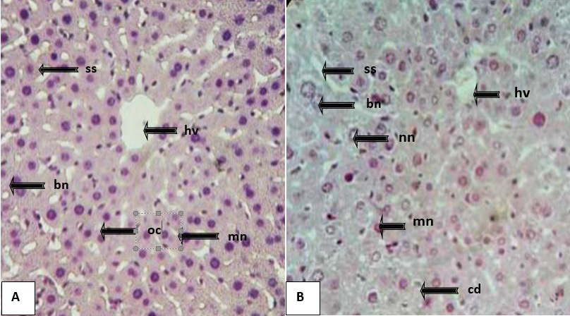
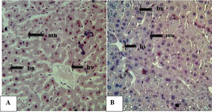
Histological analysis of liver slides of Cnt showed normal anatomical structures. In the Cnt group’s liver sections,a continuous array of one-cell thick hepatocytes that form the hepatic cord around compact central veins were seen (Figure 1). Hepatocytes are arranged in trabecularrunning radiantly from the central vein and are separated by sinusoids free of any cellular population (Figure 1A and 2A). Hepatocytes with granular cytoplasm were homogeneously distributed around the nucleus. Large well stained spherical nucleiwerepresent almost in the centre. The nucleus contains distinctly marked nucleolus and chromatin material. A compact organization was observed inperipheral hepatic portal triads or tetrads embedded in connective tissues. Binucleated were lesser in number than mono-nucleated hepatocytes, as shown in Figure 5. Kupffer cells were elongated in shape, lined on both sides of the hepatocytes. Between the hepatic cords, sinusoidal usual size spaces were observed where deposition of oval cells was obvious.
Histological observation of the liver after ATZ treatment showed marked alteration. In the ATZ treated group, loosen hepatic tissues, vacuolar degeneration of hepatocytes, diffuse necrosis, and the hepatic portal vein’s degeneration was visible. The number of hepatocytes with deformed nuclei was greatly reducedcompared to the Cnt group (Figure 1B - 2B). ATZ treatment resulted in massive degeneration of hepatocytes.The mean number of mononucleated and binucleated cells, per unit area, was affected adversely (Figures 1 - 2). ATZ treated group showed increased hepatocyte size and a highly significant increase in hepatocytes’ nuclear diametercompared to theCnt. Highly disordered trabecular arrangement (Figure 1B) and a significant reduction in the mean number of oval cells per unit area were also obvious in ATZ treated sections compared to the Cnt group.
Bodyweight
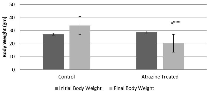
At the start of the experiment,the mean initial body weight of animals belonging to both experimental groups showed non-significant variations. At the end of the experiment ATZ treatment showed a highly significant (p<0.001) decrease in mean body weight of treated animals compared to the Cnt group. Data analysis by unpaired t-test revealed the toxic effect of atrazine on animals’ body weight at the given dose level.
Liver weight
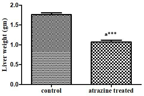
Unpaired T-test showed a highly significant (p˂0.001) reduction in mean liver weight in the ATZ treated group compared toCnt. This indicated toxicity of herbicide to the general health of animals.
Micrometric results
Mean number of mono-nucleated cells per unit area (7.62cm2)
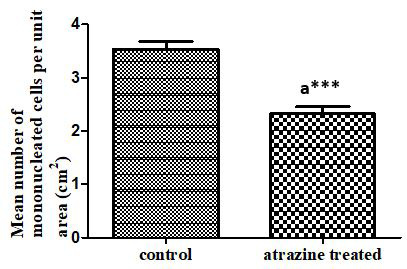
An unpaired t-test revealed that the number of mononucleated cells was highly significantly (p<0.001) reduced by 28 days treatment of atrazine compared to the Cnt group. These parenchymal cells were noticed to be the targeted cellular population of the herbicide.
Mean number of binucleated cells per unit area (7.62 cm2)
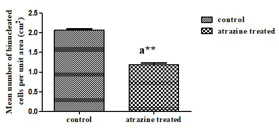
An unpaired t-test revealed that the mean number of binucleated cells was significantly (p<0.001) reduced by 200 mg/kg atrazine treatment for 28 days. Hepatocytes number either uninucleated or binucleated both were highly decreased by atrazine treatment reflecting toxicity of atrazine tothe liverat the cellular level.
Mean number of oval cells per unit area (7.62cm2)
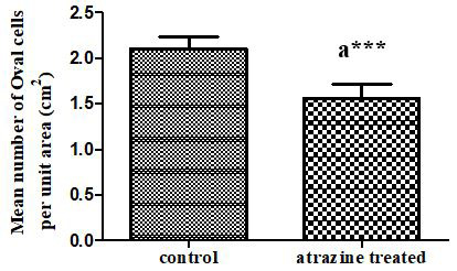
An unpaired t-test revealed that the mean number of oval cells was significantly (p<0.01) reduced by 4 weeks of ATZ compared to the Cnt group.
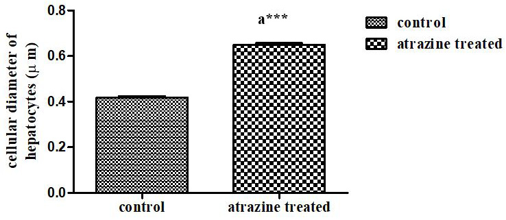
Cellular diameter of hepatocytes (µm)
An unpaired t-test revealed that hepatocytes’ cellular diameter was highly significantly (p<0.001) reduced by atrazine treatment compared to theCntgroup.
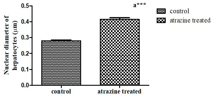
Nuclear diameter of hepatocytes (µm)
An unpaired t-test revealed that hepatocytes’ nuclear diameter was highly significantly (p<0.001) reduced by ATZ treatment compared to the Cnt group.
The liver is a critical organ in the human body that is responsible for an array of functions that help support metabolism, detoxification, digestion, immunity,vitamin storage, among other functions, soit is prone to a lot of environmental toxicants as well (Kalra et al., 2021; . In the present study, atrazine’s effects, a major water pollutant on male mice liver were explored.
The mean body and liver weight in ATZ treated mice showed a highly significant reduction (p<0.001) compared to the Cnt group. Decreased body and liver weight in mice might be attributed to the atrazine-generated oxidative stress that affected animals’ general body health .
In the histological examination of the present study, treatment of ATZ (200mg/kg) severely affected liver anatomy. In ATZ treated group, the arrangement of hepatocytes was distorted. The number of mononucleated and binucleated hepatocytes was highly decreased as compared to the Cnt group. ATZ treated sinusoidal group spaces were affected by the ATZ treatment and became disorganized, wider, and irregular (Figures 1 - 2). The number of oval cells also decreased in the treated mice liver (Figure 6). In the ATZ treated group, the hepatocytic nuclei size was highly significantly increased compared to Cnt (Figure 8). Congestion in the bile duct vein was prominent with a 200 mg per kg dose of ATZ (Figure 2). Exposure to ATZ resulted in pronounced histopathological abnormalities such as the expansion of sinusoids, dilation of hepatocytes, nuclear necrosis, emptied hepatic portal vein, reduced cytoplasm, and vacuolation in hepatocytes (Figure 1). In another study, similar findings as vacuolar degeneration of hepatocytes, diffuse necrosis and degeneration of the hepatic portal vein were also reported after administration of the oral dose of 25% (124 mg/kg/body weight) of LD50 (3090 mg/kg/body weight) of the Atrazine dissolved in water for 120 days to male Wistar Albino rats .Our findings were in agreement with the report of Senarat,who observed histopathological alterations of the liver in freshwater catfish collected from Tapee River, which is vulnerable to pesticides pollution, as it receives agricultural runoff from paddy fields, Indian rubber plantations, and vegetable and fruit crops . Histopathological alterations in the liver of catfish after exposure to pesticides consisted of cellular swelling, eosinophilic cytoplasm of hepatocytes, and constriction of sinusoidal capillaries damage of endothelial cells of blood vessels. Some liver areas showed focal necrosis and contained severe infiltration of macrophage, pyknotic nuclei, and large lipid vacuoles in the cytoplasm of hepatocytes (Figures 1 - 2).
One liver injury mechanism is mitochondrial dysfunction through free radicals generation .These radicals damage mitochondrial DNA . Consequently, some chemicals may cause hepatocellular necrosis, rapid disorganization of the hepatic architecture, breakdown of sinusoidal structures and pooling of blood in the liver through these mechanisms . It is reported that ATZ induces oxidative stress in rat tissues and that the oxidative stress was associated with increased lipid peroxidation and changes in the anti-oxidative system. These findings indicated that ATZ exposure caused various anatomical derangements in male mice’s liver possibly due to the generation of excessive reactive oxygen species .
In conclusion, results revealed that all liver cell types are highly sensitive to atrazine exposure. So, some measures should be taken to avoid atrazine exposure at the individual level to all possible extents.
Acknowledgements
We are thankful to the University of Sargodha for providing funding and all the necessary facilities for this research work. We are very obliged to provide all the key research equipment and chemicals for this research project.
Conflict of interest
The authors have declared no conflict of interest.
Figures
Figure 1:.
Photomicrographs from representing sections of the liver of Cnt and ATZ treated groups (H and E staining at 400×). Cnt: (A) liver exhibited a well-organized structure of trabeculae radiating from hepatic veins (hv) having mononucleated (mn) and binucleated (bn) hepatocytes separated by regular sinusoidal spaces (ss). ATZ treated group (B): liver showed the disorganized structure of trabeculae arising from the hepatic vein (hv) with deformed shape mononucleated hepatocytes (mn), binucleated hepatocytes with swollen nuclei (bn), cellular degeneration (cd), dilated sinusoidal spaces (ss), hollow and necrotic nuclei (nn) were also observed in the treated liver.
Figure 2:.
Photomicrographs of liver sections of Cnt and ATZ treated groups, showing distribution, size and nuclear structure of the hepatocytes (HandE staining at 400×).Cnt (A) showed well-oriented mono (mn) and binuclear hepatocytes (bn) of similar size and regular shape, nuclei darkly stained with condensed chromatin. ATZ (B) treated sections revealed a decreased number of mono (mn) and binuclear (bn) hepatocytes of variable size, mostly of deformed shape. Normal liver showed compact hepatic vein (hv) while treated liver showed narrow irregular hepatic vein (hv). Sections of ATZ treated group also revealed vacuolization in the hepatocytes.
Figure 3:.
Mean body weight of male albino mice belonging to Cnt and ATZ treated groups. Values are expressed in Mean ± SEM. a=Cnt group vs ATZ treated group. ***p Ë‚ 0.001.
Figure 4:.
Effect of atrazine treatment (200 mg/kg) for 28 days on male albino mice’s mean liver weight. Data represented as mean of 10 mice ±SE, a = Cnt vs atrazine treated group, ***p Ë‚ 0.001.
Figure 5:.
Alteration in the mean number of mononucleotide cells per unit area in the liver of atrazine treated male albino mice after 28 days of exposure at a dose of 200 mg/kg. Data represented as mean of 10 mice ±SE, a = Cnt vs ATZ group, ***p Ë‚ 0.001.
Figure 6:.
Mean number of binucleated cells per unit area in the liver of Cnt and ATZ treated male albino mice. Data represented as mean of 10 mice ±SE, a = Cnt vs ATZ treated group, ***p Ë‚ 0.001.
Figure 7:.
Effect of 4 weeks atrazine treatment on the mean number of oval cells per unit area in male albino mice’s liver. Data represented as mean of 10 mice ±SE, a = Cnt Vs ATZ treated group, ***p Ë‚ 0.001.
Figure 8:.
Cellular diameter of hepatocytes in the liver of Cnt and ATZ treated albino mice. Data represented as mean of 10 mice ±SE, a = Cnt vs ATZ treated group, ***p Ë‚ 0.001.
Figure 9:.
Effect of ATZ treatment (200mg/kg for 28 days) on the mean nuclear diameter of hepatocytes in male albino mice’s liver. Data represented as mean of 10 mice ±SE, a = CntvsATZ treated group, ***p Ë‚ 0.001.
References
Abarikwu, S.O., Duru, Q.C., Njoku, R.C.C., Amadi, B.A. and Tamunoibuomie, A., 2017. Effects of co-exposure to atrazine and ethanol on the oxidative damage of kidney and liver in Wistar rats. Renal Failure, 39: 588-596. https://doi.org/10.1080/0886022X.2017.1351373[CrossRef]
Almberg, K.S., Turyk, M.E., Jones, R.M., Rankin, K., Freels, S. and Stayner, L.T., 2018. Atrazine contamination of drinking water and adverse birth outcomes in community water systems with elevated atrazine in Ohio, 2006-2008. Int. J. Environ. Res. Publ. Hlth., 15: 1889. https://doi.org/10.3390/ijerph15091889[CrossRef]
Chen, Y.P., Liu, Q., Ma, Q.Y., Maltby, L., Ellison, A.M. and Zhao, Y., 2018. Environmental toxicants impair liver and kidney function and sperm quality of captive pandas. Ecotoxicol. Environ. Saf., 162: 218–224. https://doi.org/10.1016/j.ecoenv.2018.07.008[CrossRef]
Deshmukh, U.S. and Ramteke, P.M., 2015. Histophysiological alterations in some tissues of male wistar albino rats exposed to atrazine. Int. J. Fauna Biol. Stud., 2: 59-61.
Destro, A., Silva, S.B., Gregório, K.P., de Oliveira, J.M., Lozi, A.A., Zuanon, J., Salaro, A.L., da Matta, S., Gonçalves, R.V. and Freitas, M.B., 2021. Effects of subchronic exposure to environmentally relevant concentrations of the herbicide atrazine in the Neotropical fish Astyanax altiparanae. Ecotoxicol. Environ. Saf., 208: 111601. https://doi.org/10.1016/j.ecoenv.2020.111601[CrossRef]
Gely-Pernot, A., Saci, S., Kernanec, P.Y., Hao, C., Giton, F., Kervarrec, C. and Smagulova, F., 2017. Embryonic exposure to the widely-used herbicide atrazine disrupts meiosis and normal follicle formation in female mice. Sci. Rep., 7: 1-14. https://doi.org/10.1038/s41598-017-03738-1[CrossRef]
Juhel, G., Bayen, S., Goh, C., Lee, W.K. and Kelly, B.C., 2017. Use of a suite of biomarkers to assess the effects of carbamazepine, bisphenol A, atrazine, and their mixtures on green mussels, Perna viridis. Environ. Toxicol. Chem., 36: 429–441. https://doi.org/10.1002/etc.3556[CrossRef]
Kalra, A., Yetiskul, E., Wehrle, C.J. and Tuma, F., 2021. Physiology, liver. In: Stat pearls. Stat Pearls Publishing. Bookshelf ID: NBK535438. PMID: 30571059.
Lizotte, R., Locke, M., Bingner, R., Steinriede, R.W. and Smith, S., 2017. Effectiveness of integrated best management practices on mitigation of atrazine and metolachlor in an agricultural lake watershed. Bull. Environ. Contamin. Toxicol., 98: 447-453. https://doi.org/10.1007/s00128-016-2020-3[CrossRef]
Michael, P.O., Veronica, O.B. and Priscilla, E., 2018. Response of Clarias gariepinus (juveniles) exposed to sublethal concentrations of atrazine. Aquacult. Stud., 18: 19-26. https://doi.org/10.4194/2618-6381-v18_1_03[CrossRef]
Neequaye, P., 2019. Effect of chronic exposure of the herbicide atrazine on high fat diet-induced obesity and type 2 diabetes (Doctoral dissertation, North Carolina Central University).
Nödler, K., Licha, T. and Voutsa, D., 2013. Twenty years later–atrazine concentrations in selected coastal waters of the Mediterranean and the Baltic Sea. Mar. Pollut. Bull., 70: 112-118. https://doi.org/10.1016/j.marpolbul.2013.02.018[CrossRef]
Opute, P.A. and Oboh, I.P., 2021. Hepatotoxic effects of atrazine on clarias gariepinus (Burchell, 1822): biochemical and histopathological studies. Arch. Environ. Contamin. Toxicol., 80: 414-425. https://doi.org/10.1007/s00244-020-00792-1[CrossRef]
Rasgele, P.G., Oktay, M., Kekecoglu, M.F.D.G. and Muranli, F.D.G., 2015. The histopathological investigation of liver in experimental animals after short-term exposures to pesticides. Bulg. J. Agric. Sci., 21: 446-453.
Sagarkar, S., Gandhi, D., Devi, S.S., Sakharkar, A. and Kapley, A., 2016. Atrazine exposure causes mitochondrial toxicity in liver and muscle cell lines. Indian J. Pharmacol., 48: 200. https://doi.org/10.4103/0253-7613.178842[CrossRef]
Sena, L.R., 2017. Hepatotoxic and nephrotoxic effects of atrazine on adult male xenopus laevis frogs: A laboratory study (Doctoral dissertation).
Senarat, S., Kettratad, J., Poolprasert, P., Yenchum, W. and Jiraungkoorskul, W., 2015. Histopathological finding of liver and kidney tissues of the yellow mystus, Hemibagrus filamentus (Fang and Chaux, 1949), from the Tapee River, Thailand. Songklanakarin J. Sci. Technol., 37: 1-5.
Singh, S., Kumar, V., Chauhan, A., Datta, S., Wani, A.B., Singh, N. and Singh, J., 2018. Toxicity, degradation and analysis of the herbicide atrazine. Environmen. Chem. Lett., 16: 211-237. https://doi.org/10.1007/s10311-017-0665-8[CrossRef]
Toughan, H., Khalil, S.R., El-Ghoneimy, A.A., Awad, A. and Seddek, A.S., 2018. Effect of dietary supplementation with Spirulina platensis on Atrazine-induced oxidative stress-mediated hepatic damage and inflammation in the common carp (Cyprinus carpio L). Ecotoxicol. Environ. Saf., 149: 135-142. https://doi.org/10.1016/j.ecoenv.2017.11.018[CrossRef]
Zhang, C., Li, X.N., Xiang, L.R., Qin, L., Lin, J. and Li, J.L., 2017. Atrazine triggers hepatic oxidative stress and apoptosis in quails (Coturnix C. coturnix) via blocking Nrf2-mediated defense response. Ecotoxicol. Environ. Saf., 137: 49-56. https://doi.org/10.1016/j.ecoenv.2016.11.016[CrossRef]