
Pneumonitis, another name for lung inflammation, can be caused by respiratory infections, exposure to pollutants or allergens from the air, and lung conditions such as chronic bronchitis or asthma. An inhibitor of phosphodiesterase enzymes 3 and 5, dipyridamole is a conventional antiplatelet drug that increases adenosine. The purpose of this study was to investigate whether dipyridamole plays a role as an anti-inflammatory to improve airway inflammation. A total of 24 healthy rats (albino, male), were weighted (150-300 g), were divided into four groups, each group consisting of six rats; Group A rats were given oral distal water and was considered a negative control group. Group B rats received distal water orally with ovalbumin (ova) sensitization, which was considered a positive control group. Group C rats were given dipyridamole (26.4 mg / kg) orally with sensitization to the ova. Group D rats received prednisolone (4.12mg/kg) orally with sensitization to the ova. Subsequently, the right lung was lavage, by insertion of a tracheal cannula, three times with 10 ml of normal saline (NS 0.9%) at 37 °C and the broncho alveolar lavage fluid (BALF) was sent for laboratory evaluations and the left lung was incised for histopathological evaluation. Data analysis indicated that a substantial decrease in total white blood cell counts (WBC) was observed (p-value <0.05) and cells including (neutrophils, mononuclear) in BALF of rats treated with dipyridamole (group C) and prednisolone (group D) compared to positive control (group B), but reduction in eosinophils cells in dipyridamole treated group C was non significant (p-value>0.05) there was also an improvement in histological characteristics regarding bronchial dilatation and improvement in the alveolar sac and modification of inflammatory cells in lung tissues in dipyridamole treated rats (group C) and prednisolone treated (group D) was observed in contrast to ovalbumin (ova) sensitized rats (group B). Collectively, dipyridamole has the ability to decrease inflammation to a lower limit in the airways in rats through its ability to decrease total and differential WBCs in BALF.
Research Article · Volume 12, Issue 2, 5 June 2024 · Open Access
Study of Anti-Inflammatory Effect of Dipyridamole by Evaluation Inflammatory Cells and Histopathology in Rat: Airway Models
· ·
Affiliations & Notes ⌄
- 1Department of Pharmacology, College of Medicine, University of Basra, Basra, Iraq
- 2Department of Pharmacology and Toxicology, College of Pharmacy, University of Basra, Basra, Iraq. *Correspondence | Ali D Nashmi, Department of Pharmacology, College of Medicine, University of Basra, Basra, Iraq
- 3Email: Basratoxo@gmail.com
Article Info ⌄
- Journal:
- Journal of Animal Health and Production
- Published:
- 5 June 2024

From the article
- When looking at Table 1 Figure 4 the level of mononuclear cells (lymphocytes and monocytes) in BALF of dipyridamole-treated rats (group C) decreased significantly (P < 0.05) in contrast to positive ova sensitized (group B).
- The eosinophil count in the BALF of prednisolone-treated rats (group D) decreased significantly in this investigation and increased in ova sensitized (group B), as indicated in Table 1 Figure 2, which is consistent with the study mentioned.
- In BALF animal, dipyridamole exhibited a good anti-inflammatory effect by reducing total WBC, eosinophil, neutrophil, and mononuclear cells.
- Additionally, dipyridamole and prednisolone exhibit a marked improvement in inflammatory cells and the histological properties of the bronchial and alveolar sacs.
Selected from the article's own text.
Abstract
Introduction
Pneumonitis, characterised by the lung inflammation, can be caused by respiratory infections, exposure to airborne pollutants or irritants, and lung conditions such as chronic bronchitis or asthma . Chronic lung inflammation can alter airway content, thickness, or volume over time, resulting in bronchiectasis. . These changes can also result in hypercapnia .
Regarding eosinophils, which are significant inflammatory cells that play a pathogenic role in the pathobiology of airway disorders; in fact, their recruitment, maturation, activation, and survival within the airway lumen and bronchial wall are important pathogenic events ; . Inhaled allergens can activate several recognizing receptors once they penetrate the airway epithelium. This triggers a series of events that culminate in the production of Th2 cytokines .
Prolonged infiltration and degranulation of eosinophils leads to the continuous release of cytotoxic products, such as eosinophil cationic protein, main basic protein, neurotoxin derived from eosinophils, and eosinophil peroxidase. These factors play an important role in airway epithelium damage, goblet cell mucus overproduction, bronchial hyper responsiveness, and impaired ciliary beating ; Peters-Golden and Henderson Jr., 2007; ; . Furthermore, growth factors, including transforming growth factor alpha (TGF-a) are produced by eosinophils . In response to neutrophilic airway inflammation, T-helper1(Th1) cells, and particularly Th17 lymphocytes, are the initiators. (Aujla and Alcorn, 2011; . Specifically, the unique airway environment, which is composed of specialized costimulatory molecules and cytokines that drive the various Th cells, is vital for the commitment to the multiple Th lineages. . Neutrophil-induced airway inflammation is frequently associated with the most severe asthmatic phenotypes .
Interlukin-23 (IL-23) cytokine is crucial to preserve Th17 cell capacity to operate . Bronchial neutrophilia has been associated with cigarette smoking. ; , Airborne pollutants could trigger severe asthma . The innate immune response to inflammation, on the other hand, is highly dependent on monocytes, which circulate in the blood . Monocytes migrate to areas of inflammation, so it is essential for host defense . The release of monocytes from the bone marrow into the circulation depends on the chemokine receptor 2 and monocyte chemoattractant protein-1/monocyte chemoattractant protein-3. ;
Macrophages function as sentinels in tissues, detecting harm and infections. Macrophages have the ability to self-renew, examples of tissue-resident are alveolar macrophages in the lung and Kupffer cells in the liver (Yona et al., 2013; . Alveolar macrophages and interstitial macrophages that exist in the lung seem to be two different populations with different ancestry in the lung . Alveolar macrophages in the lung act as “first responders” to tissue damage, particles, and infections. Together with alveolar epithelial cells, they make up the mucosal barrier of the lung. Therefore, alveolar macrophages have been associated with inflammatory and fibrotic lung conditions. Host defense is the important function macrophages play in, this is demonstrated by the fact that reduction of lung damage is associated with depletion of alveolar macrophages while simultaneously being linked to poor pathogen clearance .
Adenosine receptors are found in T lymphocytes . Furthermore, T cells produce large amounts of chemokines and cytokines , which leads to increased proliferation of T cells and smooth muscle contraction in the airways in asthma .
Dipyridamole is a long-standing antiplatelet and coronary vasodilator drug; By blocking platelet phosphodiesterase, it reduces platelet aggregation and causes accumulation of cyclic adenosine monophosphate (cAMP) and cyclic guanine monophosphate (cGMP) .
The vasculature, particularly the endothelium, can benefit from dipyridamole’s direct and indirect actions due to its anti-inflammatory, antioxidant, and proliferation-inhibiting qualities .
Materials and Methods
The drugs that were used in this study include dipyridamole tablets 75 mg (Medocheme Ltd- Cyprus, prednisolone tablets 5 mg (The State company for the pharmaceutical industry and medical appliances- Iraq), phenobarbital ampule 200 mg ml (IBN HAYYAN Pharmaceutical co.- Syria), 0.9% sodium chloride solution (Pharmaceutical solution industry- Saudi Arabia), whereas other substances were Ovalbumin powder (OVA) (RIEDEL-DE HAEN AG,SEELZE- HANNOVER-Germany), aluminum hydroxide powder (MERK Darmstadt, Germany), Formaldehyde 37% (Aqua medical- Turkey).
Animals
Twenty-four albino male rats were obtained from Nahrain Collage, Biotechnology Research Center, Animal facilities of Baghdad University and Faculty of Pharmacy, Basra University; they were kept under normal conditions of temperature (21 ± 4 ° C), humidity, and light-dark cycle (12 l/12 d) and received pelleted feed and reverse osmosis water. The Research Ethics Committee of the Faculty of Medicine of the University of Basra approved the research protocol.
Experimental design
The animals were divided into four groups, each group containing six male rats:
Group (A) represents the negative control group, rats were administered distal water without sensitization for 14 days. Group (B) represents the positive control group; rats were administered distal water with ova sensitization. Group (C) treated group, rats were administered orally dipyridamole (26.4 mg kg - day) with ova sensitization. Group (D) treated group, rats were administered orally prednisolone (4.12 mg kg - day) with ova sensitization.
Groups B, C, and D rats were exposed to ova sensitization ; as follows:
Intraperitoneal (IP) injection of 1 milligrams of ova and 100 mg of Al (OH)3 dissolved in 1 ml of 0.9% sodium chloride solution (N/S) is used to sensitize rats. This procedure is repeated from day one until the third day. On the sixth day, rats receive an IP injection of 100 mg of ova and 100 mg of AL (OH) 3 dissolved in 1 ml of 0.9%N/S. On day nine, rats in groups B, C and D received 1% ova (1g ova in 100 ml 0.9% N - S) by nebulization every day for six days, lasting 30 minutes each. The animal is placed in a glass chamber 20 cm by 30 cm by 40 cm, and the mixture is nebulized using an electric nebulizer through a hole in the glass chamber. After that, all animals in groups A, B, C and D received an IP injection of phenobarbital at a dose of 800mg/kg to induce anesthesia after 24 hours of the previous dose .
A computerized auto-analysis equipment was used to perform a differential WBC analysis in the BALF of the right lung after it had been lavaged, by insertion of a tracheal cannula, three times using 10 ml N - S 0.9% at 37 ° C, then it was collected and stored in simple tubes within the laboratory for evaluation.
A histological examination of the left lung was performed after making an incision to the left lung preserved in formaldehyde 37% with dilution, embedded in paraffin, and a series of micro sections (5 um) were cut on a microtome, stained with Hematoxylin and eosin (H&E), then preparing glass slides, these glass slides were then examined under a microscope developed by GENEX Laboratories, which could capture a picture of the area under examination on the glass slides at a magnification of 40x. The microscope examined the alveoli, bronchus, bronchioles, and inflammatory cells.
Statistical Analysis
The study used the standard error of the mean (SEM) ± mean to express the data. Analysis of was done to compare different groups. Variance (ANOVA) and the unpaired student t-test were used to determine the significance between the two groups. P-values less than 0.05 (P-value<0.05) were considered significant for this work; otherwise, they were considered nonsignificant.
Results
Impact of dipyridamole on the total WBC level in BALF rats
The total count of WBCs (mean ± SEM) in BALF of rats in the positive group sensitized by ova (group B) was substantially higher (P-value<0.05) than that of the normal control group (group A), with values of 52.08±18.22 and 1.46±0.25 *109cells\l, respectively. These findings are presented in Figure 1 and Table 1. Furthermore, total WBC counts in the dipyridamole (group C) and prednisolone (group D) treated groups were significantly lower than those of the positive control group (group B), at 2.86±1.43 and 1.02±0.162 *109cells\l, respectively but the reduction in eosinophils cells was non significant in dipyridamole treated group C (p-value> 0.05).
Impact of dipyridamole on the level of eosinophils in BALF rats
The total eosinophil count (mean ± SEM) in the BALF of rats in the positive group sensitized to ova (group B) was substantially higher (P-value<0.05) than that of the normal control (group A), with values of 6.89±1.99 and 0 *109 cells\ l, correspondingly. These findings are shown in Figure 2 and Table 1. Gleichzeitig, there was a notable reduction in overall Eosinophil counts in groups C and D treated with dipyridamole and prednisolone, respectively, compared to group B, the positive control. These values were 5.95±2.11 and 0 *109 cells\l respectively.
Impact of dipyridamole on neutrophils in BALF rats
Table 1 and Figure 3 demonstrate that the total neutrophil count (mean ± SEM) in BALF of rats in the positive group
Table 1: Shows how well dipyridamole works on WBC, Eosinophils, Neutrophils, and mononuclear cells counts (monocytes and lymphocytes) in BALF rat following models of airway inflammation.
| Treatment group | Type of treatment | WBC count in BALF mean± SE | Eosinophil count in BALF mean± SE | Neutrophil count in BALF mean± SE | Mononuclear (monocyte +lymphocyte) count in BALF mean± SE |
| Group A | Negative control/DW | 1.46±0.25 | 0 | 0 | 0 |
| Group B | Positive control/OVA sensitization | 52.08±18.22* | 6.89±1.99* | 28.10±10.35* | 22.87±6.18* |
| Group C | Dipyridamole 26.4mg\kg\day | 2.86±1.43a | 5.95±2.11 | 1.86±1.21a | 1.10±0.35a |
| Group D | Prednisolone (4.12mg/kg/day) | 1.02±0.162a | 0a | 0.468±0.064a | 0.485±0.106a |
Standard error of means (SEM) is used to represent values as means ±. *= Significantly different from the negative control group (P-value< 0.05). In relation to group B, the values denoted by the superscript (a) are substantially different (P-value<0.05). D/W stands for distilled water; OVA stands for ovalbumin.
sensitized to ova (group B) was substantially higher (P-value<0.05) than that of the normal control (group A), at 28.10±10.35 and 0 *109 cells\l, respectively. Similarly, total neutrophil counts in groups C and D treated with dipyridamole and prednisolone, respectively, showed a substantial drop compared to group B, the positive control. These values were 1.86±1.21 and 0.468±0.064*109 cells\l, respectively.
Impact of dipyridamole on mononuclear cells (monocyte +lymphocyte) in BALF rats
The total number of mononuclear cells in BALF (mean ± SEM) of rats in the positive ova sensitized group (group B) was substantially higher (P-value<0.05) than in the normal control group (group A), with values of 22.87±6.18 and 0 *10 9 cells l, respectively. These findings are shown in Figure 4 and Table 1. At the same time, the total counts of mononuclear cells in groups C and D treated with dipyridamole and prednisolone showed a substantial decrease compared to group B, the positive control. The respective values were 1.10±0.35 and 0.485±0.106 *10 9 cells l, respectively.
Effect of dipyridamole on lung tissue histopathology in rats
Dissected tissues from the left lung were soaked in 37% with dilution formaldehyde solution (v / v), rinsed with normal saline, and then embedded in paraffin. H and E dyes were used to stain lung specimens after they had been sectioned. Images of particular slices were taken with a microscope equipped with a digital camera (GENEX Laboratories) at 40X magnifications.
Histopathological investigation revealed that the bronchioles and alveolar sacs in the negative control (group A) had a normal epithelium. This is depicted in Figure 5 (I, II). Sensitized rats of Group B showed an increased accumulation of inflammatory cells around the alveolar sac and bronchioles. Group C, who received dipyridamole treatment, did not show excaudate in their bronchioles, decreased paraplegia with dilatation, and minimal infiltration of inflammatory cells surrounding the bronchus. The alveolar sac was normal and the lumen contained few macrophages. Compared to the positive control (group B), prednisolone-treated (group D) showed an unquestionably improved histological appearance, less infiltration of inflammatory cells, and cleaner bronchial and alveolar sacs.
Discussion
Various lung conditions, such as asthma and chronic obstructive pulmonary disease (COPD), can be clinically diagnosed using lung function, which is a reflection of respiratory health . White blood cell (WBC) counts in the circulation are commonly recognized indicators of systemic inflammation and may have a harmful function throughout the pulmonary damage process ; .
According to Table 1 figure 1, rats sensitized by ova (group B) had higher total WBC counts in their BALF (P < 0.05) as compared to normal control (group A); this is consistent with the study conducted by .
According to a study by (Yu and Chen, 2018), airway inflammation was one of the distinctive characteristics of ovalbumin-induced eosinophilic asthma. Consistent with the findings of this study, Table 1 Figure 2 indicates that, compared to normal control (group A), ova sensitized (group B) rats had more eosinophils (P < 0.05) in their BALF.
It is important to say reported that the ova-sensitized group had a higher number of neutrophil cells than the normal control group. The results of this study are consistent with those reported in this research, as seen in Table 1 Figure 3, there is a significant increase in neutrophils (P < 0.05) in BALF ova sensitized (group B) compared to normal control (group A).
Another researcher studied, in rats sensitized by ova, found that the number of lymphocytes in these rats had increased significantly . Furthermore, monocytes were found to be higher in a study by .
In this study, when comparing the ova sensitized (group B) with the normal control (group A), Table 1 Figure 4 reports an increase (P < 0.05) in monocyte and lymphocyte counts in rat BALF, which is consistent with the studies previously discussed.
Dipyridamole has anti-inflammatory effects that act synergistically with glucocorticoids, making it a potential candidate drug to be added to the SLE treatment regime .
In this study Table 1 Figure 1 and 2 showed that total WBC cells in BALF of dipyridamole treated rats (group C) were markedly decreased (P < 0.05) as compared to the positive control (group B). Additionally, eosinophil cells in BALF of dipyridamole-treated rats (group C) slightly decreased but this reduction was non significant (P-value> 0.05) as compared to the positive control (group B).
Dipyridamole has also been demonstrated to prevent neutrophil adherence to the vascular endothelium in patients with ischemic stroke . In total insight to Table 1, Figure 3, the neutrophil cells in BALF of dipyridamole treated rats (group C) showed a remarkable reduction (P < 0.05) compared to ova-sensitized rats (group B).
Furthermore, the recruitment, activation, and secretion of pro-inflammatory mediators by lymphocytes is inhibited by dipyridamole . Also in a study done by , he approved that dipyridamole has anti-inflammatory effects on human monocytes. When looking at Table 1 Figure 4 the level of mononuclear cells (lymphocytes and monocytes) in BALF of dipyridamole-treated rats (group C) decreased significantly (P < 0.05) in contrast to positive ova sensitized (group B).
In terms of corticosteroids, they have the ability to prevent leukocytes from migrating from post-capillary venules into the extravascular matrix .
Table 1, Figure 1 shows a significant reduction in total leukocytes in BALF of prednisolone-treated rats (group D) compared to ova sensitized rats (group B), which is the same as the study by .
declare that a significant mean decrease in sputum eosinophils was found by pooling prednisone versus control and another drug. The eosinophil count in the BALF of prednisolone-treated rats (group D) decreased significantly in this investigation and increased in ova sensitized (group B), as indicated in Table 1 Figure 2, which is consistent with the study mentioned.
(Pauluse et al., 2013) conducted a study in which neutrophil lung protein levels were considerably reduced. In this investigation, the BALF of the prednisolone-treated rat (group D) showed a significantly lower neutrophil count than the sensitized ova (group B). Table 1 Figure 3 shows this.
In a study conducted by , the results showed that the largest reductions in lymphocyte counts relative to placebo were observed with most doses of prednisolone.
It is interesting to note that evaluated that the number of dendritic cells, T cells, natural killer cells, and monocyte were dramatically decreased by clobetasol and Prednisolone.
Compared to ova-sensitized cells (group B), mononuclear cells (lymphocytes and monocytes) in BALF decreased in this study in prednisolone treated cells (group D). This is consistent with the research mentioned ; and is clearly observed in Table 1 Figure 4.
The transcription factor NF-κB, which is dimeric, widely expressed and has evolved to be conserved, is essential for immunological and inflammatory responses . Strong anti-inflammatory chemical adenosine is essential for innate immunity .
Dipyridamole raises plasma adenosine levels by preventing erythrocytes, endothelial cells, and platelets from reabsorbing adenosine . Tissue perfusion is enhanced by dipyridamole’s vasodilator action . Thus, NF-KB will be inhibited as a result.
According to histological evaluation, the bronchioles and alveolar sac in the negative control (group A) did not show any alterations, but the positive control (group B) showed an accumulation of inflammatory cells around the bronchioles and alveolar sac.
Group C, who received dipyridamole treatment, did not show excaudate in their bronchioles, decreased paraplegia with dilatation, and minimal infiltration of inflammatory cells surrounding the bronchus. The alveolar sac was normal, and the lumen contained few macrophages. Furthermore, in (group D), prednisolone produced a similar effect, with less inflammatory cell infiltration and more clean alveolar and bronchial sacs. This aligns with a research conducted by .
Conclusions
In BALF animal, dipyridamole exhibited a good anti-inflammatory effect by reducing total WBC, eosinophil, neutrophil, and mononuclear cells. Nevertheless, prednisolone is still more effective than dipyridamole in reducing these cells. Additionally, dipyridamole and prednisolone exhibit a marked improvement in inflammatory cells and the histological properties of the bronchial and alveolar sacs. It is recommended to do more research to see whether other steroids have more or less effects than dipyridamole.
Acknowledgments
This research was taken from the master’s thesis that was turned into the Al-Basrah University Department of Pharmacology, College of Medicine. The authors express their sincere gratitude to Drs. Jawad H. Kadem, Manal A. Ibrahim, Zainab W. Khutaire, and Batool S. Hamza for helping me finish this article.
Conflict of interest
No conflict has been exists.
novelty statement
The research is a novel and there was no any previous study in Iraq dealing with the anti-inflammatory effect of Dipyridamole.
authors contribution
-Ali D. Nashmi: is responsible for writing the manuscript and all the experimental methods regarding the dealing with and administration of required dose of drugs for animal.
2-Jawad K. Hasan: is responsible for statistical analysis and add of the suitable words for the manuscript.
3-Manal A. Ibrahim: is responsible for study design and checks the correct references for manuscript.
Figures

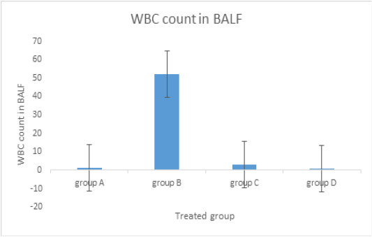
Influence of dipyridamole on BALF WBC count. Rats in Group A received distilled water as a control for a period of 14 days. Group B: rats treated solely with airway ova sensitization served as a positive control group. Group C: oral dipyridamole (26.4 mg / kg / d) in conjunction with airway sensitization by ova. Group D: received oral prednisolone (4.12 mg/kg/d) along with airway ova sensitization.
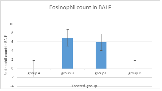
Effect of dipyridamole on BALF eosinophil count. Rats in Group A received distilled water as a control for a period of 14 days. Group B: rats treated solely with airway ova sensitization served as a positive control group. Group C: oral dipyridamole (26.4 mg / kg / d) in conjunction with airway sensitization by ova. Group D: received oral prednisolone (4.12 mg/kg/d) along with airway ova sensitization.
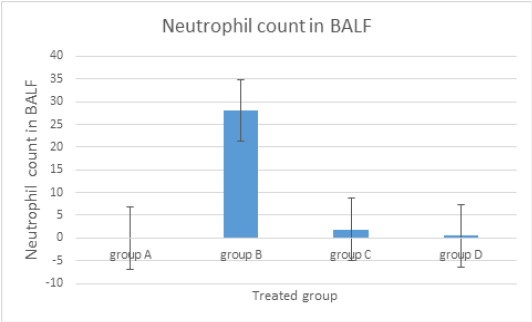
The influence of dipyridamole on the BALF neutrophil count. Rats in Group A received distilled water as a control for a period of 14 days. Group B: rats treated solely with airway ova sensitization served as a positive control group. Group C: oral dipyridamole (26.4 mg / kg / d) in conjunction with airway sensitization by ova. Group D: received oral prednisolone (4.12 mg/kg/d) along with airway ova sensitization.
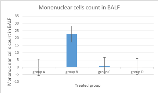
Dipyridamole influence on the count of mononuclear cells (lymphocytes + monocytes) in BALF. Rats in Group A received distilled water as a control for a period of 14 days. Group B: rats treated solely with airway ova sensitization served as a positive control group. Group C: oral dipyridamole (26.4 mg / kg / d) in conjunction with airway sensitization by ova. Group D: received oral prednisolone (4.12 mg/kg/d) along with airway ova sensitization.
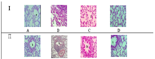
Histopathology of rat lung tissue in response to dipyridamole. Images of lung tissue taken using an X-40 light microscope. Alveolar Sac (a) in Section I. The bronchi in Section (II) (b). Group A is the negative control; Group B is the positive control; Group C is the Dipyridamole-treated group; Group D is the group treated with prednisolone.
References
- Abreu SC, Antunes MA, Mendonça L,Branco VC, de Melo EB, Olsen PC, Diaz BL, Weiss DJ, Paredes BD, Xisto DG, Morales MM, Rocco PRM (2014). Effects of bone marrow mononuclear cells from healthy or ovalbumin- induced lung inflammation donors on recipient allergic asthma mice, Stem Cell Res. Therap. 5(5):108. https://doi.org/10.1186/scrt496[CrossRef]
- Ahmed ZH, Zalzala MH, Ibrahim MA (2021). Guggulsterone Suppresses Ovalbumin-Induced Inflammation in Rat Asthmatic Model. Indian J. Forensic Med. Toxicol. 15(1): 2388-2395.
- Algaem MA, Numan IT, Hussain SA (2013). Effects of Valsartan and Telmisartan on the LungTissue Histology in Sensitized Rats. 1(4):56-60. https://doi.org/10.12691/ajps-1-4-3[CrossRef]
- Aujla SJ, Alcorn JF (2011). T𝐻17 cells in asthma and inflammation. Biochimica et Biophysica Acta.1810(11):1066–1079.
- Ayakannu R, Abdullah NA, Radhakrishnan AK,Raj VL and Liam CK (2019) .Relationship between various cytokines implicated in asthma. Human Immunol.80(9):755–763. https://doi.org/10.1016/j.humimm.2019.04.018[CrossRef]
- Bandeira-Melo C, Bozza PT, Weller PF (2002). The cellular biology of eosinophil eicosanoid formation and function, J. Allerg. Clin. Immunol.109(3):393–400. https://doi.org/10.1067/mai.2002.121529[CrossRef]
- Bharat A, Bhorade SM, Morales-Nebreda L, McQuattie-Pimentel AC, Soberanes S, Ridge K,DeCamp MM. Mestan KK,Perlman H, Budinger GRS, Misharin AV (2016). Flow Cytometry Reveals Similarities Between Lung Macrophages in Humans and Mice. Amirican Journal of Respiratory Cell Molecul. Biol.54(1):147–149. https://doi.org/10.1165/rcmb.2015-0147LE[CrossRef]
- Bours MJL, Swennen ELR, Virgilio FD, Cronstein BN, Dagnelie PC (2006). Adenosine 50 - triphosphate and adenosine as endogenous signaling molecules in immunity and inflammation. Pharmacol. Therapeutics.112(2): 358–404. https://doi.org/10.1016/j.pharmthera.2005.04.013[CrossRef]
- Brusselle G G, Maes T, Bracke K R (2013). Eosinophilic airway inflammation in nonallergic asthma,” Nat. Med.19(8):977–979. https://doi.org/10.1038/nm.3300[CrossRef]
- Buters TP, Hameeteman PW, Jansen IME, van Hindevoort FC, Voorde WT, Grievink HW, Schoonakker M, de Kam ML, Gilroy DW, Feiss G, Rissmann R, Jansen MAA, Burggraaf J, Moerland M (2022).Clinical, Cellular, and Molecular Effects of Corticosteroids on the Response to Intradermal Lipopolysaccharide Administration in Healthy Volunteers, Clin. Pharmacol. Therapeut.111(4):964-971. https://doi.org/10.1002/cpt.2516[CrossRef]
- Chakrabarti S, Freedman JE (2008). Dipyridamole, cerebrovascular disease, and the vasculature. Vascul. Pharmacol.48(4-6): 143- 149. https://doi.org/10.1016/j.vph.2007.12.004[CrossRef]
- Chakrabarti S, Vitseva O. lyu D,Varghese S, Freedman JE (2005).The effect of dipyridamole on vascular cell- derived reactive oxygen species. J. Pharmacol. Exper. Therapeutics .315(2): 494- 500. https://doi.org/10.1124/jpet.105.089987[CrossRef]
- Chanez P, Bourdin A, Vachier I, Godard P, Bousquet J, Vignola AM (2004). Effects of inhaled corticosteroids on pathology in asthma and chronic obstructive pulmonary disease. Proceed. American Thoracic Societ.1(3): 184–190. https://doi.org/10.1513/pats.200402-006MS[CrossRef]
- Chen L, Deng H, Cui H, Fang J, Zuo Z,Deng J,Li Y,Wang X, Zhao L (2018). Inflammatory responses and inflammation-associated diseases in organs. Oncotarget.9(6):7204–7218. https://doi.org/10.18632/oncotarget.23208[CrossRef]
- Defnet AE, , Hasday JD, Shapiro P (2020). Kinase inhibitors in the treatment of obstructive pulmonary diseases . Curr. Opin. Pharmacol.51:11-18. https://doi.org/10.1016/j.coph.2020.03.005[CrossRef]
- Doherty DF, Nath S, Poon J, Foronjy RF, Ohlmeyer M, Dabo AJ, Salathe M, Birrell M, Belvisi M, Baumlin N et al. , (2019). Protein phosphatase 2A reduces cigarette smoke-induced cathepsin S and loss of lung function. American J. Respirat. Crit. Care Med.200(1):51–62. https://doi.org/10.1164/rccm.201808-1518OC[CrossRef]
- Dong H, Osmanova V, Epstein PM, Brocke S (2006). Phosphodiesterase 8 (PDE8) regulates chemotaxis of activated lymphocytes. Biochem. Biophys. Res. Commun.345(2):713–719. https://doi.org/10.1016/j.bbrc.2006.04.143[CrossRef]
- Fleishaker DL, Mukherjee A, Whaley FS, Daniel S, Zeiher BG (2016). Safety and pharmacodynamics dose response of short-term prednisone in healthy adult subjects: a dose ranging, randomized, placebo-controlled, crossover study. BMC Musculoskeletal Disorders.17:293. https://doi.org/10.1186/s12891-016-1135-3[CrossRef]
- Geissmann F, Jung S, Littman DR (2003). Blood monocytes consist of two principal subsets with distinct migratory properties. Immunity.19(1):71–82. https://doi.org/10.1016/S1074-7613(03)00174-2[CrossRef]
- Ghoneim HE, Thomas PG, McCullers JA (2013). Depletion of alveolar macrophages during influenza infection facilitates bacterial superinfections. J. Immunol.191(3):1250–1259. https://doi.org/10.4049/jimmunol.1300014[CrossRef]
- Hall S, Agrawal DK (2014). Key mediators in the immunopathogenesis of allergic asthma. Int. Immunopharmacol. 23(1): 316–329. https://doi.org/10.1016/j.intimp.2014.05.034[CrossRef]
- Hallevi H, Hazan-Halevy I, Paran E (2007). Modification of neutrophil adhesion to human endothelial cell line in acute ischemic stroke by dipyridamole and candesartan. European J. Neurol. 14(9): 1002–1007. https://doi.org/10.1111/j.1468-1331.2007.01847.x[CrossRef]
- Hallstrand TS, Henderson Jr WR (2010). An update on the role of leukotrienes in asthma. Curr. Opin. Allergy Clin. Immunol. 10(1): 60–66. https://doi.org/10.1097/ACI.0b013e32833489c3[CrossRef]
- Hashimoto D, Chow A, Noizat C, Teo P, Beasley MB, Leboeuf M,Becker CD, See P, Price J, Lucas D, Greter M, Mortha A, Boyer SW, Forsberg EC,10,Tanaka M, Rooijen Nv, Garcı´a-Sastre A, Stanley ER, Ginhoux F, Frenette PS, Merad M (2013).Tissue-resident macrophages self-maintain locally throughout adult life with minimal contribution from circulating monocytes. Immunity.38(4):792–804. https://doi.org/10.1016/j.immuni.2013.04.004[CrossRef]
- Haskó G, Cronstein BC (2004). Adenosine: An Endogenous Regulator of Innate Immunity. Trends Immunol.25(1):33–39 https://doi.org/10.1016/j.it.2003.11.003.[CrossRef]
- Hill AT, Sullivan AL, Chalmers JD,De Soyza A, Elborn JS,Floto RA,Grillo L, Gruffydd -Jones K,Harvey A,Haworth CS et al.(2019). British Thoracic Society guideline for bronchiectasis in adults. Thorax.74(Suppl 1):1-69. https://doi.org/10.1136/thoraxjnl-2018-212463[CrossRef]
- Humbles AA, Lloyd CM, McMillan SJ, Friend DS, Xanthou G, McKenna EE, Ghiran S, Gerard NP, YU C, Orkin SH, Gerard C (2004).A critical role for eosinophils in allergic airways remodeling.Science.305(5691):1776–1779. https://doi.org/10.1126/science.1100283[CrossRef]
- Khaldi T, Chekchaki N, Boumendjel M, Taibi F, Abdellaoui M, Messarah M, Boumendjel A (2018). Ameliorating effects of Nigella sativa oil on aggravation of inflammation, oxidative stress and cytotoxicity induced by smokeless tobacco extract in an allergic asthma model in Wistar rats, Allergol. Immunopathol. (Madr).46(5):472-481. https://doi.org/10.1016/j.aller.2018.02.005[CrossRef]
- Kim DI, Song M-K, Lee (2019). Comparison of asthma phenotypes in OVA induced mice challenged via inhaled and intranasal routes, BMC Pulmonary Medicine.19(1):241 https://doi.org/10.1186/s12890-019-1001-9.[CrossRef]
- Kim HH, Liao JK (2008). Translational therapeutics of dipyridamole. Arteriosclerosis, Thrombosis Vascul. Biol.28(3): s39–s42. https://doi.org/10.1161/ATVBAHA.107.160226[CrossRef]
- Kyttaris VC,Zhang Z, Kampagianni O, Tsokos GC(2011). Calcium signaling in systemic lupus erythematosus t cells: A treatment target. Arthritis Rheum.63(7): 2058–2066. https://doi.org/10.1016/B978-0-12-374994-9.10059-2[CrossRef]
- Lambrecht BN, Hammad H (2015). The immunology of asthma. Nat. Immunol.16(1):45–56. https://doi.org/10.1038/ni.3049[CrossRef]
- Larché M, Robinson DS, Kay AB (2003). The role of T lymphocytes in the pathogenesis of asthma. J. Allergy Clin. Immunol. 111(3):450–463. https://doi.org/10.1067/mai.2003.169[CrossRef]
- Ley K, Laudanna C,Cybulsky MI, Nourshargh S (2007). Getting to the site of inflammation: the leukocyte adhesion cascade updated. Nat. Rev. Immunology.7(9):678–689. https://doi.org/10.1038/nri2156[CrossRef]
- McGeachy MJ, Chen Y,Tato CM, Laurence A, Joyce-Shaikh B, Blumenschein WM, McClanahan TK, O’Shea JJ, Cua DJ (2009). The interleukin 23 receptor is essential for the terminal differentiation of interleukin 17-producing effector T helper cells in vivo. Nat. Immunol. 10(3): 314–324. https://doi.org/10.1038/ni.1698[CrossRef]
- Mina S, Elfeky DM, Kabel AM and Hedya SE (2023). Ameliorative Potential of Donepezil with or without Prednisolone in Bleomycin-Induced PulmonaryFibrosis in Rats: Involvement of the Anti-Inflammatory, Antioxidant, and the Antifibrotic Pathways. Medicina (Kaunas).59(5): 980. https://doi.org/10.3390/medicina59050980[CrossRef]
- Newcomb DC, Peebles Jr RS (2013).Th17-mediated inflammation in asthma. Curr. Opin. Immunol. 25(6):755–760. https://doi.org/10.1016/j.coi.2013.08.002[CrossRef]
- Paulus P, Holfeld J, Urbschat A, Mutlak H, Ockelmann PA, Tacke S, Zacharowski K, Reissig C, Stay D, Scheller B (2013).Prednisolone as Preservation Additive Prevents from Ischemia Reperfusion Injury in a Rat Model of Orthotopic Lung Transplantation, PLOS ONE. 8(8):e73298. https://doi.org/10.1371/journal.pone.0073298[CrossRef]
- Peters-Golden M, Henderson Jr WR (2007). Mechanisms of disease: Leukotrienes. New England J. Med.357(18):1841–1854. https://doi.org/10.1056/NEJMra071371[CrossRef]
- Polosa R, Thomson NC (2013). Smoking and asthma: dangerous liaisons. Euro. Respirat. J.41(3):716–726. https://doi.org/10.1183/09031936.00073312[CrossRef]
- Roumestan C, Michel A, Bichon F, Portet K, Detoc M, Henriquet C, Jaffuel D, Mathieu M (2007). Anti-inflammatory properties of desipramine and fluoxetine. Respirat. Res.8(1):35. https://doi.org/10.1186/1465-9921-8-35[CrossRef]
- Rovina N, Koutsoukou A, Koulouris NG (2013). Inflammation and immune response in COPD: where do we stand? Mediat. Inflamm.2013:413735. https://doi.org/10.1155/2013/413735[CrossRef]
- Säfholm J, Manson ML,Bood J,Delin I,Orre AC,Bergman P,Al-Ameri M, Dahlén SE, Adner M (2015). Prostaglandin E2 inhibits mast cell-dependent bronchoconstriction in human small airways through the E prostanoid subtype 2 receptors. J. Allergy Clin. Immunol.136(5):1232–1239.e1. https://doi.org/10.1016/j.jaci.2015.04.002[CrossRef]
- Sakae TM, Maurici R, Trevisol DJ, Menezes Pizzichini MM, Pizzichini E (2014). Effects of prednisone on eosinophilic bronchitis in asthma: A systematic review and meta-analysis, J. Bras. Pneumol. 40(5):552-563. https://doi.org/10.1590/S1806-37132014000500012[CrossRef]
- Serbina NV, Pamer EG (2006). Monocyte emigration from bone marrow during bacterial infection requires signals mediated by chemokine receptor CCR2. Nature Immunology.7(3):311–317. https://doi.org/10.1038/ni1309[CrossRef]
- Shin J-W, Seol I-C, Son C-G. Interpretation of animal dose and human equivalent dose for drug sodium pentobarbital for euthanasia in laboratory rats development. J. Korean Med. 2010; 31(3):1-7.
- Soni NOP (2016). Nuclear factor-Kappa-B as molecular target in asthma , World J. Pharm. Pharmaceut. Sci. 5 (7):481-498.
- Thakur VR, Khuman V, Beladiya JV, Chaudagar KK, Mehta AA (2019). An experimental model of asthma in rats using ovalbumin and lipopolysaccharide allergens. Heliyon.5(11):e02864. https://doi.org/10.1016/j.heliyon.2019.e02864[CrossRef]
- Thomson NC, Chaudhuri R, Livingston E (2004). Asthma and cigarette smoking. European Respirat. J. 24(5):822–833. https://doi.org/10.1183/09031936.04.00039004[CrossRef]
- Trejo Bittar HE, Yousem SA, Wenzel SE (2015). Pathobiology of severe asthma. Ann. Rev. Pathol. Mechan. Dis.10:511–545. https://doi.org/10.1146/annurev-pathol-012414-040343[CrossRef]
- Tsou CL, Peters W, Si Y,Slaymaker S,Aslanian AM,Weisberg SP,Mack M and Charo IF(2007). Critical roles for CCR2 and MCP-3 in monocyte mobilization from bone marrow and recruitment to inflammatory sites. J. Clin. Investigat.117(4):902–909. https://doi.org/10.1172/JCI29919[CrossRef]
- Vogelmeier CF, Criner GJ, Martinez FJ, Anzueto A, Barnes PJ, Bourbeau J, Celli BR, Chen R, Decramer M, Fabbri LM et al.(2017). Global strategy for the diagnosis, management, and prevention of chronic obstructive lung disease 2017 report. GOLD Executive Summary. American J. Respirat. Crit. Care Med.195(5):557–582. https://doi.org/10.1164/rccm.201701-0218PP[CrossRef]
- Vroman H, den Blink Bv, Kool M (2015). Mode of dendritic cell activation: the decisive hand in Th2/Th17 cell differentiation. Implications in asthma severity? Immunobiology. 220(2):254-261. https://doi.org/10.1016/j.imbio.2014.09.016[CrossRef]
- Wang T , Yin K-s, Liu K-y, Lu G-j, Li Y-h, Chen J-d (2008).. Effect of valsartan on the expression of angiotensin II receptors in the lung of chronic antigen exposure rats Chinese Med. J. (Engl) 2008;121(22):2312-2319. https://doi.org/10.1097/00029330-200811020-00018[CrossRef]
- Weyrich AS, Denis MM, Kuhlmann-Eyre JR, Spencer ED, Dixon DA, Marathe GK, McIntyre TM, Zimmerman GA, Prescott SM (2005). Dipyridamole Selectively Inhibits Inflammatory Gene Expression in Platelet–Monocyte Aggregates. Circulation.111(5):633-642. https://doi.org/10.1161/01.CIR.0000154607.90506.45[CrossRef]
- Yona S, Kim KW, Wolf Y, Mildner A,Varol D, Breker M, Strauss-Ayali D,Viukov S, Guilliams M, Misharin A,Hume DA,Perlman H, Malissen B, Zelzer E, Jung S (2013). Fate mapping reveals origins and dynamics of monocytes and tissue macrophages under homeostasis. Immunity. 38(1):79–91. https://doi.org/10.1016/j.immuni.2012.12.001[CrossRef]
- Yu Q-L, Chen Z (2018). Establishment of different experimental asthma models in mice, Experimen. Therapeut. Med.15(3):2492–2498. https://doi.org/10.3892/etm.2018.5721[CrossRef]
- Zatroch KK,Knight CG, Reimer JN, Pang DSJ (2017). Refinement of intraperitoneal injection of sodium pentobarbital for euthanasia in laboratory rats (Rattus norvegicus). BioMed. Central Vet. Res.13(1):60. https://doi.org/10.1186/s12917-017-0982-y[CrossRef]
Services
Article usage statistics
Loading…