
The emergence of antimicrobial resistance in Staphylococcus aureus posed a major veterinary and public challenge worldwide. S. aureus being a highly versatile pathogen can quickly acquire resistance genes. The development of resistance in bacteria predates the era of antibiotic use. However, resistance developments in S. aureus have been reported since the early 1940-ties, when penicillin resistant S. aureus was first reported. Ever since, this pathogen has gain global notoriety as the most common cause of nosocomial, community and livestock associated infection. The mechanism of resistance development in bacteria involved the integration of a complex systems that included the efflux pump, alteration of drug target site, enzymatic inactivation and, mutation in drug target site and gene acquisition of resistance determinants through horizontal gene transfer. This review focused on the mechanisms of antimicrobial resistance in S. aureus. Understanding the concept of resistance development and transfer will immensely help in curtailing the global rise in antimicrobial resistance in bacteria.
Research Article · Volume 4, Issue 2, 19 October 2018 · Open Access
Staphylococcus aureus: A Review of Antimicrobial Resistance Mechanisms
· · ·
Affiliations & Notes ⌄
- 1Department of Veterinary Public Health, Faculty of Veterinary Science, Chulalongkorn University, Pathumwan, Bangkok 10330 Thailand
- 2Bacterial Research Department, National Veterinary Research Institute (NVRI) Vom, Nigeria
- 3Department of Human Physiology, College of Health Sciences, Bayero University Kano, Nigeria
- 4Department of Microbiology and Parasitology, School of Medical Sciences, Universiti Sains Malaysia Health Campus, Kubang Kerian, Kelantan Malaysia. abasinamai@gmail.com
Article Info ⌄
- Journal:
- Veterinary Sciences: Research and Reviews
- Published:
- 19 October 2018
From the article
- Over the past six decades, the use of antibiotics for a long period have been observed to ignite a number of biochemical and genetic mechanism in bacteria that allows it to maneuver the detrimental effect of antibiotics found within their immediate environment.
- Clones of bacteria with acquired or natural resistance characteristics have been used continuously as a form of evolutionary response to the use of antibiotics.
- It is a well-established fact that the acquisition of antibiotic resistance mechanism occurred because of genetic events causing changes in the primordial bacterial genome such as deletion or substitution of a single nucleotide base and multiplication of a single number of a gene.
- However, the most important means of persistence of resistance gene, is the horizontal transfer of mobile genetic elements such as transposons, integrons, and plasmids both within bacteria of the same or different species.
Selected from the article's own text.
Abstract
Introduction
Staphylococcus aureus is a ubiquitous, versatile and highly adaptive pathogen that colonizes the skin and mucous membrane of the anterior nares, gastrointestinal tracts, perineum, the genitourinary tracts and pharynx . It is the causative agent of a wide range of infections in humans and animals with a significant impact on public health . Host specialization, ability to acquire and loss resistance and virulence genes as well as its zoonotic potential posed a significant public health implication ; ; .
Clinically, S. aureus is the most pathogenic member of the genus staphylococci and the etiologic agent of a wide variety of diseases that ranges from superficial skin abscess, food poisoning and life threatening diseases such bacteremia, necrotic pneumonia in children and endocarditis . In animals, it causes mastitis in cow, botryomycosis in horses, dermatitis in dogs, septicemia and arthritis in poultry ; . The severity of the disease is due to the production of several putative virulence factors and possession of antibiotic resistance genes such as mecA, VanA, staphylococcal exotoxins and other factors that facilitates the initiation of disease process, immune evasion and host tissue destruction ; .
Antibiotics resistance development in S. aureus was first reported in the mid-1940-ties when a strain of S. aureus developed resistance against penicillin by the production of a hydrolyzing enzyme called penicillinase . Since then, S. aureus strains resistant to penicillin were widely isolated in cases of bacteremia in the UK and United States. Initially, those resistant strains were only isolated from patients and health care personnel where it derives the name nosocomial associated penicillin resistant S. aureus. However, resistant strains without apparent identifiable risk factors associated with the hospital strains were later isolated among individuals in the community . This led to a scenario where increased resistance to penicillin were observed from the late 1940s until the early 1960s when a semi-synthetic homologue of penicillin called methicillin was introduced into the clinics as a strategic drug of choice for the treatment of S. aureus infection (Jevon, 1961). However, resistance development to methicillin in S. aureus was reported within a year of its introduction as a strategic drug of choice for the treatment of S. aureus infection.
Methicillin resistant S. aureus (MRSA) arises because of the acquisition of a genomic island carrying methicillin resistance determinant, mecA. Ever since its discovery in the early 1960s in the UK, methicillin resistant S. aureus have gain global notoriety as the most common cause of human, community and livestock associated infections worldwide. Thus, leading to a reduction in the therapeutic value of many critically important antibiotics and prolonging the length of hospital admission (Purrello et al., 2011). Over the past decades, MRSA has evolved, and this could probably be due to clonal expansion of previously existing clones and from the conversion of methicillin susceptible S. aureus (MSSA) to MRSA. This is a sequel to the acquisition of a methicillin resistance determinants coding for an alternative penicillin binding protein with reduced or less susceptibility to all classes of beta lactams antibiotics . This review focused on the mechanism of antimicrobial resistance in S. aureus.
Classification of staphylococcus aureus
Staphylococcus aureus is a gram-positive non-motile, non-spore forming facultative anaerobe that is biochemically catalase and coagulase positive. It occurs as an irregularly grape-like cluster and sometimes singly or in pairs, typical colonies are smooth raised yellow to golden yellow color and hemolytic on blood agar containing 5% sheep or horse blood ; .
To date, there are about 40 Staphylococcal species that have been reported, nine of them have two subspecies while one has three subspecies (Doskar et al., 2010). The classification of Staphylococci is not complete yet; new species undergoing validation are still being reported. While some members are important to human medicine, others are relevant to veterinary medicine as they are found in animals or food. Biochemically members of the genus are grouped into two; such as coagulase positive staphylococci and coagulase negative staphylococci. Staphylococcus aureus being the most important member of coagulase positive staphylococci causing infection in both humans and animals and are considered as the most pathogenic members of the genus staphylococci ; Doskar et al., 2010). Other coagulase positive staphylococcus includes Staphylococcus intermedius, Staphylococcus hyicus, Staphylococcus pseudintermedius, Staphylococcus lutrae, Staphylococcus schleiferi subspecies coagulans, and Staphylococcus delphini which were mostly isolated in animals ; Doskar et al., 2010). Le Loir et al. (2003), reported the classification of S. aureus into six different biotypes per their source and biochemical properties these includes; human, non-β-hemolytic human, bovine, ovine, avian and nonspecific.
Morphology and biochemical characteristics of Staphylococcus aureus
The word staphylococci were derived from two Greek words staphyle which means “bunch of grapes” and coccus which means “spherical bacteria” while aureus is a Latin word that stands for “gold” and was given to these bacteria because of yellow to yellowish white colonial appearance on enriched medium . Staphylococcus aureus is a gram positive non-motile, non-spore forming, facultative anaerobe and pathogenic member of the genus staphylococci approximately 1µM in size . It forms golden colonies on rich medium and hemolysis on blood agar containing 5% sheep and horse blood due to production of carotenoids and β-hemolysin, on gram staining it appears as bluish grape-like colonies because cell division occurs at different planes . Staphylococcus aureus is catalase-positive, a unique feature that differentiates it with Streptococcus spp., it is oxidase-negative therefore requiring certain important amino acid and B vitamins for growth and can also tolerate high salt concentration. The cell wall is made up of peptidoglycan which contains crosslinks of glycine residue that allows sensitivity towards lysostaphin ; .
Adaptation of Staphylococcus aureus
Members of the genus Staphylococci are ubiquitous and highly versatile, they are found on the skin, mucous membranes, skin glands, soil, water and air . Staphylococcus aureus is a very hardy organism and can survive on dry surfaces over a long period; it is resistant to desiccation and can survive high level of salt concentration a basis for selection on growth media from other bacteria ; . The bacteria can grow on a varying range of temperature from 15 to 45 ºC. Being a facultative anaerobe, they are capable of oxidative fermentation to produce energy and lactic acid. It is one of the most important pathogenic members of the genus Staphylococci and a leading cause of nosocomial, community and livestock associated infection .
The stability and worldwide spread of this pathogen is due to its’ ability to rapidly acquire and loss resistance and virulence determinants from other members of the genus Staphylococci through horizontal transfer of mobile genetic elements (MGEs) ; ; . Studies on whole genome sequence has revealed that the S. aureus genome is divided into a relatively stable core genome which is about 75-80% of the entire genome and a relatively less stable mobile genetic element (MGE) consisting of transposons, pathogenicity island, Staphylococcus cassette chromosomes, plasmids, bacteriophage and insertion sequence ; . The MGEs in S. aureus are lineage specific and freely integrate, recombine and transfer in and out of genome via horizontal transfer . They encode a wide array of resistance and virulence gene and immune evasion genes, thus facilitating successful adaptation of MRSA and emergence of new and highly resistant and pathogenic clones.
Development of antimicrobial resistance in Staphylococcus aureus
Staphylococcus aureus offers a better and more robust model to understanding the complexity of the adaptive advancement of bacteria in the face selective antibiotic pressure. These pathogens have manifested a novel ability to speedily respond to the challenges posed by new antibiotics via the evolution of new antimicrobial resistance mechanisms. Resistance developments in these pathogens occur via alteration of the drug target site, enzymatic inactivation of the antimicrobial agent, efflux pump and sequestration of the antimicrobial agent (Figure 1). Other resistance mechanisms have developed through acquisition of resistance determinants, position selection and spontaneous mutation (Pantosti et al., 2007; .
Staphylococcus aureus have a highly clonal core genome that is categorized into lineages characterized by clonal complexes. The pathogens are also categorized based on their epidemiological features as nosocomial, community and livestock associated S. aureus. In addition, to the core genome, the pathogen possesses a highly divergent and remarkably variable mobile genetic element. More than 15% of the S. aureus genome is made of up mobile genetic elements (MGEs) such as staphylococcus cassette chromosomes (SCCs), bacteriophages, integrons, integrative conjugative plasmids, transposons and pathogenicity island. All these MGEs but, bacteriophages may carry antimicrobial resistance genes. Majority of S. aureus clinical isolates possesses a plasmid that ranges from 1 to 60kb in size and these plasmids are known to carry variable numbers of resistance genes. Resistance to tetracycline, chloramphenicol and erythromycin are carried by small plasmids while, large plasmids carry multiple drug resistance genes to aminoglycosides, beta-lactams and macrolides. Additionally, larger plasmids also integrate with other MGEs such as transposons and confer resistance to spectinomycin, trimethoprim, erythromycin, beta lactams and vancomycin ; ; ;
Antibiotic resistance in S. aureus predates the era of antibiotics use in clinical practice. Prior to introduction of penicillin, mortality because of invasive S. aureus infection was very high. However, penicillin had a significant effect in reducing the rate of mortality because of S. aureus infection, not until 1942 when a strain of S. aureus resistant to penicillin was identified first in the hospital and then from the community . The use of penicillin as a drug of choice in the treatment of S. aureus infection was very effective until the mid-1950s when the number of S. aureus resistant to penicillin significantly increased leading to a decrease in the therapeutic value of penicillin . Freeman-cook and Freeman-cook, (2006) reported that about 90% of S. aureus are penicillin resistant. Resistance to penicillin was acquired via acquisition of plasmids coding for beta lactam resistance . The greatest challenge to the treatment of S. aureus infection is in the selection of the appropriate therapeutic agent. This is because the pathogens have the potentials of developing resistance to almost all classes of antibiotics (Figure 1). The understanding that antibiotic resistance in S. aureus predates the era of antibiotics use in the clinic validates the challenges experienced because of resistance development in recent times. Prior to introduction of penicillin, mortality because of invasive S. aureus infection was very high. However, with the introduction of penicillin into clinical practice in the 1940s there was a significant reduction in the rate of mortality because of S. aureus infection . This was however short-lived in 1942 when a strain of S. aureus resistant to penicillin was identified first in the hospital and then from the community . Resistance to penicillin is mediated by blaZ gene which codes beta lactamase enzymes. Beta lactamase are extracellular enzymes synthesized on exposure to beta lactams class of antibiotics, it hydrolyses the beta lactam ring thereby reducing the therapeutic effect of penicillin .
Methicillin resistant determinant mecA is located on large 25-65kb mobile cassette chromosomes called SCCmec that facilitates the horizontal transfer of resistance determinants in and out of the bacteria . In addition, it was reported that the acquisition of mecA seems to have occurred independently in several S. aureus strains, with some clonal lineages having the propensity to colonize specific species and may be adapted to either humans or animals. Other lineages have less host-specificity and can infect a wide variety of species . Moreover, transfer and worldwide dissemination of antibiotic resistance determinants among clinically important bacteria and their mobile genetic element have long been observed to have occurred between bacteria of the same and different clusters ; Wielders et al., 2001; ; Bitrus et al., 2016a). Some studies have also demonstrated the role of horizontal gene transfer in rapid acquisition and dissemination of antibiotics resistance determinants in S. aureus ; Barlow, 2009; ; . The report of Huddleston, (2014) and Lindsay, (2014) further gives credit to these findings where they reported the role of horizontal gene transfer events in ensuring wide genetic variability as well as successful adaptation between bacteria through high transfer frequency of resistance determinants.
The evolutionary origin as well as detailed mechanism of transfer of mecA is not fully understood (Barlow, 2009; Hanssen et al; 2004). However, studies on Staphylococcus sciuri and Staphylococcus hominis have revealed the presence of methicillin resistant determinant mecA with 88% similarity in sequence of amino acid and 80 % DNA sequence identity to the mecA gene of MRSA . In addition, transfer of methicillin resistance has been observed to have occurred both in vitro and in vivo from Staphylococcus epidermidis to S. aureus indicating the role of coagulase negative Staphylococci serving as reservoirs of mecA ; . Furthermore, it has been observed that, the most common pathway of gene transfer events in S. aureus is generalized transduction, however transformation and conjugative plasmid transfer have been observed to have occurred too ; ; ; . Similarly, only in vivo conjugative plasmid transfer has been reported to be significant . Conjugative transfer of resistance determinants in S. aureus is known to be mediated by conjugative plasmids; however, transfer of resistance determinants in the absence of conjugative plasmids have been reported to have occurred .
Most studies on transfer of antibiotic resistance in human S. aureus strains have indicated coagulase negative staphylococci (CoNS) as reservoirs of resistant determinants ; ; . Similarly, studies on antibiotic resistance transfer between human and animal isolates were reported to occur, indicating the importance of resistance transfer in the dissemination and successful adaptation of methicillin resistant S. aureus ; . The rapid spread of resistance between bacteria has been one of the factors limiting the production of new antibiotics to curb the increasing impact of antibiotics resistance on healthcare cost (Barlow, 2009).
Resistance to β-lactams
The common most important inhibitory target site for beta lactams antimicrobials in S. aureus is the two-way functional transglycolylase-transpeptidase PBP2. The domain containing the transglycosylase of the enzyme coordinates the transfer of disaccharide pentapeptide raw material of peptidoglycan from membrane-bound lipid II to budding polysaccharide chains. The domain containing the transpeptidase helps to connects to the glycine cross-bridge of the fourth D-alanine of a chain adjacent to it . Members of this class of antibiotics includes, penicillin, oxacillin, methicillin and cephalosporin. They act by inhibiting the transpeptidation step of the peptidoglycan synthesis, which they achieve by binding and inactivation of the penicillin binding proteins in the bacterial cell wall . Resistance development in S. aureus to beta lactams occurs through the acquisition of a genomic island called staphylococcus cassette chromosome (SCCmec) carrying methicillin resistance determinant mecA ; . This in turn codes for an alternative penicillin binding protein with reduced or less susceptibility to methicillin. In addition, resistance to penicillin was acquired via acquisition of plasmids coding for beta lactam resistance . Penicillin resistance is mediated by blaZ gene which codes for beta lactamase enzymes. These genes are regulated by two differently transcribed genes known as blaI and blaRI . Beta lactamase are extracellular enzymes synthesized on exposure to beta lactams class of antibiotics, it hydrolyses the beta lactam ring thereby reducing the therapeutic effect of penicillin.
Resistance to vancomycin
Vancomycin is considered as a strategic drug in the treatment of S. aureus infection (Bitrus et al., 2016a). It acts by inhibiting the transpeptidation of the peptidoglycan layer in the bacterial cell wall by binding to the C-terminal D-ala-D-ala of the peptidoglycan stem pentapeptide, resulting in the prevention of interaction between the penicillin binding proteins and their substrate. Staphylococcus aureus develop resistance to vancomycin through two unique independent mechanisms; this includes: VanA mediated resistance and resistance due to thickened cell wall .
Resistance development mediated by VanA is represented by a high level of inducible resistance to vancomycin and is carried by transposon Tn1546 and closely related elements. This type of resistance development is well established in Enterococcus species (Weigel et al., 2003). The role of VanA ligase is to connect the D-ala and D-lac by esterification with resultant replacement of the D-ala-D-ala terminal of the pentapeptide stem by depsipeptide formation. Furthermore, since vancomycin has reduced affinity for the D-ala-D-lac terminal, it does not prevent the incorporation of the substrates into the bacterial cell wall. In either case, the concurrent formation of the D-ala-D-ala and D-ala-D-lac pentapeptide stem is not sufficient enough to initiate resistance development to vancomycin (Weigel et al., 2003). However, resistance development occurs when VanX hydrolyses the D-ala-D-ala dipeptide and VanY removes the C-terminal D-ala residue of the pentapeptide stem when hydrolysis of VanX is incomplete, leading to the formation of a modified less susceptible target molecule with simultaneous cleavage of either of the existing D-ala-D-ala pentapeptide stem in the cell wall S. aureus .
On the other hand, the mechanism of resistance development as a result of a thickened bacterial cell wall is mostly associated with S. aureus with intermediate resistance to vancomycin ; . Vancomycin intermediate resistant S. aureus (VISA) do not contain the Van gene or any other known determinants of vancomycin resistance but possesses a common phenotype of a thickened cell wall and a ratio of high cell wall to cell wall volume ; . These types of phenotypes have a cell wall with a characteristically low level of peptidoglycan cross-link as compared with the normal staphylococcal cell wall (Courvalin, 2006). The formation of a thickened cell wall as well as reduced formation of peptidoglycan cross-links results in the production of an increased volume of D-ala-D-ala peptide stem outside the cell wall leading to reduced uptake of vancomycin into the cell and subsequently resistance .
Resistance to aminoglycosides
Aminoglycosides are bactericidal antimicrobial agents that act by interfering with protein synthesis when it binds to the 30S ribosomal subunit. Resistance development to aminoglycoside occur through in vitro mutation in the ribosomal subunit. Similarly, acquisition of aminoglycoside modifying enzyme have been reported to serve as a medium for the development of resistance to aminoglycosides ; ; .
Resistant development to fluoroquinolones
Antibiotics under this group act by inhibiting transcription and replication of DNA by targeting DNA gyrase enzymes (Topoisomerase II and IV). Studies have shown that resistance development to quinolone derivatives occurs via two pathways that included mutation of the target Topoisomerase II and IV or through efflux pump system. In addition, it has been established that a single mutation in the target does not confer resistance to quinolones, rather it involves a cascade of mutation associated with increased minimum inhibitory concentration (MIC) of fluoroquinolones ; Courvalin, 2006). Findings have it that for resistance development to fluoroquinolones to occur; there must be mutation in the genes regulating DNA gyrase (gyrA and B) and Topoisomerase (ParC and ParE). Similarly, for resistance development mediated by the efflux pump to occur in S. aureus, it requires a multidrug efflux pump system coordinated by NorA ; .
Resistance to chloramphenicol, rifampin and mupirocin
This group of antibiotic drugs, functions by interfering with protein synthesis in bacteria through different pathways. While Rifampin inhibit transcription by binding to RNA polymerase, Chloramphenicol acts by binding to 50S ribosomal subunits and blocking the action of peptidyl transferase. Mupirocin however, functions by inhibiting isoleucine tRNA synthetase ; ; ; . Resistance development to mupirocin by S. aureus occur through acquisition of mupA gene which codes for a less sensitive tRNA synthetase while resistance to rifampin and chloramphenicol occurs through mutation in the rpoB gene that codes for the Beta subunit of RNA polymerase and action of an inactivating enzyme called chloramphenicol transferases which inactivates the drug .
Resistance to linezolid and tetracycline
Linezolid is a synthetic antimicrobial agent that belongs to the oxazolidinone family and act by interfering with protein synthesis by binding to 50s ribosomal subunits to inhibit the formation of 70s ribosomal initiation complex. It is one of the few antibiotics whose resistance in S. aureus is rare and is considered as a strategic drug of choice for the treatment of S. aureus infection. Resistance development rarely occur but, when it does it is through mutation of the chromosomal gene coding for the 23s rRNA .
Tetracycline on the other hand, is bacteriostatic in nature and acts by inhibiting the formation of protein by binding to 30s ribosomal subunits and blocking of the tRNA from moving into the acceptor site. Resistance development by S. aureus occur via two pathways which includes, ribosomal protection or efflux pump system. The protection of the ribosome is encoded by tetM, while tetK codes for the efflux pump system ; ; .
Resistance to macrolide, lincosamides and streptogramins-B
The mechanism of antibiotic resistance development in S. aureus to macrolide, lincosamides and Streptogramins-B occur via the methylation of their receptor binding site on the ribosomes. It is important to note that even though these classes of antibiotics have similar receptor binding site, they are structurally unrelated. Furthermore, the methylation that happems at their binding site is catalyzed by a methylases enzymes which is encoded by erythromycin methylases enzyme ermA, B and C whose expression is either inducible or constitutive. All the three classes of antibiotics are constitutive but only macrolide can induce expression of gene coding for erythromycin methylases erm and is also mediated by an efflux pump system encoded by mrsA. This however, does not lead to resistance development to Streptogramins or lincosamides ; ; .
Methicillin resistant Staphylococcus aureus (MRSA)
The developments of antibiotics resistance in bacteria were reported even before the era of antibiotic use in the treatment of infection . Antibiotics resistance development in S. aureus was first reported in the mid-1940s when a strain of S. aureus developed resistance against penicillin by the production of a hydrolyzing enzyme called penicillinase .
Methicillin resistant Staphylococcus aureus (MRSA) is an important human pathogen responsible for hospital, community and livestock acquired infection . It is also a leading cause of skin and soft tissue infections in both humans and animals ; and the second most common cause of blood stream infections in nosocomial associated outbreaks with high mortality and increased or prolonged hospital stay (Purrello et al., 2014).
Resistance to methicillin was first reported in the United Kingdom in 1961 not long after the introduction of methicillin for clinical use . Within a few years, outbreaks of methicillin resistance S. aureus were recorded in the United Kingdom and some part of Europe . In the mid-1970s, MRSA was reported to be a significant problem in health care hospitals in the United States. These resistant organisms are now commonly recovered in virtually every large hospital in the United States and other hospitals worldwide and have become a significant infection control problem in nursing homes and other chronic healthcare facilities .
Staphylococcus aureus acquired methicillin resistance through horizontal transfer of mecA which codes for a modified penicillin binding protein (PBP’) with low or reduced affinity to beta-lactam antibiotics. Methicillin resistant determinant, mecA is located on the staphylococcal cassette chromosome mec (SCCmec), a large 20 to 65kb mobile element in S. aureus that mediates the horizontal transfer of methicillin resistance ; ; Stojanov et al., 2012). Resistance acquisition in MRSA occurs through mutation of the target gene in the chromosomes, through efflux pump system, horizontal transfer of MGEs or enzymatic action of drugs as in the case of penicillin . Emergence of bacterial resistance to multiple antibiotics worldwide have made treatment of MRSA infections difficult, although attributed to mutation on the chromosomes, resistance is most commonly associated with extra-chromosomal elements acquired from other bacteria in the environment. However, intrinsic mechanisms not commonly specified by mobile elements such as efflux pumps that expel multiple classes of antibiotics are now recognized as major contributors to multidrug resistance in bacteria. Once established, multidrug-resistant organisms persist and spread worldwide, resulting in failures to treatment of infection . High prevalence of MRSA infection is attributed to toxin production, the ability for rapid spread between humans and animals and its ability to acquire resistance determinants to multiple antibiotics leading to an increased burden on healthcare setting due to a limited treatment options. Because of its frequent association with mobile genetic elements, natural resistance genes can be spread rapidly among pathogenic strains and therefore impedes the clinical value of many drugs .
MRSA is thought to be restricted to the hospital setting, not until the late 1990s when MRSA infection among healthy individuals in the community with no history of hospitalization, intravenous drug use, prior antimicrobial use, and underlying illnesses such as cardiovascular and pulmonary disease, diabetes, malignancy, and chronic skin diseases was reported ; . This new strain called community acquired MRSA were found to be susceptible to only beta lactams antibiotics, harbor different SCCmec class (IV and V SCCmec) and a phage-borne pantone valentine leucocidin (PVL) toxin incriminated in skin and soft tissue infection in healthy children and adults (Grundmann et al., 2006). Reported a relatively high incidence of community associated methicillin resistant S. aureus with SCCmec type IVa or V among healthy carrier patients as in the case with penicillin, methicillin resistance S. aureus were identified among individuals in the community and more recently in livestock . In addition, S. aureus strain showing low level resistance to vancomycin have also been observed .
Conclusion
The number of mechanisms inherent in pathogenic bacteria that makes it resilient or hardy in the presence of extreme conditions and confers it with the ability to resist quite a large compendium of important antibiotics and other toxic compounds are becoming extremely interesting. Over the past six decades, the use of antibiotics for a long period have been observed to ignite a number of biochemical and genetic mechanism in bacteria that allows it to maneuver the detrimental effect of antibiotics found within their immediate environment. Clones of bacteria with acquired or natural resistance characteristics have been used continuously as a form of evolutionary response to the use of antibiotics. It is a well-established fact that the acquisition of antibiotic resistance mechanism occurred because of genetic events causing changes in the primordial bacterial genome such as deletion or substitution of a single nucleotide base and multiplication of a single number of a gene. However, the most important means of persistence of resistance gene, is the horizontal transfer of mobile genetic elements such as transposons, integrons, and plasmids both within bacteria of the same or different species.
Authors’ contribution
AAB and MOP conceived the research review, gathered relevant materials and wrote the first draft of the manuscript, MAA and MDG proof read the manuscript. All authors approved the final draft of this manuscript.
Figures
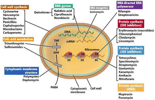
Schematic representation of antibiotic class and mechanism of antibiotic resistance in bacteria (Adopted from Labnotesweek4, 2013).
References
- • Aklilu, E., Zunita, Z., Hassan L., Cheng, C.H. 2013. Molecular epidemiology of methicillin-resistant Staphylococcus aureus (MRSA) among veterinary students and personnel at a veterinary hospital in Malaysia. Veterinary Microbiology. 164(3–4): 352-358. https://doi.org/10.1016/j.vetmic.2013.02.030[CrossRef]
- • Alekshun, M.N., Levy, S.B. 2007. Molecular mechanisms of antibacterial multidrug resistance. Cell, 128(6): 1037-1050. https://doi.org/10.1016/j.cell.2007.03.004[CrossRef]
- • Barlow, M. What antimicrobial resistance has taught us about horizontal gene transfer Horizontal Gene Transfer. (pp. 397-411): Springer.
- • Basset, P., Feil, E.J., Zanetti, G., Blanc, D.S. , 2011. 25 - The Evolution and Dynamics of Methicillin-Resistant Staphylococcus aureus. In M. Tibayrenc (Ed.), Genetics and Evolution of Infectious Disease; (pp. 669-688). London: Elsevier. https://doi.org/10.1016/B978-0-12-384890-1.00025-X[CrossRef]
- • Bitrus, A.A., Zakaria, Z., Bejo, S.K., Othman, S. 2016a. Persistence of Antibacterial Resistance and Virulence Gene Profile of Methicillin Resistant Staphylococcus Aureus (MRSA) Isolated From Humans and Animals. Pakistan Veterinary Journal. 36(1).
- • Bitrus, A.A., Zunita, Z., Bejo, S.K., Othman, S., Nadzir, N.A. 2016b. Detection of virulence genes and antibiotic resistance profiles of Staphylococcus aureus isolated from animals. Malaysian Journal of Microbiology.12(6):408-17.
- • Bitrus, A.A, Zunita, Z., Bejo, S.K., Othman, S., Nadzir, N.A. 2017. In vitro transfer of methicillin resistance determinants mec A from methicillin resistant Staphylococcus aureus (MRSA) to methicillin susceptible Staphylococcus aureus (MSSA). BMC microbiology. 17(1):83. https://doi.org/10.1186/s12866-017-0994-6[CrossRef]
- • Bitrus, A.A., Zunita, Z., Khairani-Bejo, S., Othman, S., Nadzir, N.A. 2018. Staphylococcal cassette chromosome mec (SCCmec) and characterization of the attachment site (attB) of methicillin resistant Staphylococcus aureus (MRSA) and methicillin susceptible Staphylococcus aureus (MSSA) isolates. Microbial pathogenesis. 123: 323-9. https://doi.org/10.1016/j.micpath.2018.07.033[CrossRef]
- • Bitrus, A.A., Zunita, Z., Goni, M.D. and Mshelia, I.T. 2018. Dissemination of resistance and virulence determinants in methicillin-resistant Staphylococcus aureus during colonization and disease. A Review. Advances in Animal and Veterinary Sciences. 6(1): 44-54. https://doi. org/10.17582/journal.aavs/2018/6.1.44.54
- • Bloemendaal, A.L.A., Brouwer, E.C., Fluit, A.C. 2010. Methicillin resistance transfer from Staphylocccus epidermidis to methicillin-susceptible Staphylococcus aureus in a patient during antibiotic therapy. PLoS One, 5(7): e11841. https://doi.org/10.1371/journal.pone.0011841[CrossRef]
- • Bosch, T., Verkade, E., van Luit, M., Landman, F., Kluytmans, J., Schouls, L.M. 2015. Transmission and Persistence of Livestock-Associated Methicillin-Resistant Staphylococcus aureus among Veterinarians and Their Household Members. Applied and Environmental Microbiology, 81(1): 124-129. https://doi.org/10.1128/AEM.02803-14[CrossRef]
- • Bremer, P.J., Fletcher, G.C., Osborne, C. 2004. Staphylococcus aureus. New Zealand Institute for crop and Food Research limited. Available at: http://www.commonwealthofnations.org/organisations/new_zealand_institute_for_crop_and_food_research_ltd/ Last accessed 28/8/2018
- • Bugg, T.D.H. and Brandish, P.E. 1994. From peptidoglycan to glycoproteins: common features of lipid-linked oligosaccharide biosynthesis. FEMS Microbiology Letters, 119(3): 255-262. https://doi.org/10.1111/j.1574-6968.1994.tb06898.x[CrossRef]
- • Bugg, T.D.H., Wright, G.D., Dutka-Malen, S., Arthur, M., Courvalin, P., Walsh, C.T. 1991. Molecular basis for vancomycin resistance in Enterococcus faecium BM4147: biosynthesis of a depsipeptide peptidoglycan precursor by vancomycin resistance proteins VanH and VanA. Biochemistry, 30(43): 10408-10415. https://doi.org/10.1021/bi00107a007[CrossRef]
- • Chambers, H.F. 1997. Methicillin resistance in staphylococci: molecular and biochemical basis and clinical implications. Clinical Microbiology Reviews, 10(4), 781-791
- • Charlebois, E.D., Perdreau-Remington, F., Kreiswirth, B., Bangsberg, D.R., Ciccarone, D., Diep, B.A., Ng, V.L., Chansky, K., Edlin, B., Chambers, H.F. 2004. Origins of community strains of methicillin-resistant Staphylococcus aureus. Clinical Infectious Diseases, 39(1):47-54. https://doi.org/10.1086/421090[CrossRef]
- • Chuang, Y-Y.,Huang, Y-C. 2013. Molecular epidemiology of community-associated meticillin-resistant Staphylococcus aureus in Asia. The Lancet Infectious Diseases, 13(8): 698-708. https://doi.org/10.1016/S1473-3099(13)70136-1[CrossRef]
- • Courvalin, P. Vancomycin resistance in gram-positive cocci. Clinical Infectious Disease, 42: S25–34. https://doi.org/10.1086/491711[CrossRef]
- • Cox, G., Wright G.D. 2013. Intrinsic antibiotic resistance: Mechanisms, origins, challenges and solutions. International Journal of Medical Microbiology, 303(6–7):287-292.
- • den Heijer, C.D.J., van Bijnen, E.M.E., Paget, W.J., Pringle, M., Goossens, H., Bruggeman, C.A., Stobberingh, E.E. 2013. Prevalence and resistance of commensal Staphylococcus aureus, including meticillin-resistant S aureus, in nine European countries: a cross-sectional study. The Lancet Infectious Diseases.13(5); 409-415. https://doi.org/10.1016/S1473-3099(13)70036-7[CrossRef]
- • Deurenberg, R.H., Vink, C., Kalenic, S., Friedrich, A.W., Bruggeman, C.A., Stobberingh, E.E. 2007. The molecular evolution of methicillin‐resistant Staphylococcus aureus. Clinical Microbiology and Infection, 13(3): 222-235. https://doi.org/10.1111/j.1469-0691.2006.01573.x[CrossRef]
- • Doškař, J., Pantůček, R., Růžičková, V., Sedláček, I. 2010. Molecular Diagnostics of Staphylococcus aureus Detection of Bacteria, Viruses, Parasites and Fungi, Springer, 139-184.
- • Forbes, B.A. and Schaberg, D.R. 1983. Transfer of resistance plasmids from Staphylococcus epidermidis to Staphylococcus aureus: evidence for conjugative exchange of resistance. Journal of Bacteriology,153(2):627-634.
- • Foster, T.J. 2017. Antibiotic resistance in Staphylococcus aureus. Current status and future prospects. Fems Microbiology Reviews. 41(3):430-49. https://doi.org/10.1093/femsre/fux007[CrossRef]
- • Freeman-Cook, L., Freeman-Cook, K.D., Alcamo, I.E. and Heymann, D.L. 2006. Staphylococcus aureus infections: Infobase Publishing.
- • Gorak, E.J., Yamada, S.M., Brown, J.D. 1999. Community-acquired methicillin-resistant Staphylococcus aureus in hospitalized adults and children without known risk factors. Clinical Infectious Diseases, 29(4): 797-800. https://doi.org/10.1086/520437[CrossRef]
- • Grundmann, H., Aires-de-Sousa, M., Boyce, J. and Tiemersma, E. 2006. Emergence and resurgence of meticillin-resistant Staphylococcus aureus as a public-health threat. The Lancet, 368(9538): 874-885. https://doi.org/10.1016/S0140-6736(06)68853-3[CrossRef]
- • Haaber, J., Penadés, J.R. and Ingmer, H. 2017. Transfer of antibiotic resistance in Staphylococcus aureus. Trends in Microbiology. 893-905. https://doi.org/10.1016/j.tim.2017.05.011[CrossRef]
- • Hanssen, A-M., Kjeldsen, G. and Sollid, J.U.E. 2004. Local variants of Staphylococcal cassette chromosome mec in sporadic methicillin-resistant Staphylococcus aureus and methicillin-resistant coagulase-negative Staphylococci: evidence of horizontal gene transfer? Antimicrobial Agents and Chemotherapy, 48(1): 285-296. https://doi.org/10.1128/AAC.48.1.285-296.2004[CrossRef]
- • Hiramatsu, K. 1998. Vancomycin resistance in staphylococci. Drug Resistance Updates, 1(2): 135-150. https://doi.org/10.1016/S1368-7646(98)80029-0[CrossRef]
- • Hiramatsu, K. 2004. Elucidation of the mechanism of antibiotic resistance acquisition of methicillin-resistant Staphylococcus aureus (MRSA) and determination of its whole genome nucleotide sequence. Japan Medical Association Journal, 47(4): 153-159.
- • Holden, M.T.G., Feil, E.J., Lindsay, J.A., Peacock, S.J., Day, N.P.J., Enright, M.C., Atkin, R. 2004. Complete genomes of two clinical Staphylococcus aureus strains: evidence for the rapid evolution of virulence and drug resistance. Proceedings of the National Academy of Sciences of the United States of America, 101(26): 9786-9791. https://doi.org/10.1073/pnas.0402521101[CrossRef]
- • Huddleston, J.R. 2014. Horizontal gene transfer in the human gastrointestinal tract: Potential spread of antibiotic resistance genes. Infection Drug Resistance, 7: 167-176. https://doi.org/10.2147/IDR.S48820[CrossRef]
- • Ito, T., Kuwahara, K. and Hiramatsu, K. 2007. Staphylococcal cassette chromosome mec (SCCmec) analysis of MRSA Methicillin-Resistant Staphylococcus aureus (MRSA) Protocols. Springer, 87-102.
- • Jansen, W.T.M., Beitsma, M.M., Koeman, C.J., Van Wamel, W.J.B., Verhoef, J., Fluit, A.C. 2006. Novel mobile variants of staphylococcal cassette chromosome mec in Staphylococcus aureus. Antimicrobial Agents and Chemotherapy, 50(6): 2072- 2078. https://doi.org/10.1128/AAC.01539-05[CrossRef]
- • Jenner, L., Starosta, A.L., Terry, D.S., Mikolajka, A., Filonava, L., Yusupov, M., Blanchard, S.C., Wilson, D.N. and Yusupova, G. 2013. Structural basis for potent inhibitory activity of the antibiotic tigecycline during protein synthesis. Proceedings of the National Academy of Sciences. 110(10):3812-6. https://doi.org/10.1073/pnas.1216691110[CrossRef]
- • Jevons, M.P. 1961. ‘Celbenin’–resistant staphylococci, British Medical Journal, 124: 124–5. https://doi.org/10.1136/bmj.1.5219.124-a[CrossRef]
- • Khan, S.A., Nawaz, M.S., Khan, A.A.,Cerniglia, C.E. 2000. Transfer of erythromycin resistance from poultry to human clinical strains of Staphylococcus aureus. Journal of Clinical Microbiology, 38(5): 1832-1838.
- • Lacey, R.W. 1975. Antibiotic resistance plasmids of Staphylococcus aureus and their clinical importance. Bacteriological Reviews, 39(1): 1.
- • Lamy, B., Laurent, F., Gallon, O., Doucet-Populaire, F., Etienne, J., Decousser, J-W. 2012. Antibacterial resistance, genes encoding toxins and genetic background among Staphylococcus aureus isolated from community-acquired skin and soft tissue infections in France: a national prospective survey. European Journal of Clinical Microbiology and Infectious Diseases, 31(6): 1279-1284. https://doi.org/10.1007/s10096-011-1441-5[CrossRef]
- • Le Loir, Y., Baron, F. and Gautier, M. 2003. Staphylococcus aureus and food poisoning. Genetics and Molecular Research, 2(1): 63-76.
- • Lindqvist, M. 2014. Epidemiological and molecular biological studies of multi-resistant methicillin-susceptible Staphylococcus aureus. Linköping University Medical Dissertations, ISSN 0345-0082; 1386.
- • Lindsay, J.A. 2014. Staphylococcus aureus genomics and the impact of horizontal gene transfer. International Journal of Medical Microbiology, 304: 103-109. https://doi.org/10.1016/j.ijmm.2013.11.010[CrossRef]
- • Lowy, F.D. 2003. Antimicrobial resistance: the example of Staphylococcus aureus. The Journal of Clinical Investigation. 111(9):1265-73. https://doi.org/10.1172/JCI18535[CrossRef]
- • Luzzago, C., Locatelli, C., Franco, A., Scaccabarozzi, L., Gualdi, V., Viganò, R., Sironi, G., Besozzi, M., Castiglioni, B., Lanfranchi, P. and Cremonesi, P. 2014. Clonal diversity, virulence-associated genes and antimicrobial resistance profile of Staphylococcus aureus isolates from nasal cavities and soft tissue infections in wild ruminants in Italian Alps. Veterinary Microbiology, 170(1):157-61. https://doi.org/10.1016/j.vetmic.2014.01.016[CrossRef]
- • McAleese, F., Wu, S.W., Sieradzki, K., Dunman, P., Murphy, E., Projan, S. and Tomasz, A. 2006. Overexpression of genes of the cell wall stimulon in clinical isolates of Staphylococcus aureus exhibiting vancomycin-intermediate Staphylococcus aureus type resistance to vancomycin. Journal of Bacteriology, 188(3): 1120-1133. https://doi.org/10.1128/JB.188.3.1120-1133.2006[CrossRef]
- • McCarthy, A.J. and Lindsay, J.A. 2012. The distribution of plasmids that carry virulence and resistance genes in Staphylococcus aureus is lineage associated. BMC microbiology. 12(1):104. https://doi.org/10.1186/1471-2180-12-104[CrossRef]
- • Morton, T.M., Johnston, J.L., Patterson, J. and Archer, G.L. 1995. Characterization of a conjugative staphylococcal mupirocin resistance plasmid. Antimicrobial Agents and Chemotherapy, 39(6), 1272-1280. https://doi.org/10.1128/AAC.39.6.1272[CrossRef]
- • Mukhtar, T.A., Koteva, K.P, Hughes, D.W. and Wright, G.D. 2001. Vgb from Staphylococcus aureus inactivates streptogramin B antibiotics by an elimination mechanism not hydrolysis. Biochemistry. 40(30):8877-86. https://doi.org/10.1021/bi0106787[CrossRef]
- • Musser, J.M. and Kapur, V. 1992. Clonal analysis of methicillin-resistant Staphylococcus aureus strains from intercontinental sources: association of the mec gene with divergent phylogenetic lineages implies dissemination by horizontal transfer and recombination. Journal of Clinical Microbiology, 30(8): 2058-2063.
- • Nguyen, F., Starosta, A.L., Arenz, S., Sohmen, D., Dönhöfer, A. and Wilson, D.N. 2014. Tetracycline antibiotics and resistance mechanisms. Biological chemistry. 395(5):559-75. https://doi.org/10.1515/hsz-2013-0292[CrossRef]
- • Noto, M.J., Fox, P.M. and Archer, G.L. 2008. Spontaneous deletion of the methicillin resistance determinant, mecA, partially compensates for the fitness cost associated with high-level vancomycin resistance in Staphylococcus aureus. Antimicrobial Agents and Chemotherapy, 52: 1221-1229. https://doi.org/10.1128/AAC.01164-07[CrossRef]
- • Noto, M.J., Kreiswirth, B.N., Monk, A.B. and Archer, G.L. 2008. Gene acquisition at the insertion site for SCCmec, the genomic island conferring methicillin resistance in Staphylococcus aureus. Journal of Bacteriology, 190(4): 1276-1283. https://doi.org/10.1128/JB.01128-07[CrossRef]
- • Nowrouzian, F.L., Karami, N., Welinder-Olsson, C., Åhrén, C. 2013. Virulence gene typing of methicillin-resistant Staphylococcus aureus as a complement in epidemiological typing. Journal of Microbiological Methods, 93(3): 173-176. https://doi.org/10.1016/j.mimet.2013.03.020[CrossRef]
- • Oliveira, D.C. and de Lencastre, H. 2002. Multiplex PCR strategy for rapid identification of structural types and variants of the mec element in methicillin-resistant Staphylococcus aureus. Antimicrobial Agents Chemotherapy, 46(7): 2155-2161. https://doi.org/10.1128/AAC.46.7.2155-2161.2002[CrossRef]
- • Page, M.G.P. 2012. Beta-lactam antibiotics Antibiotic Discovery and Development. Springer, 79-117.
- • Pantosti, A., Sanchini, A. and Monaco, M. 2007. Mechanisms of antibiotic resistance in Staphylococcus aureus. Future Microbiology, 2 (3): 323-334. https://doi.org/10.2217/17460913.2.3.323[CrossRef]
- • Planet, P.J., Narechania, A., Chen, L., Mathema, B., Boundy, S., Archer, G., Kreiswirth, B. 2017. Architecture of a species: phylogenomics of Staphylococcus aureus. Trends in microbiology. 25(2):153-66. https://doi.org/10.1016/j.tim.2016.09.009[CrossRef]
- • Plata, K., Rosato, A.E. and Wegrzyn, G. 2009. Staphylococcus aureus as an infectious agent: overview of biochemistry and molecular genetics of its pathogenicity. Acta Biochimica Polonica, 56(4): 597.
- • Purrello, S.M., Daum, R.S., Edwards, G.F.S., Lina, G., Lindsay, J., Peters, G., Stefani, S. 2012. Meticillin-resistant Staphylococcus aureus (MRSA) update: New insights into bacterial adaptation and therapeutic targets. Journal of Global Antimicrobial Resistance, 2(2): 61-69. https://doi.org/10.1016/j.jgar.2014.02.003[CrossRef]
- • Reynolds, P.E., Depardieu, F., Dutka-Malen, S., Arthur, M. and Courvalin, P. 1994. Glycopeptide resistance mediated by enterococcal transposon Tn 1546 requires production of VanX for hydrolysis of D‐alanyl‐D‐alanine. Molecular Microbiology, 13 (6): 1065-1070. https://doi.org/10.1111/j.1365-2958.1994.tb00497.x[CrossRef]
- • Sabet, N.S., Subramaniam, G., Navaratnam, P. and Sekaran, S.D. 2014. In vitro mecA gene transfer among Staphylococcus aureus in Malaysian clinical isolates. African Jouranl of Biotechnology, 11: 385-390.
- • Saleha, A., Zunita, Z. 2010. Methicillin resistant Staphylococcus aureus (MRSA): An emerging veterinary and zoonotic pathogen of public health concern and some studies in Malaysia. Journal Animal Veterinary Advances, 9(7): 1094-1098.
- • Schwarz, S., Shen, J., Kadlec, K., Wang, Y., Michael, G.B., Feßler, A.T. and Vester, B. 2016. Lincosamides, streptogramins, phenicols, and pleuromutilins: mode of action and mechanisms of resistance. Cold Spring Harbor perspectives in medicine. a027037. https://doi.org/10.1101/cshperspect.a027037[CrossRef]
- • Shaw, L., Golonka, E., Potempa, J. and Foster, S.J. 2004. The role and regulation of the extracellular proteases of Staphylococcus aureus. Microbiology, 150(1): 217-228. https://doi.org/10.1099/mic.0.26634-0[CrossRef]
- • Srinivasan, A., Dick, J.D., Perl, T.M. 2002. Vancomycin resistance in staphylococci. Clinical Microbiology Reviews, 15(3): 430-438. https://doi.org/10.1128/CMR.15.3.430-438.2002[CrossRef]
- • Stojanov, M., Sakwinska, O. and Moreillon, P. 2013. Expression of SCCmec cassette chromosome recombinases in methicillin-resistant Staphylococcus aureus and Staphylococcus epidermidis. Journal of Antimicrobial Chemotherapy, 68(4): 749-757. https://doi.org/10.1093/jac/dks494[CrossRef]
- • Tenover F.C. 2003. Mechanisms of antimicrobial resistance in bacteria. American Journal of Infection Control, 34(5): 3-10. https://doi.org/10.1016/j.ajic.2006.05.219[CrossRef]
- • Toh, S,M., Xiong, L., Arias, C.A., Villegas, M.V., Lolans, K., Quinn, J. and Mankin, A.S. 2007. Acquisition of a natural resistance gene renders a clinical strain of methicillin-resistant Staphylococcus aureus resistant to the synthetic antibiotic linezolid. Molecular Microbiology, 64(6): 1506-1514. https://doi.org/10.1111/j.1365-2958.2007.05744.x[CrossRef]
- • Turnidge, J., Chang, F.Y., Fowler, V.G. and Rao, N. 2008. Staphylococcus aureus. Updated December. Guided Medline Search. http://www.antimicrobe.org/sample_staphylococcus.asp Last accessed August 29, 2018.
- • Walsh, C.T., Wencewicz, T.A. 2016. Antibiotics: Challenges, Mechanisms, Opportunities. Washington, DC: ASM Press, pp 477. http://www.asmscience.org/content/book/10.1128/9781555819316 Last accessed August 29, 2018.
- • Weigel, L.M., Clewell, D.B., Gill, S.R, Clark, N.C., McDougal, L.K., Flannagan, S.E., Kolonay, J.F., Shetty, J., Killgore, G.E. and Tenover, F.C. 2003. Genetic analysis of a high-level vancomycin-resistant isolate of Staphylococcus aureus.Science, 302(5650): 1569-71. https://doi.org/10.1126/science.1090956[CrossRef]
- • Wielders, C.L.C., Vriens, M.R., Brisse, S., de Graaf-Miltenburg, L.A.M., Troelstra, A., Fleer, A., Fluit, A.C. 2001. Evidence for in-vivo transfer of mecA DNA between strains of Staphylococcus aureus. The Lancet, 357(9269): 1674-1675. https://doi.org/10.1016/S0140-6736(00)04832-7[CrossRef]
- • Wilkinson, B.J. Biology. In: Crossley, KB and Archer, GL. (eds). 1997. The Staphylococci in Human Disease. (pp 1-38). Churchill Livingstone, London.
- • Wilson, D.N. 2014. Ribosome-targeting antibiotics and mechanisms of bacterial resistance. Nature Reviews in Microbiology,12:35–48. https://doi.org/10.1038/nrmicro3155[CrossRef]
- • Wilson, D.N. 2009. The A-Z of bacterial translation inhibitors. Critical Reviews in Biochemical Molecules.; 44: 393–433. https://doi.org/10.3109/10409230903307311[CrossRef]
- • Woodford, N. 2005. Biological counterstrike: antibiotic resistance mechanisms of Gram positive cocci. Clinical Microbiology and Infection, 11(3): 2-21. https://doi.org/10.1111/j.1469-0691.2005.01140.x[CrossRef]
- • Wu, S., de Lencastre, H. and Tomasz, A. 1998. Genetic Organization of the mecA Region in Methicillin-Susceptible and Methicillin-Resistant Strains of Staphylococcus sciuri. Journal of Bacteriology, 180: 236-242.
- • Zeng, D., Debabov, D., Hartsell, T.L., Cano, R.J., Adams, S., Schuyler, J.A., McMillan, R., Pace, J.L. 2016. Approved glycopeptide antibacterial drugs: mechanism of action and resistance. Cold Spring Harbor perspectives in medicine. a026989. https://doi.org/10.1101/cshperspect.a026989[CrossRef]
- • Zunita, Z., Bashir, A., Hafizal, A. 2008. Occurrence of Multidrug Resistant Staphylococcus aureus in horses in Malaysia. Veterinary World, 1(6): 165-167.
Cited by 1 article
In journals published by ResearchersLinks.
Services
Article usage statistics
Loading…