Special Issue: Emerging and Re-Emerging Animal Health Challenges in Low and Middle-Income Countries · Volume 13, Issue 1, 14 October 2025 · Open Access
Investigation of Antimicrobial Resistance and Virulence Genes among Methicillin-Resistant Staphylococcus aureus from Subclinical Mastitic Cows
· ·
Affiliations & Notes ⌄
1Department of Microbiology, College of Veterinary Medicine, University of Basrah, Basrah, Iraq. , *Correspondence | Zainab Abdulameer Farhan, Department of Microbiology, College of Veterinary Medicine, University of Basrah, Basrah, Iraq
Consequently, (100%) of S. aureus isolates in the recent study were resistant to the cefoxitin disk.
S. aureus isolates were resistant to Vancomycin (80%) and 83% susceptible to Sulfamethoxazole and Chloramphenicol.
Few studies investigated hemolysin genes in S. aureus strains isolated from mastitis milk of dairy species.
Our findings emphasize the critical presence of S. aureus on a wider scale and propose the importance of monitoring antibiotic resistance profiles before the application of antibiotics in animals.
Selected from the article's own text.
Abstract
Subclinical mastitis is one of the most common forms of mastitis at dairy farms. In addition to their economic importance, there is serious zoonotic potential due to the spread of bacteria and toxins through milk. The present study aims to determine Methicillin-Resistant Staphylococcus aureus (MRSA) antimicrobial resistance and virulence genes isolated from cases of cattle mastitis. A total of 260 milk samples were collected from apparently normal cows, and analysis indicated that 120 (46%) were positive in the California Mastitis Test (CMT). Specifically, we further aimed to apply various techniques, including conventional microbiological tests and molecular methods (amplification by nuc gene using polymerase chain reaction) to detect the presence of Staphylococcus aureus. Application of these techniques indicated that 41% The results of these techniques indicated that 41% were S. aureus. All isolates were subjected to cefoxitin, a surrogate marker for detecting the mecA gene. Phenotypically, all isolates were resistant to the cefoxitin disk, while genotypically, 30 (73%) carried the mecA gene and was considered MRSA. A total of 80% of isolates were resistant to Vancomycin, whereas 83% susceptible to sulfamethoxazole and Chloramphenicol. All 30 isolates were verified for the presence of virulence genes, including hla, hlb, and eta. The presence of hla, hlb, and eta was identified with a rate of 63.3%, 70 %, and 100%, respectively. Taken together, these findings revealed the MRSA-carrying bacteria in mastitis cases in cows, which could potentially transfer to humans and may pose antibiotic resistance or intolerance.
Bovine mastitis continues to be the most frequent and costly disease affecting dairy cattle due to its detrimental impact on health, welfare, and productivity (Naranjo-Lucena and Slowey, 2023).
Two forms of mastitis, including subclinical and clinical, are frequently reported; however, overt subclinical mastitis (SCM) signs are rarely seen in the milk or udder. Mastitis led to decreased milk output and an increase in somatic cells counts Williamson et al. (2022). On the other hand, clinical mastitis (CM) is characterized by quick onset, changed milk content, reduced milk production, and the emergence of basic, immediately identifiable signs of inflammation in the infected udder . One of the primary bacteria that causes mastitis in dairy cattle is S. aureus . S. aureus is a facultative anaerobic, gram-positive bacteria that is coagulase-positive, catalase-positive, and oxidase-negative. It is a spherical, non-spore-forming, non-motile bacteia with a diameter of 0.5 to 1.5 µm. S. aureus is a significant infectious agent affecting the population and the health field . Methicillin-resistant Staphylococcus aureus (MRSA), also referred to as resistant staph or superbug, is regarded as one of the primary bacteria that infect patients in hospitals and the general public.
The World Health Organization ( WHO ) has designated MRSA as a high-priority bacterium for additional study and management (World Health Organization, 2017).
Methicillin-resistant Staphylococcus aureus (MRSA) appeared, probably due to acquiring the mecA gene . However, S. aureus pathogenicity involves the production of virulence factors that help the bacterium evade host defenses, allowing the microorganism to colonize the mammary glands of ruminants . The prevalence of (MRSA) is the most significant issue, making S. aureus infections especially problematic . Cell wall-associated adhesins and toxins are just two of many virulence factors that Staphylococci have that help the bacteria evade the immune system and make infections more severe. However, most of these factors were originally identified in S. aureus . Hemolysins are among the several exoproteins produced by S. aureus, facilitating the organism to proliferate and cause disease in its mammalian hosts . While most strains identified from intramammary infection in bovine express β-hemolysin, sphingomyelinase, and α-hemolysin is cytotoxic, both toxins are known to promote S. aureus adherence to mammary gland epithelial cells ; .
Two exfoliative toxins (ETs) with different immunological profiles were found to create some strains of S. aureus, namely eta and etb. Additionally, they have been linked to a group of impetiginous staphylococcal infections known as staphylococcal scalded skin syndrome. Many molecular epidemiological studies have already been conducted on enterotoxigenic S. aureus isolated from sheep milk and food .
The class of serine proteases that includes exfoliative toxins exhibits remarkable substrate selectivity. This hydrolysis causes keratinocytes to dissociate in human and animal skin . This study aims to identify the nature and genetics of S. aureus in mastitis cases and to underpin the genes driving the antibiotic resistance. The finding would be valuable information for both animal handlers and milk consumers.
Materials and Methods
Samples collection
California Mastitis Test (CMT) was used to screen all 260 raw milk cows in Basrah province for subclinical mastitis from 21 November 2023 to 28 February. The milk was evaluated for Staphylococcus aureus using conventional and molecular methods.
Microbiological techniques
Bacterial isolation and identification
All samples were primarily submitted to the (CMT ). The test relies on the reagent’s interaction with the DNA of somatic cells found in milk . Briefly, a tiny sample of milk was extracted from each udder quadrant using the white plastic paddle and gently mixed with 2 mL of the scam reagent. Depending on the degree of gel, within 20 seconds, based on how much gel formation has occurred, the test is concluded positive. The positive CMT samples were transported immediately to the Central Research Unit utilizing an icebox.
Upon laboratory arrival, the milk samples were inoculated in brain heart infusion broth and incubated overnight at 37°C.
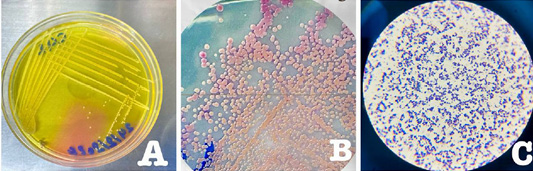
Following preincubation, the samples were subcultured on mannitol salt agar (Himedia, India) and Chromogenic media (Pioneer/France). The plates were incubated aerobically for 24 hours at 37°C. Gram’s staining was used to identify the suspected colonies on chromogenic agar.
Molecular techniques
The suspected isolates were confirmed by amplifying the nuc gene using PCR. By the instructions provided by the bacterial extraction kit manufacturer (Genaid, Korea), bacterial DNA was extracted. The primers were procured from Bioneer, Korea, and were specific to the nuc gene, which encodes a particular thermostable nuclease, Table 1.
Table 1: Sequence of primer for nuc gene with their manufacture.
Gene
Primer sequences (5→3)
Length
Product size
Source
Manufacturer
nuc
F: 5´- GCTTGCTATGATTGTGGTAGCC 3'
22
423 bp
Microgen/ Korea
R: 5´- TCTCTAGCAAGTCCCTTTTCCA 3'
22
Table 2: Sequence of primer for mecA gene with their manufacturer.
Gene
Primer sequences (5→3)
Length
Product size
Source
Manufacturer
mecA
F:5’-TCCAGATTACAACTTCACCAGG-3’
22
162bp
Stegger et al. (2012)
Microgen/ Korea
R:5’-CCACTTCATATCTTGTAACG-3
20
Table 3: Sequence of primers for hla,hlb, eta genes and their expected products.
Gene
Oligonucleotide sequences (5→3)
Length
Amplicon size (bp)
References
hla
F:5- CTGATTACTATCCAAGAAATTCGATTG-3’
27
209bp
Khodabux et al. (2023)
R: 5-CTTTCCAGCCTACTTTTTTATCAGT-3
26
hlb
F: 5- GTGCACTTACTGACAATAGTGC-3
22
309 bp
R: 5-GTTGATGAGTAGCTACCTTCAGT-3
23
eta
F5-CGCTGCGGACATTCCAACATGG-3
20
676bp
R-5-TACATGCCCGCCACTTGCTTGT-3
20
For the nuc gene, a total of 20μl of the PCR reaction mixture was prepared. Ten microliters of Green Master Mix, one microliter of each oligonucleotide primer, one microliter of DNA template, and seven microliters of nuclease-free water was used .PCR program was applied with 35 cycles of PCR conducted at 94°C for 7 minutes and 58°C for 30 seconds ,72°C for 45 seconds and 72°C for 7 minutes).
Phenotypic and genotypic detection of MRSA
To identify the MRSA phenotype, all identified S. aureus isolates were evaluated for antimicrobial sensitivity to cefoxitin (10μg) (NCCLS, 2023). The mecA gene was then found to molecularly validate MRSA isolates, Table 2.
Molecular detection of Methicillin-resistant staphylococcus aureus (MRSA) by (mecA) gene
Oligonucleotide primers for PCR amplification
By identifying the mecA gene using primer sequences shown in Table 2, MRSA isolates were molecularly confirmed.
The PCR reaction mixture for the mecA gene was prepared in a total amount of 20μl. One microliter of each oligonucleotide primer, one μl of DNA template, ten microliters of Green master mix, and seven microliters of nuclease-free water were combined. The thermocycling includes: 94°C for 5 minutes and 30 cycles of (94°C for 1 minute, 59°C for 1 minute, 72 °C for 1 minute, and final 72 °C for 7 minutes).
Antimicrobial-susceptibility testing
Eight antibiotics were chosen for the antibiotic sensitivity assays based on their common use in the field. This test was conducted according to the method described earlier.. The antibiotic discs were provided by (Bioanalyse/ Turkey), including Erythromycin (30 μg), Gentamycin (30 μg), Ciprofloxacin (5 μg), Penicillin (10 μg), Tetracycline (10 μg), Vancomycin (10 μg), Sulfamethoxazole (10 μg), and Chloramphenicol(10 μg).
Molecular detection of virulence genes among MRSA isolates
Primers were employed to identify the virulence genes (including hla, hlb, and eta) in MRSA-positive isolates (Table 3).
A total of 20μl of the PCR reaction mixture for the hla and hlb genes was produced. With each of the oligonucleotide primers (0.5 μl), DNA template (2 μl), Green master mix (10 μl), and nuclease-free water (6 μl).
For the hla, hlb gene, a total of 20μl of the PCR reaction mixture was prepared, carrying 2 μl of DNA template, 0.5 μl of each oligonucleotide primer, 10 μl of Green Master Mix, and 6 μl of nuclease-free water.
The reaction mixture of (eta) consisted of 1 microliter of DNA template, 0.5 microliter of each oligonucleotide primer, 10 microliters of Green master mix, and 8 microliters of nuclease-free water.
The hla and hlb genes were amplified under the following conditions: 94°C for 5 minutes, 35 cycles (94°C for 30 seconds, 56°C for 30 seconds, 72°C for 30 seconds, and 72°C for 5 minutes). Whereas, the (eta) gene was subjected to 30 cycles of PCR conditions, which included 95 °C for 5 minutes, 54 °C for 30 seconds, 72 °C for 30 seconds, and 72 °C for 5 minutes.
Results
Subclinical mastitis detection
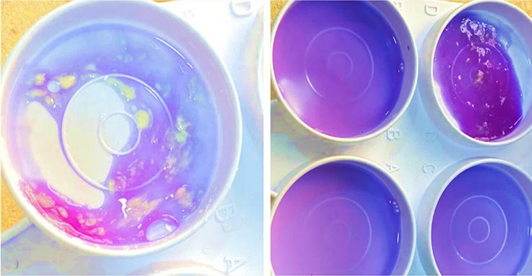
Based on CMT, a total of 120 (46.15%) samples showed positive results (Table 4; Figure 1).
Table 4: Percentage of subclinical mastitis according to CMT.
No. of samples
Positive results (%)
Trace (%)
Weak (%)
Distinct (%)
Strong (%)
260
120 (46.15)
8 (6.7%)
39(32.5%)
50(41.7%)
23 (19.1%)
Chi-square: 33.80, P-value < 0.001. Not all categories have the same distribution.
Table 5: Illustrate the number of S. aureus isolates detected by molecular detection, traditional microbiological methods, and cultural methods.
Total number of milk samples
Number of suspected isolates of Staphylococcus aureus
Confirmed isolates by using Molecular detection of nuc gene
Mannitol salts agar
CHROMagar™ Staph aureus
No
No
%
No
%
No
%
260
110
53
80
31
41
16
Chi-square: 57.17, P < 0.001
Staphylococcus aureus identification using PCR and standard microbiological methods
The percentage of suspected S. aureus isolates was confirmed using mannitol salts agar and CHROMagar™ Staph aureus was positive in 140 (53%) and 80 (31%), respectively.
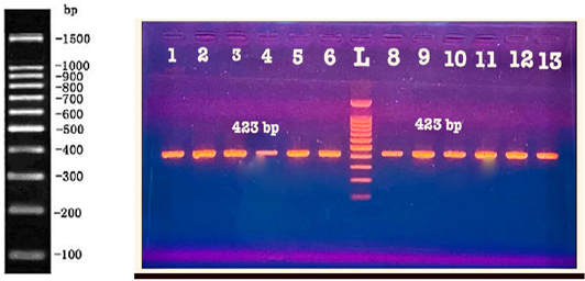
Table 3 all suspected isolates were subjected to PCR targeting the nuc gene (Table 5, Figures 2 and 3).The molecular technique-based assay characterized S. aureus in 41 (16%) of samples.
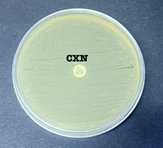
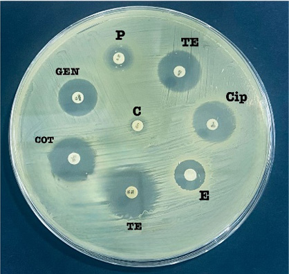
Methicillin-resistant Staphylococcus aureus can be identified phenotypically utilizing cefoxitin antimicrobial disk. All verified S. aureus isolates were resistant to a cefoxitin disk 41/41 (100 %) (Figure 4).
Methicillin-resistant staphylococcus aureus (MRSA) isolates were detected using the mecA gene
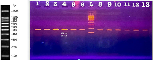
All S. aureus isolates were submitted to molecular detection for the (mecA) gene using gene specific primers of 41 S.aureus isolates, only 30 isolates were mecA positive. The results are shown in Table 6 and Figure 5.
Table 6: Percentage of (mec A) gene in S. aureus isolates among subclinical mastitis.
No. of S. aureus
No. of mec A positive
No
No
%
41
30
73
Antimicrobial-susceptibility testing
Eight antimicrobials were used to assess antibiotic sensitivity of S. aureus isolates, as shown in Table 7 and Figure 6. The isolates exhibited resistant to Vancomycin, Gentamycin, Penicillin, Erythromycin, and Tetracycline with percentages of ( 80%, 63.3%, 37%, 20 %, and 17%), respectively. In contrast, the isolates were vulnerable to Sulfamethoxazole, Chloramphenicol, Tetracycline, Penicillin, Ciprofloxacin, and Gentamycin, with a ratio of 83%, 83%, 67%, 60%, 47%, and 27%, respectively.
Molecular detection of virulence genes (hla, hlb and eta)
Molecular detection of hemolysin alpha (hla), beta toxin (hlb) and Exfoliative toxin (eta)
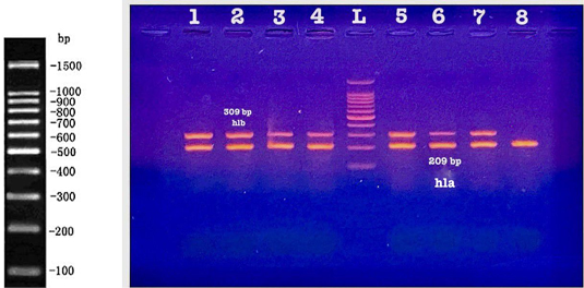
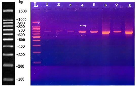
The majority of prevalent virulence gene found in this investigation was hlb 70% (21/30), which was followed by hla 63.3% (19/30), as shown in Figures 7 and 8 the eta gene percentage in this investigation was (30/30) 100%.
Table 7: Antibiogram of S. aureus isolates (n=30) against (8 antimicrobial agents).
Antimicrobial class
Antimicrobial agent
Code
Quality (μg)
S. aureus antibiotic resistance pattern
Resistant
Intermediate
Sensitive
Aminoglycosides
Gentamycin
GEN
30μg
19 (63.3%)
3 (10%)
8 (27%)
Amphenicol
Chloramphenicol
C
10μg
3(10%)
2 (7%)
25 (83.3%)
Fluoroquinolones
Ciprofloxacin
CIP
5μg
1 (3.3%)
15 (50%)
14 (47%)
Glycopeptides
Vancomycin
VA
10 μg
24 (80%)
4 (13.3%)
2 (7%)
Macrolides
Erythromycin
E
30μg
6 (20 %)
18 (60%)
6 (20%)
Sulfonamides
Sulfamethoxazole
COT
10μg
2 (7%)
3 (10%)
25(83.3%)
Tetracyclines
Tetracycline
TE
10μg
5 (17%)
5 (17%)
20 (67%)
β–lactam
Penicillin
P
10μg
11 (37%)
1 (3.3)
18 (60%)
Discussion
The infection of the mammary gland, known as bovine mastitis, is one of the most serious diseases affecting dairy herds globally because of its financial implications. It causes considerable losses in decreased production and culling rates ; . Based on clinical features, clinical and subclinical mastitis can be differentiated. The former is characterized by watery discharges, clots, or flakes in milk, and the affected areas are usually swollen, hot, and unpleasant.
It is more difficult to diagnose subclinical mastitis because there are no outward signs in animals or milk. Its primary signs are decreased milk supply and an increased somatic cell count. Subclinical duration lasts longer than clinical, and infection allows the spread of pathogens within the herd . In this study, the California mastitis test results revealed that 140 (41.3%) samples tested positive for CMT.
In Iraq, studies including recorded that the incidence of subclinical mastitis among cattle was 38.89% in Al-Sulaimaniyah Province. Compared with other studies conducted in Basrah, the detection rate of SCM using CMT ranged from 38% to 56.6% ; . The traditional microbiological methods used for S. aureus detection rely on enriching the milk sample in Brain heart infusion broth (BHI), differentiating it on Mannitol salts agar, and subculturing on Chromogenic agar. According to several studies, the pathogens most frequently present in mastitis cases are Staphylococcus aureus, Streptococcus agalactiae, Streptococcus uberis, Escherichia coli, and Klebsiella pneumonia . The S. aureus isolation rate in this study from SCM was 16%. This rate was in line with a previous study in Nepal, which reported that 15.2% of CMT-positive milk was S. aureus . S. aureus in our study is higher than in a study conducted by , 6(2.8%) cows with SCM were positive for S. aureus. Variations in the sample’s attributes (size, season, type), isolation technique, and geographic locations may account for the discrepancy in S. aureus prevalence between this study and earlier research.
MRSA strains have emerged as formidable nosocomial pathogens and have disseminated globally due to their ability to develop antimicrobial chemotherapy resistance . Although using antibiotics in modern medicine to treat infections caused by bacteria has changed the field, the indiscriminate, improper, and frequently abusive use of antibiotics has led bacteria to develop drug resistance . Most of the world is plagued with bacteria and superbugs resistant to many drugs. Pathogens with antibiotic resistance dramatically increase the rates of morbidity and death.
Antimicrobial resistance is growing quickly and threatens to outpace the introduction of new antimicrobials . Cefoxitin is phenotypically an alternative marker and potentially more sensitive for detecting methicillin resistance, as recorded in .
Consequently, (100%) of S. aureus isolates in the recent study were resistant to the cefoxitin disk. This result is higher than reported previously. Mohammed and Al-Iedani (2019), who clarified that the detection rate of MRSA using the cefoxitin disc diffusion method was 66.66%.
When it comes to MRSA isolation, one of the best methods is the detection of mecA. The mecA encodes the lowest β-lactam potential alters protein (PBP2a) found on SCCmec-resistant genomes . In the current study, the MRSA ratio according to the mecA detected by PCR was 73%. This rate agrees with a previous study , where it is reported that the rate of MRSA according to the mecA detected by PCR was 86.66%.
By applying the disc diffusion technique, 30 S. aureus isolates were submitted to antimicrobial susceptibility tests against eight antimicrobial agents. According to the findings of the current study. S. aureus isolates were resistant to Vancomycin (80%) and 83% susceptible to Sulfamethoxazole and Chloramphenicol. These results are higher than reported earlier , where it was reported that S. aureus isolates were resistant to Vancomycin in percentage (56.8%). Our findings are in agreement with another study Hoque et al. (2023), where it is reported that S. aureus isolates were susceptible to sulfamethoxazole in a higher percentage (77%).
Furthermore, this study found resistance against Gentamycin, Penicillin, and Erythromycin with decreasing rates of (63.3%, 37%, and 20%), respectively. These results are lower than those reported before Tassew et al. (2017), who have found resistance to penicillin (95.6 %), and in line with gentamycin (59.4%), On the other hand, the isolates were susceptible to Sulfamethoxazole, Chloramphenicol, Tetracycline, Penicillin, Ciprofloxacin and Gentamycin, with a ratio of 83%, 83%, 67%, 60%, and 47% and 27%, respectively.
These results are higher than those reported earlier , who found that Tetracycline was sensitive ( 48.3%).
In contrast, it has been found that maximum susceptibility to Ciprofloxacin is higher than in this study (92% ) and similar to the percentage of Gentamycin in this study (69.23%) . The source of isolates may cause minor variations in resistance, and usage of these antibiotics may be the cause of the ongoing rise in resistance.
Few studies investigated hemolysin genes in S. aureus strains isolated from mastitis milk of dairy species. . In the current study, hla (70.3%) and hlb (78%) genes in S. aureus isolates are lower than reported before , with a higher percentage of hla (95.93% ) and hlb 93.50%. In contrast, another study. Moraveji et al. (2014) reported the presence of hlb only in one of 20 isolates of S. aureus from bovine mastitis milk. Moreover, the percentage of exfoliative toxin (eta) in this study was 100%. This result disagrees with a previous study Yang et al. (2020), where it is clarified that none of the MRSA isolates carried the eta gene.
Conclusion
Our findings emphasize the critical presence of S. aureus on a wider scale and propose the importance of monitoring antibiotic resistance profiles before the application of antibiotics in animals.
Acknowledgments
I would like to express my sincere gratitude to my supervisors for their invaluable guidance, support, and encouragement throughout this research. I also appreciate the support of my family and friends, for their patience and motivation.
Novelty Statement
The nature and genetics of S. aureus in mastitis cases, as well as the presence of virulence genes, underpin the driving of antibiotic resistance. The finding would be valuable information for both animal handlers and milk consumers.
Author’s Contribution
NNJ, RAF: Supervision of writing review and editing.
ZAF: Data collection, formal analysis, writing original draft.
All authors contributed significantly to the research, discussed the results, and approved the final version of the manuscript.
Generative AI and AI-assisted technology statement
The authors have carefully reviewed all AI-assisted outputs to ensure accuracy, integrity, and compliance with the ethical standards of research and publication. Responsibility for the content of this manuscript remains entirely with the authors.
Conflict of interest
The author declares that there are no conflicts of interest related to this research. The study was conducted independently, without any financial, commercial, or personal relationships that could be construed as potential sources of bias. All experimental design, data collection, analysis, and interpretation were carried out solely by the author and collaborators within the academic framework.
Figures
Figure 1:.
Results of California mastitis test based on the degree of gel formation.
Figure 2:.
A: Staphylococcus aureus appeared as Golden yellow colonies on mannitol salts agar, B: Chromogenic agar: colonies seemed mauve (pink), C: Gram-positive cocci.
Figure 3:.
Electropherogram of nuc gene amplification. The PCR product was run on 1 % agarose gel, stained with ethidium bromide. L: mean Ladder and nuc gene product size approximately (423 bp).
Figure 4:.
Cefoxitin disk test on muller Hinton medium.
Figure 5:.
Electropherogram of amplification of mecA gene. The PCR products were run on a 1 % agarose gel stained with ethidium bromide. L: mean Ladder (Solarbio100 bp), mecA approximately (162 bp).
Figure 6:.
A: Antimicrobial susceptibility test on Muller Hinton medium.
Figure 7:.
Electropherogram of amplification of hla and hlb genes. The PCR products were run on a 1 % agarose gel stained with ethidium bromide. L: mean Ladder (Solarbio100 bp), (hla 209 bp, hlb:309 bp approximately).
Figure 8:.
Electropherogram of amplified eta gene. The PCR products were run on a 1 % agarose gel stained with ethidium bromide. L: mean Ladder (Solarbio100 bp), (eta: 676 bp ) approximately.
References
Acosta AC, Oliveira PRF, Albuquerque L, Silva IF, Medeiros ES, Costa MM, Mota RA (2018). Frequency of Staphylococcus aureus virulence genes in milk of cows and goats with mastitis. Pesquisa Vet. Brasil., 38: 2029-2036. https://doi.org/10.1590/1678-5150-pvb-5786[CrossRef]
Algammal AM, Enany ME, El-Tarabili RM, Ghobashy MO, Helmy YA (2020). Prevalence, antimicrobial resistance profiles, virulence, and enterotoxins-determinant genes of MRSA isolated from subclinical bovine mastitis in Egypt. Pathogens, 9(5): 362. https://doi.org/10.3390/pathogens9050362[CrossRef]
Ali T, Kamran N, Raziq A, Wazir I, Ullah R, Shah P, Liu G (2021). Prevalence of mastitis pathogens and antimicrobial susceptibility of isolates from cattle and buffaloes in Northwest of Pakistan. Front. Vet. Sci., 8: 746755. https://doi.org/10.3389/fvets.2021.746755[CrossRef]
Al-Iedani AA (2016). Phenotypic study on the capacity of biofilm production in S. aureus isolated from bovine subclinical mastitis and their impact on resistance to antimicrobials. Basrah J. Vet. Res., 15(2): 111–127. https://doi.org/10.33762/bvetr.2016.124293[CrossRef]
Al-Iedani AA, Ghazi SS (2016). Evaluation of raw milk from local markets and milk samples taken directly from cows in Basrah-Iraq. J. Agric. Vet. Sci., 9(12): 59-64.
Al-Jebouri MM, Mdish SA (2013). Antibiotic resistance pattern of bacteria isolated from patients of urinary tract infections in Iraq. Open J. Urol., 3(7): 124–131. https://doi.org/10.4236/oju.2013.32024[CrossRef]
Aslantas O, Keskin O, Yucetepe AG (2022). Molecular characterization of Staphylococcus aureus from clinical sheep mastitis cases. Israel J. Vet. Med., 77: 99-105.
Azooz MF, El-Wakeel SA, Yousef HM (2020). Financial and economic analyses of the impact of cattle mastitis on the profitability of Egyptian dairy farms. Vet. World, 13(9): 1750. https://doi.org/10.14202/vetworld.2020.1750-1759[CrossRef]
Bauer A, Kirby W, Sherris J, Turk A (1966). Antibiotic susceptibility testing by a standardized single disk method. Am. J. Clin. Pathol., 45: 493-496. https://doi.org/10.1093/ajcp/45.4_ts.493[CrossRef]
Bitrus AA, Peter OM, Abbas MA, Goni MD (2018). Staphylococcus aureus: A review of antimicrobial resistance mechanisms. Vet. Sci. Res. Rev., 4(2): 43-54. https://doi.org/10.17582/journal.vsrr/2018/4.2.43.54[Full text][CrossRef]
Boss R, Cosandey A, Luini M, Artursson K, Bardiau M, Breitenwieser F, Graber HU (2016). Bovine Staphylococcus aureus: Subtyping, evolution, and zoonotic transfer. J. Dairy Sci., 99(1): 515-528. https://doi.org/10.3168/jds.2015-9589[CrossRef]
Bude SA, Mengesha AK (2021). Isolation and identification of Staphylococcus aureus from dairy farms in Bishoftu Town, Ethiopia. https://doi.org/10.21203/rs.3.rs-311125/v1[CrossRef]
Chakraborty N, Jha D, Roy I, Kumar P, Gaurav SS, Marimuthu K, Gautam HK (2022). Nanobiotics against antimicrobial resistance: Harnessing the power of nanoscale materials and technologies. J. Nanobiotechnol., 20(1): 375. https://doi.org/10.1186/s12951-022-01573-9[CrossRef]
Christaki E, Marcou M, Tofarides A (2020). Antimicrobial resistance in bacteria: mechanisms, evolution, and persistence. J. Mol. Evol., 88(1): 26-40. https://doi.org/10.1007/s00239-019-09914-3[CrossRef]
Cobirka M, Tancin V, Slama P (2020). Epidemiology and classification of mastitis. Animals, 10(12): 2212. https://doi.org/10.3390/ani10122212[CrossRef]
Dinges MM, Orwin PM, Schlievert PM (2000). Exotoxins of Staphylococcus aureus. Clin. Microbiol. Rev., 13(1): 16-34. https://doi.org/10.1128/CMR.13.1.16[CrossRef]
Dive G, Bouillon C, Sliwa A, Valet B, Verlaine O, Sauvage E, Marchand-Brynaert J (2013). Macrocycle-embedded β-lactams as novel inhibitors of the Penicillin Binding Protein PBP2a from MRSA. Eur. J. Med. Chem., 64: 365-376. https://doi.org/10.1016/j.ejmech.2013.03.052[CrossRef]
Abril GAG, Villa T, Barros-Velázquez J, Cañas B, Sánchez-Pérez A, Calo-Mata P, Carrera M (2020). Staphylococcus aureus exotoxins and their detection in the dairy industry and mastitis. Toxins, 12(9): 537. https://doi.org/10.3390/toxins12090537[CrossRef]
González-Martín M, Corbera JA, Suárez-Bonnet A, Tejedor-Junco MT (2020). Virulence factors in coagulase-positive staphylococci of veterinary interest other than Staphylococcus aureus. Vet. Quart., 40(1): 118-131. https://doi.org/10.1080/01652176.2020.1748253[CrossRef]
Hammadi KM, Yousif AA (2013). Prevalence of clinical and subclinical ovine mastitis caused by Staphylococcus aureus. Al-Anbar J. Vet. Sci., 6(1).
Hoque MF, Fazal MA, Chowdhury EMS, Hossain H, Imranuzzaman M, Islam MN, Ahad A (2023). Prevalence and antibiotic susceptibility profile of Staphylococcus aureus in clinical and subclinical mastitis milk samples. Bangladesh J. Vet. Med., 21(1): 27-37. https://doi.org/10.33109/bjvmjj2023fam2[CrossRef]
Ibrahim HK, Madhi KS, Baqer GK, Gharban HA (2023). Effectiveness of raw bacteriocin produced from lactic acid bacteria on biofilm of methicillin-resistant Staphylococcus aureus. Vet. World, 16(3): 491. https://doi.org/10.14202/vetworld.2023.491-499[CrossRef]
Jani M, Sengupta S, Hu K, Azad RK (2017). Deciphering pathogenicity and antibiotic resistance islands in methicillin-resistant Staphylococcus aureus genomes. Open Biol., 7(12): 170094. https://doi.org/10.1098/rsob.170094[CrossRef]
Khodabux RMJ, Mariappan S, Sekar U (2023). Spectrum of virulence factors in clinical isolates of Staphylococcus aureus and prevalence of SCCmec types in methicillin-resistant Staphylococcus aureus in a tertiary care center. J. Lab. Phys., 15(3): 450-461. https://doi.org/10.1055/s-0043-1764483[CrossRef]
Király J, Hajdučková V, Gregová G, Szabóová T, Pilipčinec E (2024). Resistant S. aureus isolates and is capable of producing biofilm from the milk of dairy cows with subclinical mastitis in Slovakia. Agriculture, 14(4): 571. https://doi.org/10.3390/agriculture14040571[CrossRef]
Mohammed AL, Al-Iedani AA (2019). Phenotypic study of the effects of environmental factors on the biofilm formation by staphylococcus aureus isolates. Basrah J. Vet. Res., 18(2): 258-277.
Mohammed AL, Al-Iedani AA (2020). Molecular detection of ica gene and some surface proteins in biofilm producer of methicillin-resistant and methicillin-sensitive Staphylococcus aureus. Biochem. Cell. Arch., 20.
Morales-Ubaldo AL, Rivero-Perez N, Valladares-Carranza B, Velázquez-Ordoñez V, Delgadillo-Ruiz L, Zaragoza-Bastida A (2023). Bovine mastitis, a worldwide impact disease: Prevalence, antimicrobial resistance, and viable alternative approaches. Vet. Anim. Sci., pp. 100306. https://doi.org/10.1016/j.vas.2023.100306[CrossRef]
Moraveji Z, Tabatabaei M, Aski HS, Khoshbakht R (2014). Characterization of hemolysins of Staphylococcus strains isolated from human and bovine, southern Iran. Iran. J. Vet. Res., 15(4): 326.
Naranjo-Lucena A, Slowey R (2023). Invited review: Antimicrobial resistance in bovine mastitis pathogens: A review of genetic determinants and prevalence of resistance in European countries. J. Dairy Sci., 106(1): 1-23. https://doi.org/10.3168/jds.2022-22267[CrossRef]
National Committee for Clinical Laboratory Standards (2023). Performance Standards for Antimicrobial Disk SusceptibilityTests. Eighth Edition; Approved Standard M2-A8. NCCLS, Wayne, PA, USA.
Nishifuji K, Sugai M, Amagai M (2008). Staphylococcal exfoliative toxins: molecular scissors of bacteria that attack the cutaneous defense barrier in mammals. J. Dermatol. Sci., 49(1): 21-31 https://doi.org/10.1016/j.jdermsci.2007.05.007.[CrossRef]
Philpot WN, Nickerson SC (1991). Counter attack: A strategy to combat mastitis. Badson Brothers Co., Illinois.
Sangappa M, Thiagarajan P (2012). Methicillin-resistant Staphylococcus aureus: Resistance genes and their regulation. Int. J. Pharma. Pharmaceut. Sci., 4(1): 658-667.
Scherrer D, Corti S, Muehlherr JE, Zweifel C, Stephan R (2004). Phenotypic and genotypic characteristics of Staphylococcus aureus isolates from raw bulk-tank milk samples of goats and sheep. Vet. Microbiol., 101(2): 101-107. https://doi.org/10.1016/j.vetmic.2004.03.016[CrossRef]
Sharun K, Dhama K, Tiwari R, Gugjoo MB, Iqbal YM, Patel SK, Chaicumpa W (2021). Advances in therapeutic and managemental approaches of bovine mastitis: A comprehensive review. Vet. Quart., 41(1): 107-136. https://doi.org/10.1080/01652176.2021.1882713[CrossRef]
Shittu AO, Okon K, Adesida S, Oyedara O, Witte W, Strommenger B, Nübel U (2011). Antibiotic resistance and molecular epidemiology of Staphylococcus aureus in Nigeria. BMC Microbiol., 11: 1-8. https://doi.org/10.1186/1471-2180-11-92[CrossRef]
Shrestha A, Bhattarai RK, Luitel H, Karki S, Basnet HB (2021). Prevalence of methicillin-resistant Staphylococcus aureus and pattern of antimicrobial resistance in mastitis milk of cattle in Chitwan, Nepal. BMC Vet. Res., 17: 1-7. https://doi.org/10.1186/s12917-021-02942-6[CrossRef]
Stegger Á, Andersen PS, Kearns A, Pichon B, Holmes MA, Edwards G, Larsen AR (2012). Rapid detection, differentiation and typing of methicillin-resistant Staphylococcus aureus harbouring either mecA or the new mecA homologue mecALGA251. Clin. Microbiol. Infect., 18(4): 395-400. https://doi.org/10.1111/j.1469-0691.2011.03715.x[CrossRef]
Tassew A, Aki A, Legesse K (2017). Isolation, identification and antimicrobial resistance profile of Staphylococcus aureus and occurrence of methicillin resistant S. aureus isolated from mastitic lactating cows in and around assosa town, Benishangul Gumuz region, Ethiopia. J. Dairy Vet. Anim. Res., 6(3): 308-314. https://doi.org/10.15406/jdvar.2017.06.00180[CrossRef]
Turner NA, Sharma-Kuinkel BK, Maskarinec SA, Eichenberger EM, Shah PP, Carugati M, Fowler Jr VG (2019). Methicillin-resistant Staphylococcus aureus: An overview of basic and clinical research. Nat. Rev. Microbiol., 17(4): 203-218. https://doi.org/10.1038/s41579-018-0147-4[CrossRef]
Williamson J, Callaway T, Rollin E, Ryman V (2022). Association of milk somatic cell counts with bacteriological cure of intramammary infection review. Agriculture, 12(9): 1437. https://doi.org/10.3390/agriculture12091437[CrossRef]
Wongboot W, Chomvarin C, Engchanil C, Chaimanee P (2013). Multiplex PCR for detecting superantigenic toxin genes in methicillin-sensitive and methicillin-resistant Staphylococcus aureus isolated from patients and carriers of a hospital in northeast Thailand. S. Asian J. Trop. Med. Publ. Health, 44(4): 660-671.
World Health Organization (2017). WHO publishes list of bacteria for which new antibiotics are urgently needed.
Yang F, Zhang S, Shang X, Li H, Zhang H, Cui D, Sun Y (2020). Detection and molecular characterization of methicillin-resistant isolated from subclinical bovine mastitis cases in China. J. Dairy Sci., 103(1): 840-845. https://doi.org/10.3168/jds.2019-16317[CrossRef]
Zhang L, Gao J, Barkema HW, Ali T, Liu G, Deng Y, Han B (2018). Virulence gene profiles: Alpha-hemolysin and clonal diversity in Staphylococcus aureus isolates from bovine clinical mastitis in China. BMC Vet. Res., 14: 112. https://doi.org/10.1080/21505594.2018.1491256[CrossRef]
Investigation of Antimicrobial Resistance and Virulence Genes among Methicillin-Resistant Staphylococcus aureus from Subclinical Mastitic Cows · ResearchersLinks