
The outer surfaces of scales have provided significant adaption and functional value in lizards. In this paper, we examined the microornamenation of the outer surface of the dorsal scales from the mid-body region of Acanthodactylus opheodurus (Lacertidae), Mesalina guttulata guttulata (Lacertidae) and Trapelus ruderatus blanfordi (Agamidae). Skin specimens were prepared and investigated by using scanning electron microscopy. The microornamenation of the examined species exhibited different pattern in the same microhabitat. Variety was observed in the two related species, having a common family, Lacertidae. Whereas, Agamaidae sp. showed different pattern of microstructures. So, we conclude that there are other factors, which influence scale surface structures not only with microhabitat.
Research Article · Volume 54, Issue 1, 17 November 2021 · Open Access
Scale Microornamentation in Some Lizard Species
· · ·
Affiliations & Notes ⌄
- 1Department of Biology, Faculty of Education, University of Majmaah, Al Majma’ah 15341, KSA , * Corresponding author: a.elhady@mu.edu.sa
Article Info ⌄
- Journal:
- Pakistan Journal of Zoology
- Published:
- 17 November 2021

From the article
- The dorsal scale surface appears graded (strap-shaped) (Fig. 1B).
- In M. guttulata guttulata, the dorsal scales are smooth, polygonal and Juxtaposed in regular transverse rows without raised posterior edge (Fig. 1D).
- In T. ruderatus blanfordi and A. opheodurus, the scale surface is rough and keeled with strap-shaped cells to limit the shine.
- The latter two species are found on ground not in holes so pitting is dispersed on the surface of the scales where adhesion is less.
Selected from the article's own text.
Abstract
Introduction
The scale morphology of reptiles varies greatly among species. In crocodilian, keeled scales with a central, elevated corneous ridge showed minor overlapping , 2001, 2002; , 2006a, b; . Keeled scales were also observed in some armored agamid lizard, Lacerta angilis, L. viridis, L. praticola and Cerastes cerastes ; Rocha-Barbosa and Moraes a Silva, 2009; . In squamates, the non-overlapping scales are present on the heads of snakes and lizards although, the most frequently occurring scales are the overlapping scales, which have distinct outer and inner surfaces. Scales with ridges are found on the back of skink or the neck of anole, while the round scales (tuberculate scales) are present on the sides of the body of the green iguana ; .
The scales of squamates are covered by the oberhaϋhtchen forming the outer surface of the scale. The outer layer of the oberhaϋhtchen showed a complex microscopical structures (Leyding, 1872, 1873). The oberhaϋhtchen are folded structures producing ridges on the scale surface . The overall structures and features of the oberhaϋhtchen surface and epidermal folding is termed microornamentation ; or microstructure ; ; .
Many authors have studied the microornamentation of squamate scales on the dorsal body with the help of scanning electron microscopy showing their functional significance ; ; ; ; ; ; ; ; ; ; ; ; ; ; ; ; ; .
Although microornamentation did not correlate closely with known environmental parameters ; Peterson, 1984a, b), several studies correlated the function of the microornamentation with ecological variation. Gower (2003) found close relationship of microornamentation with general ecology. Crowe-Riddell et al. (2016) concluded that the microstructure features of the scales may be the result of direct adaptation pressures and could be reliable indicators of interspecific relationships. On the contrary, Price (1982) reported that microornamenation structures reflect the phylogenetic relationship, rather than environmental or habitat impacts and there was no evidence of correlation between microornamenation and habitat or environment.
In spite of these extensive studies, no assessment of the evolution of the different patterns of microornamenation have been made. In lacertid, there are only a few studies on microornamenation ; Peterson, 1984a; . Harvery and Gutberlet (1995) supported some phylogenetic utility of outer surface of scales. Arnold (2002) investigated briefly the microornamenation of some lacertid lizard explaining the variation in microornamenation morphology through the phylogenetic and functional analysis. The present study was aimed to show the variation and adaptation of microornamenation of the superficial surface of scales of three different lizard species inhabiting similar habitat establishing the hypothesis that the pattern of lizard scales have no association with habitat.
Materials and methods
The procedures conducted were in accordance with the standards set forth in the guidelines for the care and use of experimental animals by the Committee for the Purpose of Control and Supervision of Experiments on Animals by The National Institutes of Health (NIH). Three adult specimens of three lizard species were investigated. The specimens captured during spring of 2015-2017 from Majmaaha district, Kingdom of Saudi Arabia (KSA). The specimens were anesthetized by inhalation anesthetics . Skin samples from the mid-dorsal region were washed with distilled water to remove any impurities. The skin samples were left to dry at room temperature. Samples were fixed in 4% glutaraldehyde and washed in 0.1 M cacodylate buffer and postfixed in a solution of 1% osmium tetraoxide at 37°C for 2 h. This procedure followed by dehydration, critical point drying and platinum-palladium ion sputtering. The specimens were examined under a scanning electron microscope JEOL JSM 6510 lv using different magnifications.
Acanthodactylus opheodurus , the striped fringe-toed lizard is widespread in Kingdom of Saudi Arabia inhabiting open desert rocky terrain. These lizards have a basic back pattern consisting of five dark stripes. The dark vertebral stripe extends simple (unforked) from hind limbs to occiput in females. Mesalina guttulata guttulata (Lichtenstein, 1823), seen on the lower slopes of rocky escarpments. It has two disconnected dorsal lines on both sides of vertebral line; possessing irregular black blotches with white ocelli. The ventral side is bluish gray. Tail may have dark vertebral bars on the sides. The female small-spotted lizard has a proportionately longer body and slightly smaller head than the male. Preanal plate are large in males, smaller in females, bordered by two semicircles of small plates. Trapelus ruderatus blanfordi (Blanford, 1881), Anderson’s Agama, is common in different habitats in KSA. When approached, these lizards displayed their blue chin. It has a large triangular head. It appears light gray with dark specific lines on the back. Dark brown rings characterize the tail which are interrupted by light vertebral spots. This pattern is sometimes indistinct in males. Males have light blue cast on chin (at least seasonally). Throat seems pink in females though males have longitudinal gray stripes.
Results
In A. opheodurus, the dorsal scales are tough and keeled. They are triangular arranged in longitudinal rows with dorsal and lateral overlap. The posterior edge is raised (Fig. 1A). The dorsal scale surface appears graded (strap-shaped) (Fig. 1B). Its posterior border is slightly raised without denticulation (the posterior border is straight). In addition, large number of pits observed. At high magnification, the scale surface display indefinite structures, hair-like and papilla (Fig. 1B, C).
In M. guttulata guttulata, the dorsal scales are smooth, polygonal and Juxtaposed in regular transverse rows without raised posterior edge (Fig. 1D). The dorsal scale surface is broad graded (strap-shaped) (Fig. 1E). Its hind margin projects backward to overlap the cell behind. The posterior edge of the strap-shaped appears notched (Fig. 1F). At high magnification, many “minute” pits observed (Fig. 1F).
In T. ruderatus blanfordi, the dorsal scales are rough and keeled (Fig. 1G). They arrange in oblique rows with lateral and dorsal overlapping. The posterior edge margin projects upward at a steeper angle (Fig. 1J). The dorsal scale surface is graded (strap-shaped) (Fig. 1H). Its posterior border is wavy. At high scale, micro-villi, papillae, pustules and multiple deep pits observed (Fig. 1J, K).
Discussion
In lizards and snakes the microstructure and microornamenation plays an important role in intraspecific and interspecies variations associated with the ontogeny, scales and habitat ; Roch-Barbosa and Moraes e Silva, 2009; .
T. ruderatus blanfordi feeds on insects and invertebrates inhabiting different habitats. The micro-villi, papillae, pustules and multiple deep pits are observed on the dorsal scale surface. In contrast no microstructures or microornamenation was found on the S. stellio a primitive agama lizard which lives in mountainous area of the desert ; .
A. opheodurus, inhabits open desert rocky terrain. The superficial layer of scales appear (strap-shaped) and wavy without denticulation. In addition, large number of pits, indefinite structures, hairs and papilla were observed. In A. boskianus a hay-like structures and a large number of pits were seen. The hay-like structures enable it to live under the hay where it is densely distributed in vegetated deserts .
M. guttulata guttulata is usually seen on the lower slopes of rocky escarpments. Its dorsal scale surface is broad graded (strap-shaped) with posterior notched border. In addition, pits were observed. The dorsal scale surface of M. guttulata guttulata resembles with those of Lacerta monticola cantabrica which inhabit the rocky habitat but has no pits on the scale surface.
Generally, the scale surface of M. guttulata guttulata is smoother than the two other species which are without microvilli, papillae, hair-like and deep pits. The differences in microornamenation allowed a functional interpretation . In uropeltid snakes, the smooth scales minimized the friction when burrowing . The smooth scales of laticaudine (sea snakes) reduced the possibility of the skin being colonized by marine algae and other organisms . Whereas, the very rough scale of surface on the tail of uropeitid snakes encouraged the accumulation of a plug of earth which helps in preventing predators following the snakes in their burrows . Conversely, a complex microornamenation on the body and tail were potentially likely to increase locomotry friction. The dorsal body scales of lizard appeared smoother in their exposed areas ; . As strong microornamenation were absent on the most exposed parts of the body scales of lacertids, it is unlikely to have much importance in gaining creep because it depends on limbs for locomotion. However, general smoothness may permit significant reduction in friction when passing through vegetation or through narrow cavities. In most skinks, which frequently retreats into very narrow crevices relatively smooth scales were noticed .
In squamata, skin roughness creates more friction allowing undulating locomotion ; ; which requires ventral skin to provide high, directional friction in order to support forward motion, and slide along the substrate . Baeckens et al. (2019) demonstrated that the roughness increases with body size in A. cristatellus.
A relatively smooth scale surface limits such adhesion and permits dirt to wipe off easily. The scales in Adolfus alleni and Holapis allows them to brush against objects in their environment. In contrast, dirt particles are likely to settle down in the concavities of complex microornamenation in Psammodromus algirus, Ichnotropis and Ophisops . In geckos, the rough surfaces enable self-cleaning .
A complex microornamenation were found on the dorsal scales of three different species of lizards Takydromus, Gastropholis tropidopholis and Poromera which climbe extensively in vegetation matrices and are out of contact with ground much of the time . In Mauritian skink, more three- dimensional microornamenations tend to produce coherent reflection .
In the three examined species, the surface of dorsal scales appeared as strap-shaped as shown by Stewart and Daniel (1973) in some Anguids; by Stewart and Daniel (1975) and Peterson (1984a) in Sphenodon; by Peterson and Bezy (1985) in Xantusiid lizard; by Harvey and Gutberlet (1995) in gerrhosaurids; by Maderson et al. (1998) in Lepidosaurian, and by Arnold (2002) in Lacerta monticola cantabrica.
In the present study, the pits are minute and highly dispersed in M. guttulata guttulata whereas, in A. opheodurus and T. ruderatus blanfordi are large and deep. Many investigations have revealed that the low level of pitting is a primitive state in the lacertidae ; Peterson, 1984a; ; . Some partly aquatic natricine snakes, have pores on their dorsal body scales that exude lipids that collect in hollows in the scale helping the skin waterproof . In Pseuderemias and Pedioplanis undata, the dense pitting is mostly found in dry habitat where adhesion is less of a problem because pitted surface are more prone to hold dirt Arnold (2002). Moreover pitting makes epidermis less producer by reducing the amount of B-keratin needed.
In the strap-shaped cells, the hind cell margin projects backward to overlap the cell behind in M. guttulata guttulata while in A. opheodurus it is slightly raised but projects upward at a steeper angle in T. ruderatus blanfordi. Lizards occupying relatively mesic area showed strong raised posterior cell edge , 1989, 2002), whereas, in Teiioidea which live in moderate temperature areas the posterior cell edges are not markedly raised ; Peterson, 1984a; ; . The shine is greatly reduced at steeper angles compared with forms with primitive microornamenation in many predator lizards .
In the present study, the scales of T. ruderatus blanfordi and A. opheodurus are rough and keeled whereas in M. guttulata guttulata, the scales are smooth without keeling. In Lacerta angilis, L. viridis and L. praticola (lacertidae lizard), the keeled scales were detected on the dorsal surface, while they disappeared on the sides of the body to limit shine . The keeled scales could be adaptation linked to the stress produced by desert habitat (Rocha-Barbosa and Moraes a Silva, 2009). In addition, the rough and keeled scales were detected in Cerastes cerastes inhabiting dry, sandy areas with sparse rock outcroppings areas .
In gecko which is nocturnal feeding on insects, hairs, papilla and microvilli-like structures were observed on the dorsal scale surface . In Algyroides, the pustules interfere with coherent reflection from scale surface. Denticulation abundantly developed in Gallotia stehlini showing the same effect of pustules in Algroids .
Conclusion
In conclusion, the smooth surface of scales in Mesalina guttulata guttulata permits hiding in shallow holes in the hard ground and easy escape from spiders (Latrodectus, Theridiidae) preying on lizards and Psammophis schokari which are considered as possible predators. The predation by a shrike (Lanius sp.) on the lizard M. adramitana is being reported for the first time. In T. ruderatus blanfordi and A. opheodurus, the scale surface is rough and keeled with strap-shaped cells to limit the shine. The latter two species are found on ground not in holes so pitting is dispersed on the surface of the scales where adhesion is less. In addition, papillae, pustules and indefinite structures tend to produce coherent reflection.
Acknowledgment
The authors would like to thank the Deanship of Scientific Research at Majmaah University for funding this work through the research groups program no (R -1441-159).
Statement of conflict of interest
The authors declare no conflict of interest.
Figures
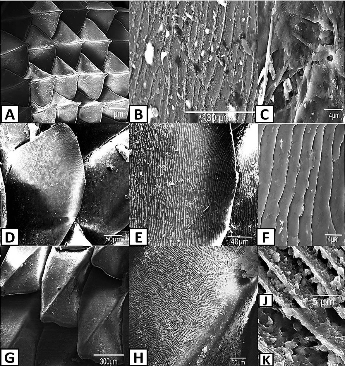
Scanning electron photomicrographs of scale ornamentation in lizards. A, shows the keeled scales of mid-dorsal trunk skin of Acanthodactylus opheodurus (15x); B, shows the pits on the outer surface of mid-dorsal trunk skin scales of A. opheodurus (3000x); C, shows indefinite structures, hair-like and papilla on the outer surface of mid-dorsal trunk skin scales of A. opheodurus (10000x); D, shows the outer surface of mid-dorsal trunk skin scales of Mesalina guttulata guttulata (500x); E, shows the outer surface of mid-dorsal trunk skin scales of M. guttulata guttulata (1000x); F, shows the minute pits on the outer surface of mid-dorsal trunk skin scales of M. guttulata guttulata (10000x); G, shows the keeled scale of mid-dorsal trunk skin of Trapelus ruderatus blanfordi (150x); H, shows the outer surface of mid-dorsal trunk skin scales of T. ruderatus blanfordi (500x); J, shows the microstructure of mid-dorsal trunk skin scales of T. ruderatus blanfordi (5000x); K, shows the microstructure of mid-dorsal trunk skin scales of T. ruderatus blanfordi (30000x).
References
- Alibardi, L., 1996. Scale morphogenesis during embryonic development in the lizard Anolis lineatopus. J. Anat., 188: 713-725.
- Alibardi, L., 2003. Adaptation to the land: The skin of reptiles in comparison to that of amphibians and endotherm amniotes. J. exp. Zool. Part B: Mol. develop. Evolut., 298: 12-41. https://doi.org/10.1002/jez.b.24[CrossRef]
- Alibardi, L., 2006a. Structural and immunocytochemical characterization of keratinization in vertebrate epidermis and epidermal derivatives. Int. Rev. Cytol., 259: 177-253. https://doi.org/10.1016/S0074-7696(06)53005-0[CrossRef]
- Alibardi, L., 2006b. Cells of embryonic and regenerating germinal layers within barb ridges: implication for the development, evolution and diversification of feathers. J. Subm. Cytol. Pathol., 38: 51-76.
- Alibardi, L. and Thompson, M.B., 2000. Scale morphogenesis and ultrastructure of dermis during embryo development in the alligator (Alligator mississippiensis, Crocodilia, Reptilia). Acta Zool., 81: 325-338. https://doi.org/10.1046/j.1463-6395.2000.00063.x[CrossRef]
- Alibardi, L. and Thompson, M.B., 2001. Fine structure of the developing epidermis in the embryo of the american alligator (Alligator mississippiensis, Crocodlilia ,Reptilia). J. Anat., 198: 265-282. https://doi.org/10.1046/j.1469-7580.2001.19830265.x[CrossRef]
- Alibardi, L. and Thompson, M.B., 2002. Keratinization and ultrastructure of the epidermis of late embryonic stages in the alligator (Alligator mississippiensis). J. Anat., 201: 71-84. https://doi.org/10.1046/j.1469-7580.2002.00075.x[CrossRef]
- Alibardi, L. and Toni, M., 2006. Cytochemical, biochemical and molecular aspects of the process of keratinization in the epidermis of reptilian scales. Prog. Histochem. Cytochem., 40: 173-134. https://doi.org/10.1016/j.proghi.2006.01.001[CrossRef]
- Allam, A.A. and Abo-Eleneen, R.E., 2012. Scales microstructures of some snakes inhabited the Egyptian area. Zool. Sci., 29: 770-775. https://doi.org/10.2108/zsj.29.770[CrossRef]
- Allam, A.A., Daza, J.D. and Abo-Eleneen, R.E., 2016. Histology of the skin of three limbless squamta dwelling in mesicand arid environments. Anat. Rec., 299: 979-989. https://doi.org/10.1002/ar.23356[CrossRef]
- Allam, A.A., Abo-Eleneen, R.E. and Othman, S.I., 2017. Microstructure of scales in selected lizard species. Saudi J. biol. Sci., 26: 129-136. https://doi.org/10.1016/j.sjbs.2017.03.012[CrossRef]
- Arnold, E.N., 1980. The scientific results of the Oman flora and fauna survey 1977 (Dhofar). The reptiles and amphibians of Dhofar, southern Arabia. J. Oman Stud. Sp. Rep., 2: 273-332.
- Arnold, E.N., 1987. Resource partition among lacertid lizards in southern Europe. J. Zool. Lond. B, 1: 739-782. https://doi.org/10.1111/j.1096-3642.1987.tb00753.x[CrossRef]
- Arnold, E.N., 1989. Systematics and adaptive radiation of equatorial African lizard assigned to the genera Adolfus, Bedriagai, Gastropholis, Holaspis and Lacerta (Reptilia: Lacertidae). J. nat. Hist., 23: 525-555. https://doi.org/10.1080/00222938900770311[CrossRef]
- Arnold, E.N., 2002. History and function of scale microornamentation in lacertid lizards. J. Morphol., 252: 145-169. https://doi.org/10.1002/jmor.1096[CrossRef]
- Baeckens, S., Wainwright, D.K, Weaver, J.C., Irschick, D.J. and Losos, J.B., 2019. Ontogenetic scaling patterns of lizard skin surface. J. Anat., 235: 346-356. https://doi.org/10.1111/joa.13003[CrossRef]
- Baig, K.J., Wagner, P.P., Ananjeva, N.B. and Böhme, W., 2012. A morpholiogy-based taxonomic revision of Laudakia Gray, 1845 (Squamata: Agamidae). Verteb. Zool., 62: 213-260.
- Bea, A., 1986. A general review of the dorsal scales microornamenatio in Vipera species (Reptilia: Viperidae). In: Studies in herpetology (ed. Z. Roček), Proc. 3rd Ordinary General Meeting of the Societas Europaea Herpetologica. Charles University, Prague, pp. 367-372.
- Bertelsen, M.F., 2007. Squamates (snakes and lizards). In: Zoo animal and wildlife immobilization and anesthesia (eds. G. West, D. Heard and N. Caulkett). Blackwell, Ames, IA, pp. 233-244. https://doi.org/10.1002/9780470376478.ch19[CrossRef]
- Bezy, R.L. and Peterson, J.A., 1988. The microstructure of scale surfaces in the xantusiid lizard genus Lepidophyma. Herpetologica, 44: 281-289.
- Blanford, W.T., 1881. On a collection of Persian reptiles recently added to the British Museum. Proc. Zool. Soc. London, 1881: 671-682. https://doi.org/10.1111/j.1096-3642.1881.tb01324.x[CrossRef]
- Blondheim, S. and Werner, Y.L., 1989. Lizard prédation by the widow spiders Latrodectus pallidus and L. revivensis (Theridiidae). Br. Herpetol. Soc. Bull., 30: 26-27.
- Bowker, R.G., Spindler, H.S., Tilden, A., Bairos, V.A. and Murray, R., 1987. Reflections on lizard skin: The ultrastructure of the scales of Cnemidophorus exsanguis and Podarcis bocagei. In: Proc. 4th Ordinary General Meeting of the Societas Europaea Herpetologica (eds. J.J. Vav Gelder, H. Strijbosch and P.J.M. Bergers). Societas Europaea Herpetologica, Nijmegen, pp. 83-86.
- Bryant, S.V., Breathnach, A.S. and Bellaris, A.A., 1967. Ultrastructure of the epidermis of the lizard (Lacerta vivipara) at the resting stage of the sloughing cycle. J. Zool., 152: 209-219. https://doi.org/10.1111/j.1469-7998.1967.tb01886.x[CrossRef]
- Chang, C., Wu, P., Baker, R.E., Maini, P.K., Alibardi, L. and Chuong, C., 2009. Reptile scale paradigm Evo-Devo, pattern formation and regeneration. Int. J. dev. Biol., 53: 813-826. https://doi.org/10.1387/ijdb.072556cc[CrossRef]
- Chiasson, R.B. and Lowe, C.H., 1989. Ultrastructural scale patterns in Nerodia and Thamnophis. J. Herpetol., 23: 109-118. https://doi.org/10.2307/1564016[CrossRef]
- Crowe-Riddell, J.M., Snelling, E.P., Watson, A.P., Suh, A.K., Patrige, J.C. and Sanders, K.L., 2016. The evolution of scale sensilla in the transition from land to sea in elapid snakes. Open Biol., 6: 1-6. https://doi.org/10.1098/rsob.160054[CrossRef]
- Cogălniceanu, D., Valdeón, A., Gosa, A., Al-Hemaidi, A.A.M. and Castilla, A.M., 2015. Shrike predation on the lizard Mesalina adramitana in Qatar: A review of reported reptile and amphibian prey. QScience Connect, 1: 1-8. https://doi.org/10.5339/connect.2015.1[CrossRef]
- Gans, C. and Baic, D., 1977. Regional specialization of reptile scale surfaces: Relation of texture and biologic role. Science, 195: 1348-1350. https://doi.org/10.1126/science.195.4284.1348[CrossRef]
- Gasc, J. and Gans, C., 1990. Tests on locomotion of the elongate and limbless lizard Anguis fragilis (Squamata: Anguidae). Copeia, 1990: 1055-1067. https://doi.org/10.2307/1446489[CrossRef]
- Gower, D.J., 2003. Scale microornamentation of Uropeltid snakes. J. Morphol., 258: 249-268. https://doi.org/10.1002/jmor.10147[CrossRef]
- Harvey, M.B., 1993. Microstrucure, ontogeny and evolution of scale surfaces in Xenosaurid lizard. J. Morphol., 216: 161-177. https://doi.org/10.1002/jmor.1052160205[CrossRef]
- Harvey, M.B. and Gubtberlet, R.L., 1995. Microstrucure, evolution and ontogeny of scale surfaces in cordylid and gherrhosaurid lizard. J. Morphol., 226: 121-139. https://doi.org/10.1002/jmor.1052260202[CrossRef]
- Hazel, J., Stone, M., Grace, M.S. and Tsukruk, V.V., 1999. Nanoscale design of snakeskin for reputation locomotions via friction anisotropy. J. Biomech., 32: 477-484. https://doi.org/10.1016/S0021-9290(99)00013-5[CrossRef]
- Hu, D.L., Nirody, J., Scott, T. and Shelley, M.J., 2009. The mechanics of slithering locomotion. Proc. natl. Acad. Sci. USA, 106: 10081-10085. https://doi.org/10.1073/pnas.0812533106[CrossRef]
- Irish, F.J., Williams, E.E. and Seling, E., 1988. Scanning electron microscopy of changes in epidermal structure occurring during the shedding cycle in squamata reptiles. J. Morphol., 197: 195-206. https://doi.org/10.1002/jmor.1051970108[CrossRef]
- Jayne, B.C., 1986. Kinematics of terrestrial snake locomotion. Copeia, 1986: 915-927. https://doi.org/10.2307/1445288[CrossRef]
- Lang, M., 1989. The morphology of the oberhautchen with the description and distribution of scale organs in basiliscine iguanians. Amphibia-Reptilia, 10: 423-434. https://doi.org/10.1163/156853889X00061[CrossRef]
- Leyding, F., 1872. Die in Deutschland lebenden Arten der Saurier. Laupp, Tϋbingen.
- Leyding, F., 1873. Uber die äusseren Bedeckungen der Reptilien und Amphibien. Arch. Mikrosk. Anat. Entwick., 9: 753-794. https://doi.org/10.1007/BF02956189[CrossRef]
- Maderson, P.F.A., Rabinowitz, T., Tandler, B. and Alibardi, L., 1998. Uitrastructureal contributions to an understanding of the cellular mechanisms involved in lizard skin shedding with comments on the function and evolution of unique lepidosaurian phenomenon. J. Morphol., 236: 1-24. https://doi.org/10.1002/(SICI)1097-4687(199804)236:1<1::AID-JMOR1>3.0.CO;2-B[CrossRef]
- McCarthy, C.J., 1987. Sea snake puzzles. In: Proc. 4th Ordinary General of the Societas Europaea Herpetologica (eds. J.J. Van Gelder, H. Strijbosch and P.J.M. Bergers). Societas Europaea Herpetologica, Nijmegen, pp. 279-284.
- Lichtenstein, M., and Hinrich, C., 1823. Verzeichniss der Doubletten des zoologischen Museums der Königl. Universität zu Berlin nebst Beschreibung vieler bisher unbekannter Arten von Säugethieren, Vögeln, Amphibien und Fischen. Königl. Preuss. Akad. Wiss. T. Trautwein, 10: 118.
- Perret, J.L. and Wuest, J., 1983. La microstructure des écailles de quelques Scincidés africains et parléarctiques (Lacertilia), observe au microscope électronique á balayage, II. Rev. Suisse Zool., 90: 913-928. https://doi.org/10.5962/bhl.part.117752[CrossRef]
- Peterson, J.A., 1984a. The scale microstructure of Sphenodon punctatus. J. Herpetol., 18: 40-47. https://doi.org/10.2307/1563670[CrossRef]
- Peterson, J.A., 1984b. The scale microstructure of Iguanid lizards. J. Herpetol., 18: 437-467. https://doi.org/10.2307/1564106[CrossRef]
- Peterson, J.A. and Bezy, R.L., 1985. The microstructure and evolution of scale surfaces in xantusiid lizard. Herpetologica, 41: 298.
- Price, R.M., 1982. Dorsal snake scale microdermatoglyphics: Ecological indicator or taxonomic tool? J. Herpetol., 17: 292-294. https://doi.org/10.2307/1563721[CrossRef]
- Price, R.M. and Kelly, P., 1989. Microdermatoglyphics: Basal patterns and transition zones. J. Herpetol., 23: 244-261. https://doi.org/10.2307/1564446[CrossRef]
- Renus, S. and Gasc, J.P., 1989. Microornamentations of the skin and the spatial position of the squamata in the environment. Fortschr. Zool., 35: 597-601.
- Renus, S., Gasc, J.P. and Diop, A., 1985. Microstructure of the integumentary surface of the squamata (reptilia) in relation to their spatial position and their locomotion. Fortschr. Zool., 30: 487-489.
- Rocha-Barbosa, O. and Moraes e Silva, R.B., 2009. Analysis of the microstructure of Xenodontinae snake scales associated with different habitat occupation strategies. Braz. J. Biol., 69: 919-923. https://doi.org/10.1590/S1519-69842009000400021[CrossRef]
- Ruibal, R., 1968. The ultrastructure of the surface of lizard scales. Copeia, 1968: 698-703. https://doi.org/10.2307/1441836[CrossRef]
- Sindaco, R., Simo-Riudalbas, I.M., Sacchi, R. and Carrana, S., 2018. Systematics of the Mesalina guttulata species complex (Squamata: Lacertidae) from Arabia with the description of two new species. Zootaxa, 4429: 513-547. https://doi.org/10.11646/zootaxa.4429.3.4[CrossRef]
- Stewart, G.R. and Daniel, R.S., 1973. Scanning electron microscopy of scales from different body regions of three lizard species. J. Morphol., 139: 377-388. https://doi.org/10.1002/jmor.1051390402[CrossRef]
- Stewart, G.R. and Daniel, R.S., 1975. Microornamenta-tion of lizard scales: Some variations and taxonomic correlations. Herpetologica, 31: 117-130.
- Stille, B., 1987. Dorsal scale microdermatoglyphics and rattle-snake (Crotalus and Sistrurus) phylogeny (Reptilia: Viperdae: Crotanlinae). Herpetologica, 43: 98-104.
- Vaccaro, O., Uriondo, A. and Filipello, A.M., 1988. Microornamenataciones de las células de Oberhäutchen en Tupinambis teguixinn (Linné,1758), Tupinambis rufescens, Gϋnther, 1987) (Sauria, Teiidae). Iheringia Sér Zool., 67: 65-75.
- Watson, G.S., Schwarzkopf, L., Cribb, B.W., Myhra, S., Gellender, M. and Jolanta A. Watson, J.A., 2015. Removal mechanisms of dew via self-propulsion off the gecko skin. J. R. Soc. Interface, 12: 1396. https://doi.org/10.1098/rsif.2014.1396[CrossRef]
Services
Article usage statistics
Loading…