
To elucidate the tissue expression levels of BMPR1B, BMP15, GDF9, Smad1, Smad5, and Smad9 genes in rams with different fecundity, quantitative real-time polymerase chain reaction was used to investigate the expression level of six genes in the brain, cerebellum, hypothalamus, pituitary, testis, epididymis, vas deferens, and adrenal gland in high fecundity (Small Tail Han sheep) and low fecundity (Sunite sheep) rams. The results were as follows: BMPR1B, GDF9, Smad1, Smad5 and Smad9 were expressed in all selected tissues, but BMP15 was specifically expressed in the epididymis. Further study indicated that the expression of BMPRIB in the brain, hypothalamus, pituitary, epididymis, and adrenal gland was significantly higher in Sunite sheep than in Small Tail Han sheep (p <0.05, p <0.01); the expression of BMP15 in the epididymis was significantly higher in Sunite sheep than in Small Tail Han sheep (p <0.01); the expression of GDF9 in the cerebellum and vas deferens was significantly higher in Small Tail Han sheep than in Sunite sheep (p <0.05); the expression of GDF9 in the adrenal gland was significantly higher in Sunite sheep than in Small Tail Han sheep (p <0.01); the expression of Smad1 in the brain and adrenal gland was significantly higher in Small Tail Han sheep than in Sunite sheep (p <0.05); the expression of Smad1 in vas deferens was significantly higher in Sunite sheep than in Small Tail Han sheep (p <0.01); the expression of Smad5 in the adrenal gland was significantly higher in Small Tail Han sheep than in Sunite sheep (p <0.05); the expression of Smad9 in the brain and epididymis was significantly higher in Sunite sheep than in Small Tail Han sheep (p <0.05, p <0.01); and the expression of Smad9 in the cerebellum and hypothalamus was significantly higher in Small Tail Han sheep than in Sunite sheep (p <0.05). This is the first study to systematically analyze the BMPR1B, BMP15, GDF9, Smad1, Smad5, and Smad9 genes’ tissue expression pattern in rams.
Research Article · Volume 52, Issue 5, 6 May 2020 · Open Access
Expression Analysis of BMPR1B, BMP15, GDF9, Smad1, Smad5, and Smad9 in Rams with Different Fecundity
· · · · ·
Affiliations & Notes ⌄
- 1College of Animal Science and Technology, Yangzhou University, Yangzhou 225009, China 2Key Laboratory of Animal Genetics, Breeding and Reproduction of Ministry of Agriculture, Institute of Animal Science, Chinese Academy of Agricultural Sciences, Beijing 100193, China 3State Key Laboratory for Sheep Genetic Improvement and Healthy Production, Xinjiang Academy of Agricultural and Reclamation Sciences, Shihezi 832000, China 4Joint International Research Laboratory of Agriculture and Agri-Product S
Article Info ⌄
- Journal:
- Pakistan Journal of Zoology
- Published:
- 6 May 2020

From the article
- In conclusion, we found that BMPR1B, GDF9, Smad1, Smad5, and Smad9 are expressed in all selected tissues and are highly expressed in the epididymis, whereas BMP15 is specifically expressed in the epididymis, which indicates that they may play important roles in the ovine epididymis and promote spermatogenesis.
- Our findings of ovine BMPR1B, BMP15, GDF9, Smad1, Smad5, and Smad9 will help to further understand their expression and function, and may contribute to exploring their role in the ram reproduction system.
- This is the first study of the six genes tissue expression pattern in rams.
Selected from the article's own text.
Abstract
Introduction
Lambing is one of the most economically important traits of sheep, being closely related to the economic benefits of sheep breeding. The major genes related to the prolificacy of sheep have received much attention from researchers since the 1980s . To date, three major candidate genes for the prolificacy of sheep have been found: bone morphogenetic protein receptor 1B (BMPR1B), bone morphogenetic protein 15 (BMP15), and growth differentiation factor-9 (GDF9) .
BMPR1B is the first major candidate gene found to be related to the prolificacy of sheep and possesses a mutation (A to G), known as FecB, which results in one amino acid substitution (Q to R) increasing the ovulation rate of Booroola ewes . The FecB mutation has an additive effect on ovine ovulation number and litter size, so that one copy of the FecB mutation can increase the ovulation number by 1.3–1.6-fold and the litter size by 0.9–1.2-fold, and two copies by 2.73 and 1.1–1.7, respectively .
BMP15 also known as growth differentiation factor-9B (GDF9B), and GDF9, which both belong to the transforming growth factor-β (TGF-β) superfamily and are recognized as major candidate genes for the prolificacy of sheep—were found to regulate the growth and differentiation of follicles, the secretion of reproductive hormones, and the growth of germ cells . To date, FecXI, FecXH , FecXG , FecXL, FecXB , FecXR , FecXGr , FecXO and FecXBar mutations have been found on the sheep BMP15 gene; they strong affect the ovulation rate and prolificacy, similar to the effect of G1 , G4 , G6, FecGF (G7) , FecGH (G8) , FecGE , FecTT (FecI) , FecGV , FecGT ,and FecGSI mutations on the sheep GDF9 gene.
Many studies revealed that the prolificacy of sheep is closely related to bone morphogenetic proteins (BMPs), BMP receptors (BMPR), and Smads, a downstream signaling molecule of the TGF-β/Smad signaling pathway . Members of the BMPs initiate signaling from the cell membrane by interacting with two distinct serine/threonine kinase receptors. Ligand binding induces the formation of a complex in which the type II receptor phosphorylates and activates the type I receptor. This protein then propagates the signal by phosphorylating the Smad proteins such as Smad1, Smad5, and Smad9 . Phosphorylated Smad1/5/9 can form a complex by interacting with Smad4, which can further activate or inhibit the expression of target genes . Given its interaction with BMPs, Smad1/5/9 might be related to the prolificacy of sheep.
Small Tail Han sheep (STH) and Sunite sheep (SNT) are two Chinese local sheep (Ovis aries) breeds with different estrous modes (year-round and seasonal, respectively. Both are known for their excellent meat production performance . Significant differences between the two sheep breeds in fecundity have resulted in increasing interest in the expression pattern of major prolificacy genes in these sheep.
BMPR1B, BMP15, GDF9, Smad1, Smad5, and Smad9 are important in prolificacy. Many studies on the expression of these six genes in the tissues of ewes have been reported; however, no research has yet been reported about these genes in rams. To explore the potential role of these six genes in rams, we analyzed the tissue expression profile and the mRNA expression levels in eight prolificacy-related tissues between high fecundity sheep breed (STH) and low fecundity sheep breed (SNT) rams. Our study helps elucidate the genetic mechanism controlling high fecundity in rams.
Materials and Methods
Selection of experimental sheep and sample collection
The 3 Small Tail Han rams and 3 Sunite rams were supplied by Yuncheng Breeding Sheep Farm (Yuncheng County, China) and Sheep and Goat Breeding Farm of Tianjin, Institute of Animal Sciences (Tianjin, China). All rams were healthy, approximately 2.5 years old, and were kept in a sheltered outdoor paddock and were provided with alfalfa hay and concentrate, with clear water available ad libitum. Eight tissues (brain, cerebellum, hypothalamus, pituitary, testis, epididymis, vas deferens, and adrenal gland) were collected from each animal. All tissues were snap-frozen in liquid nitrogen and then stored at −80°C to be used for RNA extraction.
All experimental procedures mentioned in the present study were approved by the Science Research Department (in charge of animal welfare issue) of the Institute of Animal Sciences, Chinese Academy of Agricultural Sciences (IAS-CAAS) (Beijing, China). Ethical approval was provided by the animal ethics committee of IAS-CAAS (No. IASCAAS-AE-03, December 12, 2016).
Total RNA extraction and cDNA synthesis
Tissue RNA was extracted from the 8 tissues using a total RNA extraction kit for animal tissue (Tiangen, Beijing, China) and Trizol (Invitrogen Inc., Carlsbad, CA, USA) was used to dissolve the tissues (each tissue smashed, mixed, and 50–100 mg used for RNA extraction). The quantity and quality of total RNA were monitored using 1.5% agarose gel electrophoresis and ultraviolet spectrophotometry (UV-1201, Shimadzu, Kyoto, Japan), respectively. Then, the RNA was stored at −80°C until use.
The first strand of cDNA was prepared following the instructions of the PrimeScriptTM RT Reagent Kit (TaKaRa Bio Inc., Dalian, China). The reaction program was as follows: 37°C for 15 min, followed by 85°C for 5 sec, with a total volume of 20 μL that contained PrimeScript RT Enzyme 1.0 µL, Random 6 mers 1.0 µL, 5 × PrimeScript Buffer (for Real Time) 4.0 µL, total RNA 1.0 µL and RNase-free ddH2O 13 µL. Prior to storage at −80°C, the standard working concentration of cDNA is 200ng/ul, the cDNA quality was evaluated by housekeeping gene (RPL-19) amplification, and then the reverse products were stored at −20°C until use.
RNA extraction and cDNA synthesis
Total RNA samples were analyzed using 1.5% agarose gel electrophoresis (U = 150 V; I = 240 mA). Three bands were detected (28S, 18S, and 5S)—the 28S band was brighter than the 18S band, and the 5S band was unclear. The OD260 nm/OD280 nm ratios (1.8–2.0) of the RNA samples were all 1.9 to 2.0, which showed that the extracted total RNA was of sufficient purity with no contamination or degradation. Therefore, these tissue RNAs were appropriate for use in the follow-up experiment.
Primer design
A total of 7 primers were designed using the Primer Premier (version 5.0, PREMIER Biosoft Co., Palo Alto, CA, USA) software to amplify different fragments of ovine BMPR1B, BMP15, GDF9, Smad1, Smad5, Smad9, and RPL-19 genes based on their assembled sequences in GenBank. All primers were synthesized by Beijing Tianyi Biotechnology Co., Ltd. (Beijing, China). The housekeeping gene (RPL-19, accession number: XM_012186026.1) was used as an internal control to normalize the threshold cycle (Ct) values. The primers are detailed in Table I.
qPCR
Real-time polymerase chain reaction (PCR) amplification was performed in 20 μL of reaction mixture that contained 10 µL SYBR Premix EX Taq II (TaKaRa Bio Inc., Dalian, China), 0.4 µL of each forward and reverse primer, 6.4 µL RNase-Free ddH2O, and 2 µL cDNA. PCR amplification was performed in triplicate wells using the following conditions: initial denaturation at 95°C for 5 min, followed by 40 cycles of 95°C for 10 sec, and 60°C for 30 sec. The dissociation curve was analyzed after amplification. A melting temperature (Tm) peak at 85±0.8°C on the dissociation curve was used to determine the specificity of PCR amplification.
Data
The 2−ΔΔCt method was used to process the real-time PCR results. Statistical analyses were carried out using SPSS 19.0 software (IBM, Armonk, NY, USA). The levels of gene expression were analyzed for significant differences with one-way analysis of variance (ANOVA), followed by the Fisher’s least significant difference (LSD) test as a multiple comparison test. All experimental data are shown as mean ± SEM. A probability of p ≤ 0.05 was considered statistically significant, and a probability of p ≤ 0.01 was considered to be extremely statistically significant.
Table I.- Primers of studied genes.
| Gene Name | Primer sequence (5′→3′) | Length (bp) | Tm (°C) | Accession No. |
| BMPR1B | F: 5′-TGACGGACCTATACACCACA-3′ R: 5′-GTACCGAGGTCTGGCTTCTT-3′ | 121 | 60 | NM_001142888.2 |
| BMP15 | F: 5′-TGTTGGGCAAAAGCTCTGGA-3′ R: 5′-GCCATGCCACCAGAACTCAA-3′ | 106 | 60 | NM_001114767.1 |
| GDF9 | F: 5′-AACAGACGCCACCTCTACAA-3′ R: 5′-CACGATCCAGGTTAAACAGCA-3′ | 124 | 60 | NM_001009431.1 |
| Smad1 | F: 5′-TGGTTCCAAGACACAGCGAATA-3′ R: 5′-GGTGTATCTGCTGGCATCTGAA-3 | 252 | 60 | XM_015101506.1 |
| Smad5 | F: 5′-GCACAGCCTTCTGGTTCA-3′ R: 5′-GGGTAGGGACTATTTGGAG-3′ | 132 | 60 | XM_012115987.1 |
| Smad9 | F: 5′-CCAGCACTCAGATTTTCGGC-3′ R: 5′-GCACTCGGCATAGACCTCTC-3′ | 147 | 60 | XM_015098108.1 |
| RPL-19 | F: 5′-ATCGCCAATGCCAACTC-3′ R: 5′-CCTTTCGCTTACCTATACC-3′ | 154 | 60 | XM_012186026.1 |
Results
Expression levels of BMPR1B, BMP15, GDF9, Smad1, Smad5, and Smad9
The expression levels of BMPR1B, BMP15, GDF9, Smad1, Smad5, and Smad9 in eight tissues (brain, cerebellum, hypothalamus, pituitary, testis, epididymis, vas deferens, and adrenal gland) in both high fecundity breed Small Tail Han sheep and low fecundity breed Sunite sheep were measured by qPCR in this study.
As shown in Figure 1, BMPR1B is expressed in all tissues with the highest level in the epididymis, followed by the hypothalamus, brain, and cerebellum. The expression of BMPR1B in the brain, hypothalamus, pituitary, epididymis, and adrenal gland is significantly higher in SNT than in STH (p < 0.01, p < 0.05).
As shown in Figure 2, BMP15 is specifically expressed in the epididymis and the expression of BMP15 in the epididymis is significantly higher in SNT than in STH (p < 0.01).
As shown in Figure 3, GDF9 is expressed in all tissues with the highest level in the brain, followed by the testis, cerebellum, and hypothalamus. The expression of GDF9 in the cerebellum and vas deferens is significantly higher in STH than in SNT (p < 0.05). The expression of GDF9 in the adrenal gland is significantly higher in SNT than in STH (p < 0.01).
As shown in Figure 4, Smad1 is expressed in all tissues with the highest level in the epididymis, followed by the cerebellum, brain, and hypothalamus. The expression of Smad1 in the brain and adrenal gland is significantly higher in STH than in SNT (p < 0.05). The expression of Smad1 in the vas deferens is significantly higher in SNT than in STH (p < 0.01).
As shown in Figure 5, Smad5 is expressed in all tissues with the highest level in cerebellum, followed by the brain, hypothalamus, and adrenal gland. The expression of Smad5 in the adrenal gland is significantly higher in STH than in SNT (p < 0.05).
As shown in Figure 6, Smad9 is expressed in all tissues with the highest level in the brain, followed by the cerebellum, epididymis, and hypothalamus. The expression of Smad9 in the brain and epididymis is significantly higher in SNT than in STH (p < 0.05, p < 0.01, respectively). The expression of Smad9 in the cerebellum and hypothalamus is significantly higher in STH than in SNT (p < 0.05).
Discussion
BMPR1B
BMPR1B (FecB gene) is one of the major fecundity genes in female reproduction; however, not much is known about the reproductive role of the BMPR1B gene in male reproduction. Previous reports have found that BMPR1B belongs to the type I receptors of BMPs ; , which figures prominently in the directional migration and proliferation of primordial germ cells (PGCs) ; and precursor cells of sperm ; . Therefore, the BMPR1B gene has a certain impact on male reproduction.
Studies have found that BMPR1B is widely expressed in the ovary, liver, hypothalamus, pituitary, uterus, heart, and muscle of mammals ; ; . In ewes, BMPR1B is highly expressed in the reproductive tissues and moderately expressed in the brain, skeletal muscle, and kidney ; ; . In this research, BMPR1B was found to be expressed in all selected tissues and highly expressed in the brain, cerebellum, hypothalamus, and epididymis, which indicated that BMPR1B may have a role in both ewe and ram reproduction.
The expression of BMPRIB in the brain, hypothalamus, pituitary, epididymis, and adrenal gland is significantly higher in SNT than in STH. This observation was different from a previous study comparing prolific and non-prolific ewes , in which ewes with high fecundity were reported to have a higher expression of BMPR1B in the reproductive tissues, which implies that ram may have a different regulation mechanism in reproduction when compared to ewe. Considering the function of BMPR1B in the proliferation of PGCs, it seems plausible that BMPR1B may have a certain inhibitory effect on ram reproduction. Of course, further studies should be performed deeply to investigate the relationship between BMPR1B and ram reproduction.
BMP15
BMP15 is an important regulator of male germ stem cell (mGSC) proliferation and differentiation . Hu et al. (2017) reported that over-expression of BMP15 in goat mGSCs leads to the increased expression level of c-Kit, a gene that promotes spermatogonial differentiation and the proliferation of mGSCs. Thus, BMP15 is an important candidate gene in male fertility.
In 1998, Dube et al. (1998) explored the expression of BMP15 in several tissues, including ovary and testis in mice, and found that BMP15 is specifically expressed in the ovary. The expression of BMP15 in goats and pigs is similar to that in mice. In contrast, Aaltonen et al. (1999) reported the expression of BMP15 in the testis and ovary in humans. Similarly, Pennetier et al. (2004) reported the expression of BMP15 in the testis and ovary in cattle. This study suggested that BMP15 is specifically expressed in the epididymis in rams. One potential explanation is that the differences in the genetic models led to these results.
It is known that BMP15 exerts its biological effects by initially interacting with a type II receptors of BMPs, which results in the activation and phosphorylation of BMPR1B (Moore et al., 2003). We compared the expression level of BMP15 and BMPR1B, the expression of BMP15 and BMPR1B in the epididymis in SNT was found to be significantly higher than in STH which implies that the expression level of BMP15 and BMPR1B may be negatively correlated with the fecundity of rams.
GDF9
For a long time, GDF9 was considered to be specifically expressed in the ovaries of animals, until Fitzpatrick et al. (1998) reported the expression of GDF9 in non-ovarian tissues including the testis, brain, pituitary and bone marrow. Earlier, the GDF9 expression was detected in the testis in rats, mice, humans, cattle , alpacas (Guo et al., 2013) and cats . To our knowledge, no research on the expression of GDF9 in rams has ever been reported. In the present study, GDF9 was detected in all 8 tissues in rams, which implies that it plays a role in promoting the differentiation of many tissues. The highest expression of GDF9 in the epididymis further confirmed that GDF9 is associated with the epididymal function.
Because numerous studies revealed that GDF9 has promoting effects on genetic and cellular signaling levels and the mitosis of germ cells ; , we compared the expression level of GDF9 in the testis, epididymis, and vas deferens between two sheep breeds. We found no significant difference between the expression level of GDF9 in the testis and epididymis of SNT and STH, but the expression level of GDF9 in the vas deferens of STH is significantly higher than in SNT. Our findings are in agreement with Tang et al. (2017) who found exogenous GDF9 significantly promoted Sertoli cells (SCs) proliferation and inhibited the apoptosis of SCs which suggested GDF9 to have a supporting role for the maintenance of spermatogenesis. Therefore, we concluded that the prolificacy of ram might be due to the high expression level of the GDF9 gene.
Smad
Smad1/5/9 is widely expressed in mammals, especially in the brain and hypothalamus–pituitary–gonadal (HPG) axis , b; . The present study found that Smad1/5/9 is detectable in eight tissues of rams; meanwhile, the expression level of Smad1/5/9 in the brain, cerebellum, and HPG axis is higher than in other tissues, which is consistent with our expectation.
BMP type I receptors are transphosphorylated by type II receptors, resulting in cascades of Smad signaling . Shi et al. (2016) found that the deletion of BMPR1B leads to an increased phosphorylation level and decreased expression level of Smad1/5/9 in male mice. To explore the expression pattern of BMPR1B in rams, we compared the expression level of BMPR1B and Smad1/5/9. We surprisingly found that the expression level of BMPR1B in the brain, epididymis, and adrenal gland is significantly higher in SNT than in STH; meanwhile, the expression level of Smad1/5 in the brain and adrenal gland was higher in STH than in SNT. The results show that BMPR1B signaling may be involved in some of those mechanisms in the brain and adrenal gland of rams; however, the expression level of Smad1 in the epididymis is significantly higher in SNT than in STH. We speculate that BMPR1B may have a certain inhibitory effect on the spermatogenesis of rams; however, the degree of the function of BMPR1B in rams remains to be investigated.
Studies have demonstrated that the BMP/Smad signaling pathway may be associated with the spermatogenesis process . Research on mice and rats may provide an insight into the synergistic effect of BMP15 and Smad1/5/9 in male animals: the expression level of Smad1/5/9 is positively regulated by BMP signaling . Additionally, we compared the expression level of BMP15 and Smad1/5/9. The expression of BMP15 and Smad1 in the epididymis of SNT is significantly higher than in STH, but there is no significant difference between the expression of Smad5 and Smad9 in the epididymis of two sheep breeds. We provide evidence that the BMP/Smad signaling pathway may be associated with the spermatogenesis process in rams to some degree. Further research is necessary to draw conclusions.
Conclusions
In conclusion, we found that BMPR1B, GDF9, Smad1, Smad5, and Smad9 are expressed in all selected tissues and are highly expressed in the epididymis, whereas BMP15 is specifically expressed in the epididymis, which indicates that they may play important roles in the ovine epididymis and promote spermatogenesis. Our findings of ovine BMPR1B, BMP15, GDF9, Smad1, Smad5, and Smad9 will help to further understand their expression and function, and may contribute to exploring their role in the ram reproduction system. This is the first study of the six genes tissue expression pattern in rams.
Acknowledgements
This research was funded by National Natural Science Foundation of China (31772580, 31872333), Earmarked Fund for China Agriculture Research System (CARS-38), Agricultural Science and Technology Innovation Program of China (ASTIP-IAS13), China Agricultural Scientific Research Outstanding Talents and Their Innovative Teams Program, China High-level Talents Special Support Plan Scientific and Technological Innovation Leading Talents Program (W02020274), Tianjin Agricultural Science and Technology Achievements Transformation and Popularization Program (201704020), The Projects of Domesticated Animals Platform of the Ministry of Science, Key Research and Development Plan (modern agriculture) in Jiangsu Province (BE2018354), Major new varieties of agricultural projects in Jiangsu Province (PZCZ201739), Jiangsu Agricultural Science and Technology Innovation Fund (CX(18)2003), The Project Funded by the Priority Academic Program Development of Jiangsu Higher Education Institutions, Major projects of Natural Science Research of Colleges and Universities in Jiangsu Province (17KJA230001), The Project of six peak of talents of Jiangsu Province of China and Postgraduate Research and Practice Innovation Program of Jiangsu Province (SJCX18_0804).
Statement of conflicts of interest
All authors declare no conflicts of interest.
Figures
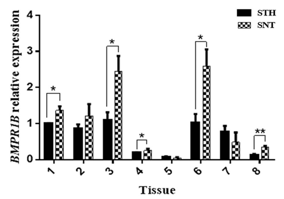
Comparison of the expression of BMPR1B in eight tissues (Tissues 1–8: brain, cerebellum, hypothalamus, pituitary, testis, epididymis, vas deferens, and adrenal gland, respectively) between Small Tail Han sheep (STH) and Sunnite sheep (SNT). Means with different superscripts are significantly different. The significant results with a p-values lower than 0.01 and 0.05 are given two asterisks (**) and one asterisk (*), respectively.
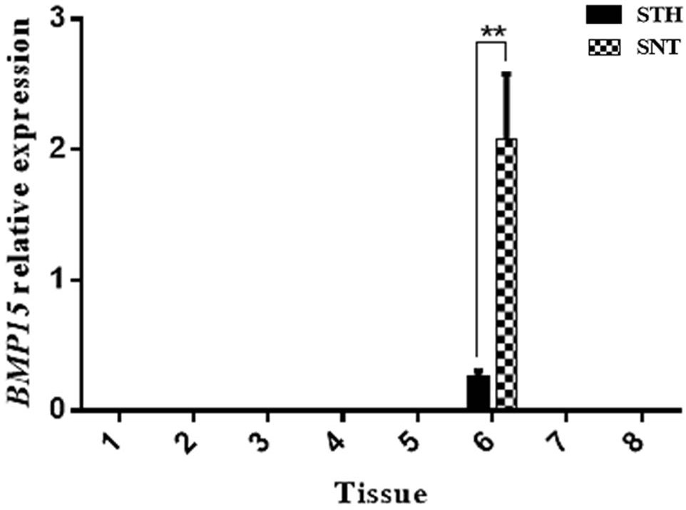
Comparison of the expression of BMP15 in eight tissues (Tissues 1–8: brain, cerebellum, hypothalamus, pituitary, testis, epididymis, vas deferens, and adrenal gland, respectively) between STH and SNT. Means with different superscripts are significantly different. The significant results with a p-values lower than 0.01 is given two asterisks (**).
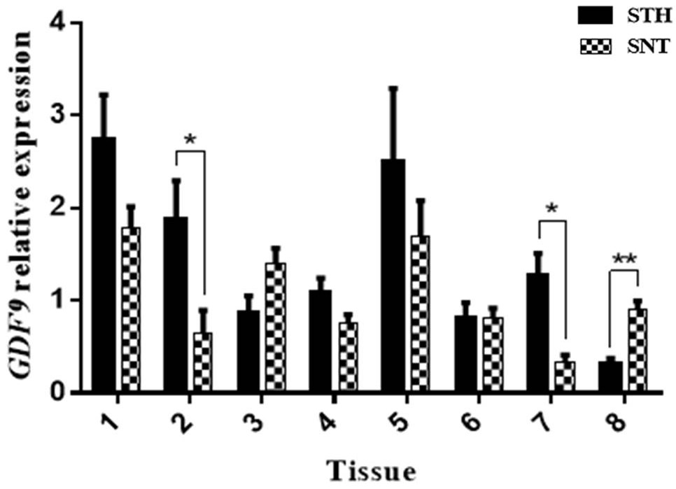
Comparison of the expression of GDF9 in eight tissues (Tissues 1–8: brain, cerebellum, hypothalamus, pituitary, testis, epididymis, vas deferens, and adrenal gland, respectively) between STH and SNT. Means with different superscripts are significantly different. The significant results with a p-values lower than 0.01 and 0.05 are given two asterisks (**) and one asterisk (*), respectively.
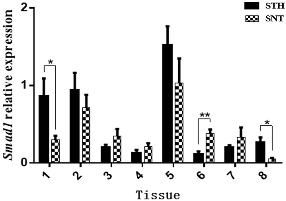
Comparison of the expression of Smad1 in eight tissues (Tissues 1–8: brain, cerebellum, hypothalamus, pituitary, testis, epididymis, vas deferens, and adrenal gland, respectively) between STH and SNT. Means with different superscripts are significantly different. The significant results with a p-values lower than 0.01 and 0.05 are given two asterisks (**) and one asterisk (*), respectively.
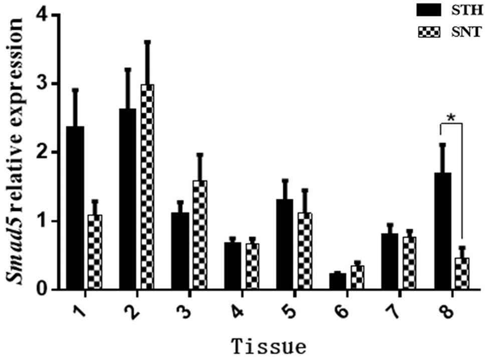
Comparison of the expression of Smad5 in eight tissues (Tissues 1–8: brain, cerebellum, hypothalamus, pituitary, testis, epididymis, vas deferens, and adrenal gland, respectively) between STH and SNT. Means with different superscripts are significantly different. The significant results with a p-values lower than 0.05 is given one asterisk (*).
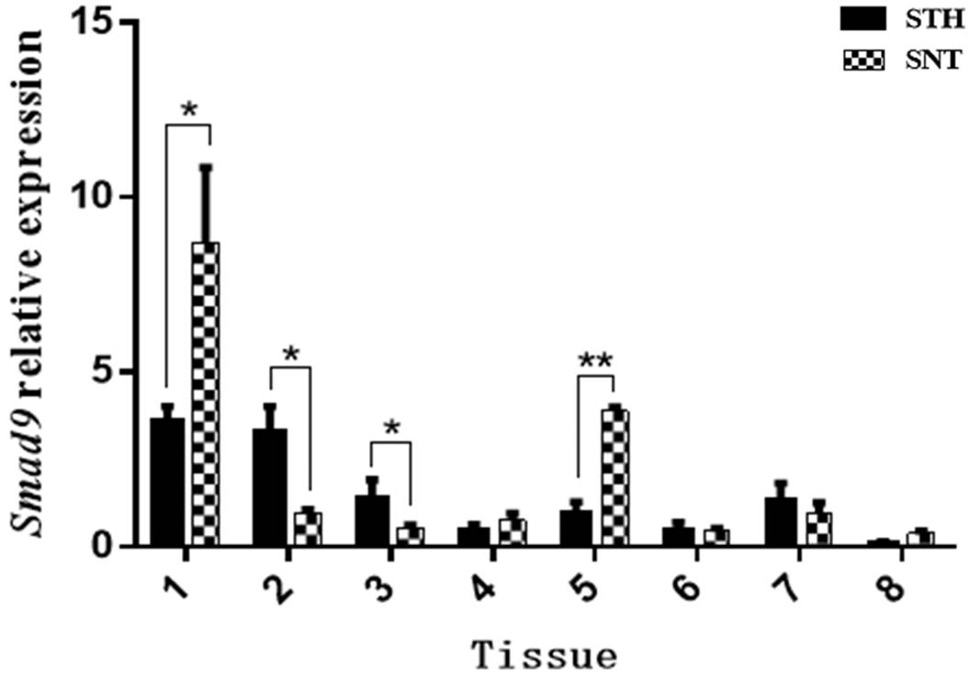
Comparison of the expression of Smad9 in eight tissues (Tissues 1–8: brain, cerebellum, hypothalamus, pituitary, testis, epididymis, vas deferens, and adrenal gland, respectively) between STH and SNT. Means with different superscripts are significantly different. The significant results with a p-values lower than 0.01 and 0.05 are given two asterisks (**) and one asterisk (*), respectively.
References
- Aaltonen, J., Laitinen, M.P., Vuojolainen, K., Jaatinen, R., Horelli, K.N., Seppä, L., Louhio, H., Tuuri, T., Sjöberg, J. and Bützow, R., 1999. Human growth differentiation factor 9 (GDF-9) and its novel homolog GDF-9B are expressed in oocytes during early folliculogenesis. J. clin. Endocrinol. Metabol., 84: 2744-2750. https://doi.org/10.1210/jc.84.8.2744[CrossRef]
- Alam, M.H., Lee, J. and Miyano, T., 2018. GDF9 and BMP15 induce development of antrum-like structures by bovine granulosa cells without oocytes. J. Reprod. Devel., 64: 423-431. https://doi.org/10.1262/jrd.2018-078[CrossRef]
- Aquino, M.R., Artigas, N., Gámez, B., Rosa, J.L. and Ventura, F., 2017. Extracellular calcium promotes bone formation from bone marrow mesenchymal stem cells by amplifying the effects of BMP-2 on SMAD signalling. PLoS One, 12: e178158. https://doi.org/10.1371/journal.pone.0178158[CrossRef]
- Belli, M. and Shimasaki, S., 2018. Molecular aspects and clinical relevance of GDF9 and BMP15 in ovarian function. Vitam. Horm, 107: 317-348. https://doi.org/10.1016/bs.vh.2017.12.003[CrossRef]
- Bodin, L., Di, P.E., Fabre, S., Bontoux, M., Monget, P., Persani, L. and Mulsant, P., 2007. A novel mutation in the bone morphogenetic protein 15 gene causing defective protein secretion is associated with both increased ovulation rate and sterility in Lacaune sheep. Endocrinology, 148: 393–400. https://doi.org/10.1210/en.2006-0764[CrossRef]
- Braun, V., Mahren, S. and Ogierman, M., 2003. Regulation of the FecI-type ECF sigma factor by transmembrane signalling. Curr. Opin. Microbiol., 6: 173–180. https://doi.org/10.1016/S1369-5274(03)00022-5[CrossRef]
- Ciller, I.M., Palanisamy, S.K., Ciller, U.A. and Mcfarlane, J.R., 2016. Postnatal expression of bone morphogenetic proteins and their receptors in the mouse testis. Physiol. Res., 65: 673-682.
- Chan, K.H., Galuska, S.P., Kudipudi, P.K., Riaz, M.A., Loveland, K.L. and Konrad, L., 2017. Signaling by TGF-betas in tubule cultures of adult rat testis. Am. J. Transl. Res., 9: 1173–1182.
- Crawford, J.L., Heath, D.A., Reader, K.L., Quirke, L.D., Hudson, N.L., Juengel, J.L. and McNatty, K.P., 2011. Oocytes in sheep homozygous for a mutation in bone morphogenetic protein receptor 1B express lower mRNA levels of bone morphogenetic protein 15 but not growth differentiation factor 9. Reproduction, 142: 53-61. https://doi.org/10.1530/REP-10-0485[CrossRef]
- Dube, J.L., Wang, P., Elvin, J., Lyons, K.M., Celeste, A.J. and Matzuk, M.M., 1998. The bone morphogenetic protein 15 gene is X-linked and expressed in oocytes. Mol. Endocrinol., 12: 1809-1817. https://doi.org/10.1210/mend.12.12.0206[CrossRef]
- Dudley, B.M., Runyan, C., Takeuchi, Y., Schaible, K. and Molyneaux, K., 2007. BMP signaling regulates PGC numbers and motility in organ culture. Mechan. Dev., 124: 68–77. https://doi.org/10.1016/j.mod.2006.09.005[CrossRef]
- El-Seedy, A.S., Hashem, N.M., El-Azrak, K.M., Nour, E.A., Ramadan, T.A., Taha, T.A. and Salem, M.H., 2017. Genetic screening of FecB, FecX(G) and FecX(I) mutations and their linkage with litter size in Barki and Rahmani sheep breeds. Reprod. Domest. Anim., 52: 1133–1137. https://doi.org/10.1111/rda.13002[CrossRef]
- Fitzpatrick, S.L., Sindoni, D.M., Shughrue, P.J., Lane, M.V., Merchenthaler, I.J. and Frail, D.E., 1998. Expression of growth differentiation factor-9 messenger ribonucleic acid in ovarian and nonovarian rodent and human tissues. Endocrinology, 139: 2571–2578. https://doi.org/10.1210/en.139.5.2571[CrossRef]
- Foroughinia, G., Fazileh, A. and Eghbalsaied, S., 2017. Expression of genes involved in BMP and estrogen signaling and AMPK production can be important factors affecting total number of antral follicles in ewes. Theriogenology, 91: 36–43. https://doi.org/10.1016/j.theriogenology.2016.12.023[CrossRef]
- Galloway, S.M., McNatty, K.P., Cambridge, L.M., Laitinen, M.P., Juengel, J.L., Jokiranta, T.S., McLaren, R.J., Luiro, K., Dodds, K.G. and Montgomery, G.W., 2000. Mutations in an oocyte-derived growth factor gene (BMP15) cause increased ovulation rate and infecundity in a dosage-sensitive manner. Nat. Genet., 25: 279–283. https://doi.org/10.1038/77033[CrossRef]
- Goyal, S., Aggarwal, J., Dubey, P.K., Mishra, B.P., Ghalsasi, P., Nimbkar, C., Joshi, B.K. and Kataria, R.S., 2017. Expression analysis of genes associated with prolificacy in FecB carrier and noncarrier Indian sheep. Anim. Biotechnol., 28: 220–227. https://doi.org/10.1080/10495398.2016.1262869[CrossRef]
- Guo, X.F., Wang, X.Y., Liang, B.M., Di, R., Liu, Q.Y., Hu, W.P., He, X.Y., Zhang, J.L., Zhang, X.S. and Chu, M.X., 2018. Molecular cloning of the B4GALNT2 gene and its single nucleotide polymorphisms association with litter size in Small Tail Han sheep. Animals, 8: 160. https://doi.org/10.3390/ani8100160[CrossRef]
- Hammoud, S.S., Low, D.H., Yi, C., Carrell, D.T., Guccione, E. and Cairns, B.R., 2014. Chromatin and transcription transitions of mammalian adult germline stem cells and spermatogenesis. Cell Stem Cell, 15: 239–253. https://doi.org/10.1016/j.stem.2014.04.006[CrossRef]
- Hanrahan, J.P., Gregan, S.M., Mulsant, P., Mullen, M., Davis, G.H., Powell, R. and Galloway, S.M., 2004. Mutations in the genes for oocyte-derived growth factors GDF9 and BMP15 are associated with both increased ovulation rate and sterility in Cambridge and Belclare sheep (Ovis aries). Biol. Reprod., 70: 900–909. https://doi.org/10.1095/biolreprod.103.023093[CrossRef]
- He, Z., Wu, Y., Xie, J., Wang, T., Zhang, L. and Zhang, W., 2012. Growth differentiation factor 9 (GDF9) was localized in the female as well as male germ cells in a protogynous hermaphroditic teleost fish, ricefield eel Monopterus albus. Gen. Comp. Endocrinol., 178: 355–362. https://doi.org/10.1016/j.ygcen.2012.06.016[CrossRef]
- Hu, S., 2017. The effects of BMP15 gene on proliferation and differentiation of cashmere goats mGSCs. Northwest A&F University: Yangling, China.
- Itman, C. and Loveland, K.L., 2010. SMAD expression in the testis: An insight into BMP regulation of spermatogenesis. Devel. Dynam., 237: 97–111. https://doi.org/10.1002/dvdy.21401[CrossRef]
- Julie, D., Stéphane, F., Julien, S., Raffaella, R., Hélène, G., Luca, P., Gwenola, T.K., Philippe, M., Zuzanna, N. and Wioleta, D., 2013. Genome-wide association studies identify two novelbmp15mutations responsible for an atypical hyperprolificacy phenotype in sheep. PLoS Genet., 9: e1003482. https://doi.org/10.1371/journal.pgen.1003482[CrossRef]
- Katakawa, Y., Funaba, M. and Murakami, M., 2016. Smad8/9 is regulated through the BMP pathway. J. Cell. Biochem., 117: 1788–1796. https://doi.org/10.1002/jcb.25478[CrossRef]
- Kaivo-oja, N., Jeffery, L.A., Ritvos, O. and Mottershead, D.G., 2006. Smad signalling in the ovary. Reprod. Biol. Endocrinol., 4: 21. https://doi.org/10.1186/1477-7827-4-21[CrossRef]
- Larriba, E., Rial, E. and Del, M.J., 2018. The landscape of mitochondrial small non-coding RNAs in the PGCs of male mice, spermatogonia, gametes and in zygotes. BMC Genom., 19: 634. https://doi.org/10.1186/s12864-018-5020-3[CrossRef]
- Lassoued, N., Benkhlil, Z., Woloszyn, F., Rejeb, A., Aouina, M., Rekik, M., Fabre, S. and Romdhani, S.B., 2017. FecXBar a novel BMP15 mutation responsible for prolificacy and female sterility in Tunisian barbarine sheep. BMC Genet., 18: 43. https://doi.org/10.1186/s12863-017-0510-x[CrossRef]
- Li, H.K., Kuo, T.Y., Yang, H.S., Chen, L.R., Li, S.S. and Huang, H.W., 2008. Differential gene expression of bone morphogenetic protein 15 and growth differentiation factor 9 during in vitro maturation of porcine oocytes and early embryos. Anim. Reprod. Sci., 103: 312–322. https://doi.org/10.1016/j.anireprosci.2006.12.017[CrossRef]
- Lin, X., Dai, Y., Xu, W., Shi, L., Jin, X., Li, C., Zhou, F., Pan, Y., Zhang, Y. and Lin, X., 2018. Hypoxia promotes ectopic adhesion ability of endometrial stromal cells via TGF-β1/Smad signalling in endometriosis. Endocrinology, 159: 1630–1641. https://doi.org/10.1210/en.2017-03227[CrossRef]
- Liu, C., Yuan, B., Chen, H., Xu, M., Sun, X., Xu, J., Gao, Y., Chen, C., Jiang, H. and Zhang, J., 2018. Effects of MiR-375-BMPR2 as a key factor downstream of BMP15/GDF9 on the Smad1/5/8 and Smad2/3 signaling pathways. Cell Physiol. Biochem., 46: 213–225. https://doi.org/10.1159/000488424[CrossRef]
- Mendis, S.H., Meachem, S.J., Sarraj, M.A. and Loveland, K.L., 2011. Activin A balances Sertoli and germ cell proliferation in the fetal mouse testis. Biol. Reprod., 84: 379–391. https://doi.org/10.1095/biolreprod.110.086231[CrossRef]
- Monteagudo, L.V., Ponz, R., Tejedor, M.T., Laviña, A. and Sierra, I., 2009. A 17 bp deletion in the Bone Morphogenetic Protein 15 (BMP15) gene is associated to increased prolificacy in the Rasa Aragonesa sheep breed. Anim. Reprod. Sci., 110: 139–146. https://doi.org/10.1016/j.anireprosci.2008.01.005[CrossRef]
- Montgomery, G.W., Lord, E.A., Penty, J.M., Dodds, K.G., Broad, T.E., Cambridge, L., Sunden, S.L., Stone, R.T. and Crawford, A.M., 1994. The Booroola fecundity (FecB) gene maps to sheep chromosome 6. Genomics, 22: 148–153. https://doi.org/10.1006/geno.1994.1355[CrossRef]
- Moore, R.K., Otsuka, F. and Shimasaki, S., 2002. Molecular basis of bone morphogenetic protein-15 signaling in granulosa cells. J. biol. Chem., 278: 304-310. https://doi.org/10.1074/jbc.M207362200[CrossRef]
- Mullen, M.P. and Hanrahan, J.P., 2014. Direct evidence on the contribution of a missense mutation in GDF9 to variation in ovulation rate of Finnsheep. PLoS One, 9: e95251. https://doi.org/10.1371/journal.pone.0095251[CrossRef]
- Nicol, L., Bishop, S.C., Pong, W.R., Bendixen, C., Holm, L., Rhind, S.M. and McNeilly, A.S., 2009. Homozygosity for a single base-pair mutation in the oocyte-specific GDF9 gene results in sterility in Thoka sheep. Reproduction, 138: 921–933. https://doi.org/10.1530/REP-09-0193[CrossRef]
- Ohyama, K., Ohta, M., Hosaka, Y.Z., Tanabe, Y., Ohyama, T. and Yamano, Y., 2015. Expression of anti-Mullerian hormone and its type II receptor in germ cells of maturing rat testis. Endocrinol. J., 62: 997–1006. https://doi.org/10.1507/endocrj.EJ15-0370[CrossRef]
- Okamura, D., Hayashi, K. and Matsui, Y., 2005. Mouse epiblasts change responsiveness to BMP4 signal required for PGC formation through functions of extraembryonic ectoderm. Mol. Reprod. Devel., 70: 20–29. https://doi.org/10.1002/mrd.20136[CrossRef]
- Pan, Z., Wang, X., Di, R., Liu, Q.Y., Hu, W.P., Cao, X.H., Guo, X.F., He, X.Y., Lv, S.J., Li, F.K. and Chu, M.X., 2018. A 5-methylcytosine site of growth differentiation factor 9 (GDF9) gene affects its tissue-specific expression in sheep. Animals, 8: 200. https://doi.org/10.3390/ani8110200[CrossRef]
- Pennetier, S., Uzbekova, S., Perreau, C., Papillier, P., Mermillod, P. and Dalbiès-Tran, R., 2004. Spatio-temporal expression of the germ cell marker genes MATER, ZAR1, GDF9, BMP15 and VASA in adult bovine tissues, oocytes, and preimplantation embryos. Biol. Reprod., 71: 1359–1366. https://doi.org/10.1095/biolreprod.104.030288[CrossRef]
- Rol, N., Kurakula, K.B., Happé, C., Bogaard, H.J. and Goumans, M.J., 2018. TGF-beta and BMPR2 signaling in PAH: Two black sheep in one family. Int. J. mol. Sci., 19: 2585. https://doi.org/10.3390/ijms19092585[CrossRef]
- Shafieiyan, Z., Mohammadi, G., Jolodarzadeh, A. and Amiri, S., 2013. No mutations of FecB and FecG(H) in Iranian Lory sheep. Vet. Res. Forum, 4: 265–268.
- Shokrollahi, B. and Morammazi, S., 2018. Polymorphism of GDF9 and BMPR1B genes and their association with litter size in Markhoz goats. Reprod. Domest. Anim., 53: 971–978. https://doi.org/10.1111/rda.13196[CrossRef]
- Shi, C., Iura, A., Terajima, M., Liu, F., Lyons, K., Pan, H., Zhang, H., Yamauchi, M., Mishina, Y. and Sun, H., 2016. Deletion of BMP receptor type IB decreased bone mass in association with compromised osteoblastic differentiation of bone marrow mesenchymal progenitors. Scient. Rep., 6: 24256. https://doi.org/10.1038/srep24256[CrossRef]
- Silva, B.D., Castro, E.A., Souza, C.J., Paiva, S.R., Sartori, R., Franco, M.M., Azevedo, H.C., Silva, T.A., Vieira, A.M. and Neves, J.P., 2011. A new polymorphism in the growth and differentiation factor 9 (GDF9) gene is associated with increased ovulation rate and prolificacy in homozygous sheep. Anim. Genet., 42: 89–92. https://doi.org/10.1111/j.1365-2052.2010.02078.x[CrossRef]
- Silva, J.R., Hurk, R., Tol, H.T., Roelen, B.A. and Figueiredo, J.R., 2005. Expression of growth differentiation factor 9 (GDF9), bone morphogenetic protein 15 (BMP15), and BMP receptors in the ovaries of goats. Mol. Reprod. Devel., 70: 11–19. https://doi.org/10.1002/mrd.20127[CrossRef]
- Song, W., Ghosh, A.C., Cheng, D. and Perrimon, N., 2018. Endocrine regulation of energy balance by drosophila TGF-beta/Activins. Bioessays, 40: e1800044. https://doi.org/10.1002/bies.201800044[CrossRef]
- Souza, C.J., McNeilly, A.S., Benavides, M.V., Melo, E.O. and Moraes, J.C., 2014. Mutation in the protease cleavage site of GDF9 increases ovulation rate and litter size in heterozygous ewes and causes infecundity in homozygous ewes. Anim. Genet., 45: 732–739. https://doi.org/10.1111/age.12190[CrossRef]
- Tang, J., Hu, W., Di, R., Liu, Q.Y., Wang, X.Y., Zhang, X.S., Zhang, J.L. and Chu, M.X., 2018. Expression analysis of the prolific candidate genes, BMPR1B, BMP15, and GDF9 in Small Tail Han ewes with three fecundity (FecB gene) genotypes. Animals, 8: 166. https://doi.org/10.3390/ani8100166[CrossRef]
- Tang, K.Q., Yang, W.C., Zhang, X.X. and Yang, L.G., 2013. Effects of polymorphisms in the bovine growth differentiation factor 9 gene on sperm quality in Holstein bulls. Genet. mol. Res., 12: 2189–2195. https://doi.org/10.4238/2013.July.4.1[CrossRef]
- Tang, K.Q., Wang, L., Jin, Y., Yang, W. and Yang, L., 2017. GDF9 affects the development and tight junction functions of immature bovine Sertoli cells. Reprod. Domest. Anim., 52: 640–648. https://doi.org/10.1111/rda.12960[CrossRef]
- Våge, D.I., Husdal, M., Kent, M.P., Klemetsdal, G. and Boman, I.A., 2013. A missense mutation in growth differentiation factor 9 (GDF9) is strongly associated with litter size in sheep. BMC Genet., 14: 1. https://doi.org/10.1186/1471-2156-14-1[CrossRef]
- Valdecantos, P.A., Bravo, Miana, R.D., García, E.V., García, D.C., Roldán, O.M. and Miceli, D.C., 2017. Expression of bone morphogenetic protein receptors in bovine oviductal epithelial cells: Evidence of autocrine BMP signaling. Anim. Reprod. Sci., 185: 89–96. https://doi.org/10.1016/j.anireprosci.2017.08.006[CrossRef]
- Wang, L., Ding, Q., Zhao, L., Pan, Y., Song, Z., Qin, Y. and Yan, X., 2018. Decreased BMP-7 and p-Smad1/5/8 expression, and increased levels of gremlin in hepatocellular carcinoma. Oncol. Lett., 16: 2113–2118. https://doi.org/10.3892/ol.2018.8918[CrossRef]
- Wang, S., Hu, S., Wang, J., Liu, Y., Zhao, R., Tong, M., Cui, H., Wu, N. and Chen, X., 2018. Conditioned medium from bone marrow-derived mesenchymal stem cells inhibits vascular calcification through blockade of the BMP2-Smad1/5/8 signaling pathway. Stem Cell Res., 9: 160. https://doi.org/10.1186/s13287-018-0894-1[CrossRef]
- Wang, X., Yang, Q., Wang, K., Yan, H., Pan, C., Chen, H., Liu, J., Zhu, H., Qu, L. and Lan, X., 2018. Two strongly linked single nucleotide polymorphisms (Q320P and V397I) in GDF9 gene are associated with litter size in cashmere goats. Theriogenology, 125: 115–121. https://doi.org/10.1016/j.theriogenology.2018.10.013[CrossRef]
- Wilson, T., Wu, X.Y., Juengel, J.L., Ross, I.K., Lumsden, J.M., Lord, E.A., Dodds, K.G., Walling, G.A., McEwan, J.C. and O’Connell, A.R., 2001. Highly prolific Booroola sheep have a mutation in the intracellular kinase domain of bone morphogenetic protein IB receptor (ALK-6) that is expressed in both oocytes and granulosa cells. Biol. Reprod., 64: 1225–1235. https://doi.org/10.1095/biolreprod64.4.1225[CrossRef]
- Xu, Y.F., Li, E.L., Han, Y.D., Chen, L. and Xie, Z.A., 2010. Differential expression of mRNAs encoding BMP/SMAD pathway molecules in antral follicles of high- and low-fecundity Hu sheep. Anim. Reprod. Sci., 120: 47–55. https://doi.org/10.1016/j.anireprosci.2010.02.009[CrossRef]
- Zhao, L., He, J., Guo, Q., Wen, X., Zhang, X. and Dong, C., 2011. Expression of growth differentiation factor 9 (GDF9) and its receptor in adult cat testis. Acta Histochem., 113: 771–776. https://doi.org/10.1016/j.acthis.2010.11.005[CrossRef]
Cited by 1 article
In journals published by ResearchersLinks.
Services
Article usage statistics
Loading…