
Slit and roundabout homologs have emerged as important players in signaling cascades of tumor metastasis. In previous reports it is stated that the interactions between Slit2 and Robo1 are facilitated by heparan sulphate that is abundantly expressed on cell surface and extracellular matrix. Slit2 reduces tumor proliferation in lung cancer and glioblastoma cells whereas TGF-β is a well-described tumor inducer. The present study was aimed at deciphering the role of heparan sulphate in Slit2 mediated inhibition of cancer metastasis. Cancer proliferation was induced by TGF-β in lung cancer cells (H1650) and glioblastoma cells (SF767) and then the anti-proliferative role of Slit2 was analyzed in presence and absence of heparan sulfate. The data revealed that, heparan sulfate plays important role in enhancing tumor inhibition by Slit2 in cancer cells as there was further reduction in cell proliferation when Slit2 was administered along with heparin.
Research Article · Volume 56, Issue 5, 13 August 2024 · Open Access
Recombinant Slit2 Requires Heparin Sulphate to Inhibit TGF- β Induced Tumor Proliferation in Lung Cancer and Glioblastoma
·
Affiliations & Notes ⌄
- 1School of Biological Sciences, University of the Punjab, Quaid-i-Azam Campus, Lahore 54590, Pakistan 2Cancer Research Centre, University of the Punjab, Quaid-i-Azam Campus, Lahore 54590, Pakistan * Corresponding author: arshakoori.sbs@pu.edu.pk, arshaksbs@yahoo.com
Article Info ⌄
- Journal:
- Pakistan Journal of Zoology
- Published:
- 13 August 2024

From the article
- Previous reports showed the crucial role of heparin in Slit-Robo complex.
- The present study was designed to determine the role of heparin in Slit2-Robo1 mediated down-regulation in cancer proliferation in TGF-β induced cancer cells.
- The cancer cells were first induced with 24 h exposure to TGF-β, then recombinant human Slit2 protein was added in cells, with and without heparin.
- The results showed that with presence of heparin, anti-proliferative effect of Slit2 was enhanced.
Selected from the article's own text.
Abstract
INTRODUCTION
Within the sites of tumor metastasis, chemokines and cytokines play pivotal role. There are a number of cytokines that are expressed on surfaces of malignant and pre-malignant tumors. TGF-β is among the well characterized cytokines that plays role in induction of fibrotic tumors by regulating various molecular programs including epithelial to mesenchymal transition (EMT) . EMT is known as among the basic phenomenon that takes place as soon as the cell decides to invade its neighborhood ; . Recent studies have also suggested that TGF-β stimulates fibroblasts differentiation by targeting Slit2 (Slit guidance ligand 2) protein expression ; . Slit2-Robo1 signaling, previously known for only neuronal cells, has been widely studied in last decade for being crucial candidates in cancer studies in both diagnostic and therapeutic prospective ; . In lung cancer Slit2 is down-regulated by hypermethylation ; . Similar epigenetic inactivation of Slit2 is known for gliomas and glioblastomas .
Heparan sulfates are the proteoglycans that are present on cell surfaces as well as the extracellular matrix, where they interact with a number of ligand molecules to mediate cellular functions . Heparin (heparan sulfate) is also the interacting partner of Slit2 and forms a ternary complex stabilizing the Slit2-Robo interactions . It is also reported that Slit2 mediated directional migrations and invasion only happens in presence of heparin. It also serves as a co-receptor in Slit-Robo signaling .
The present study was aimed at specifically addressing the role of heparin (heparan sulfate) in mediating Slit2-Robo1 signal transduction. The role of Slit2 instigated inhibition of tumor proliferation was examined in TGF-β induced lung cancer and glioblastoma cells in the presence of heparin. The data suggested that heparin facilitates the inhibitory role of Slit2 in cancer metastasis in both cancer types under study, by regulating the expression levels of genes involved in epithelial to mesenchymal transition.
MATERIALS AND METHODS
Cell culture maintenance and conditioning
Lung cancer cells (H1650) and glioblastoma cells (SF767) were maintained in DMEM (Gibco-12800-017), with addition of 10% FBS as supplement and 1% pen/strep. Cultures were incubated in standard culture conditions that were temperature of 37°C and 5% CO2 in humidified conditions. At second day of culturing cancer cells were induced by addition of 1ng/ml TGF-β (Invitrogen™ PHG9204). When cells reached at 70% confluency, media were conditioned with 1ng/ml Human recombinant Slit2 protein (Abcam-ab82131) and heparin (Fisher Scientific-BP2524100). 24 h after Slit2 and Heparin addition cells were retrieved for analysis.
Morphology analysis
After 24 h exposure to the required concentration of Slit2 protein and heparin, cells were analyzed using Nikon Eclipse TS100 microscope with Nikon ELWD 0.3/OD75 camera system to observe any changes in morphology. Cell number was analyzed by counting the cells by hemocytometer by doing Trypane blue staining.
Cell proliferation assays
Cells were cultured in 96 well plates with density of 1x10³ cells/well. After induction by TGF-β and exposure to Slit2 and heparin, different assays were performed to evaluate changes in cell proliferation, cell viability and cell metabolic activity.
MTT assay
MTT assay was done for estimation of cell metabolic activity. For MTT assay 0.1mg/ml MTT salt (Thermo fisher Scientific- M6494) in PBS was added to cells followed by incubation of 3 h. After addition of de-staining solution (DMSO), Absorbance at 570nm was analyzed by BioTek ELx808 absorbance reader.
WST-II assay
WST-II proliferation assay was performed by Quick cell proliferation assay kit (Abcam-ab65475) following manufacturer’s instructions. Briefly, after addition of 10ul WST-II solution (prepared in ECS solution), cells were incubated for 3 h. After incubation for recommended time period, stop solution was added to stop the reaction and absorbance was taken at 450 nm using BioTek ELx808 absorbance reader.
BrdU assay
BrdU assay was performed by using colorimetric BrdU cell proliferation assay kit (Exalpha-X1327K) following instructions by the manufacturer. The BrdU taken up by the cells was detected by an anti-BrdU anti-body and absorbance was taken at 450 nm using BioTek ELx808 absorbance reader.
Neutral red assay
For Neutral assay cells were incubated with final concentration of 40μg/ml neutral red (Sigma Aldrich-N4638) diluted in DMEM, followed by 1 h incubation. After de-staining (with solution of 50%: absolute ethanol, 49%: deionized water and 1%: Glacial acetic acid) absorbance was taken at 570nm.
FDA/PI staining
For analysis of cell apoptosis, differential staining of viable and non-viable cells was done by adding 5µl of FDA (5mg/ml) and PI (1mg/ml) each (Sigma-F7378). The cells were incubated for 20 min in incubator at 37°C. The cells were then analyzed by fluorescent microscope using red and green filters.
Gene expression analysis by RT-PCR
As cell migration is an important aspect of cancer metastasis, so the expression of cell adhesion proteins including epithelial and mesenchymal markers was checked. The samples for RNA isolation were stored in 1ml Trizol reagent and kept at -80°C. RNA was isolated using Trizol method following manufacturer’s instructions of extraction of RNA from the aqueous phase. After quantification using pico-drop and DNAse treatment, the extracted RNA was subjected to cDNA synthesis. cDNA synthesis was done using 1µg purified RNA using cDNA synthesis kit (Thermo-K1622). Real time PCR (RT-PCR) was performed using 5µM forward and reverse primers. Sequences are mentioned in Table I. The retrieved data from melting curve analysis and ΔCT values of test genes were compared with those of housekeeping genes (GUS B). The results were represented in graphical form.
Table I. Primer sequences.
| Genes | Sequence 5´-3´ |
| GUSB glucuronidase | F: ACCACGATGGCATAGGAATGG R: CGGCTCTTCTCTCCACAGTCAG |
| Human vimentin | F: CGGTTTCCTCGTTCCCCTTT R: ATTGCTCGTGGGTTGTGTTG |
| Human E-Cadherin | F: CTTTGACGCCGAGAGCTACA R: TCCAAGGGGTGTCGTTTGAG |
| Integrin subunit beta 1 | F: ATCTAATGTACCCCAATTCTGGCT R: TGGGTCAGTTCTGGGAAAGGT |
| N-CAD (Neuronal cadherin) | F: GCTTCAGGCGTCTGTAGAGG R: AGAGGCTGTCCTTCATGCAC |
Statistical analysis
All experiments were performed in experimental and technical triplicates. Statistical significance was estimated using one-way and two-way ANOVA by Graph pad prism. P-values less than 0.05 were considered significant and were represented in results sterically.
RESULTS
Heparin amplified Slit2 induced reduction in cell number
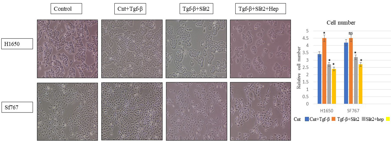
The cell number was increased in cancer cells H1650 and SF767 after 24 h induction with TGF-β. But with Slit2 administration, there was drastic decrease in cell number even in presence of TGF-β (Fig. 1). This down-regulation in cell growth was more profound when Slit2 was added in cancer cells along with heparin.
When cell morphology was examined, it was observed that SF767 cells appeared slightly longitudinal with TGF-β treatment but there was no change in morphology of H1650 cells. No evident change in cell morphology was observed after addition of Slit2 and heparin in lung cancer cells and glioblastoma cells (Fig. 1).
Heparin facilitate reduction in cell proliferation by Slit2
There was an evident up-regulation in cell metabolic activity and proliferation after TGF-β treatment but with Slit2 addition there was reduction in cell metabolic activity in TGF-β treated cells (Fig. 2). When Slit2 was added along with heparin, there was further down-regulation in cell proliferation. Cell viability however, remained un-affected by Slit2 and heparin addition.
Slit2 and heparin decreased the number of proliferating cells and caused apoptosis in cancer cells
To analyze the effect of Slit2 and heparin on viability of TGF-β induced cells, differential staining with FDA and PI was done to label the live and dead cells. Live and viable cells appeared green after FDA uptake, whereas dead and damaged cells appeared red with PI staining. After analysis of cells in fluorescent microscope it was observed that the increase in the viable cells after induction with TGF-β, decreased after addition of Slit in lung cancer cells as well as in glioblastoma cells (Fig. 3).
In contrast, the number of dead and damaged cancer cells increased after Slit2 addition along with heparin, indicating that the proliferative effect of TGF-β induction was down-regulated with Slit2 and this down-regulation was further enhanced when Slit2 was administered along with heparin (Fig. 4).
Slit2 reversed TGF-β instigated up-regulation in mesenchymal genes expression
With treatment of TGF-β, there was an up-regulated expression of N-CAD and vimentin in both cancer types, there was also up-regulation in E-CAD expression in lung cancer cells. With Slit2 and heparin addition, E-CAD expression was further up-regulated, while there was down-regulation in mesenchymal marker genes for both cancer types (Fig. 5).
Integrin beta 1 expression was increased after addition of TGF-β but it was not affected in TGF-β induced cells after Slit2 addition.
DISCUSSION
Roundabout receptor is a tumor endothelial marker, expressed in the vascular network of various tumor entities. In mammals four roundabout proteins (Robo1-4) have been recognized, that are cognate receptors for Slits that are axon guidance molecules . Slit-Robo signaling has the dual role in cancer management. As they show both tumor-genic and anti-tumor traits that is purely cancer type dependent . Table II summarizes the status of Slit-Robo signaling pathway in some of the cancer types, defining their role as onco-genic or tumor suppressor.
The leucine rich domain of Slit, that binds to the Robo, also has the binding affinity for heparan sulphate . Heparan sulphate binds to both Slit and Robo, facilitating their receptor-ligand interactions . It is also known that there is up to three folds reduction in Slit-Robo interactions by removal of heparan sulphate . It belongs to glycosaminoglycan family of polysaccharides that critically regulate several biological mechanisms including cell growth, differentiation, lipids metabolism, blood coagulation, cell to cell and cell to matrix interactions and cancer metastasis, by interacting a number of protein ligands . Previous reports showed the crucial role of heparin in Slit-Robo complex.
Table II. Oncogenic and tumor suppressive roles of Slit-Robo signaling in human carcinomas mentioned in multiple reports summarized by Jiang et al. (2019).
| Tumor type | SLIT/ROBO pathway status | Function |
| Pancreatic cancer | ROBO3↑ | Oncogenic |
| Pancreatic cancer | SLIT2↓ | Tumor-suppressor |
| Lung cancer | SLIT3↓ | Tumor-suppressor |
| Lung cancer | USP33↓ | Tumor-suppressor |
| Colorectal cancer (CRC) | SLIT2 ↓: USP33↓ | Tumor-suppressor |
| Colorectal cancer | ROBO1 ↑: ROBO4↑ | Oncogenic |
| Colorectal cancer | SLIT2 ↑: ROBO1↑ | Oncogenic |
| Colorectal cancer | SLIT2 ↑: ROBO2↑ | Oncogenic |
| Breast cancer | srGAP3↓ | Tumor-suppressor |
| Mucoepidermoid carcinoma | SLIT2 ↑: ROBO1↑ | Oncogenic |
| Ovarian cancer | SLIT2 ↓: SLIT3 ↓: ROBO1 ↓: ROBO2 ↓: ROBO4 ↓ | Tumor-suppressor |
| Oral squamous cancer | SLIT2↓ | Tumor-suppressor |
| Nasopharyngeal cancer | ROBO1↑ | Oncogenic |
| Hepatocellular cancer | SLIT1, SLIT2, and SLIT3 genes were methylated | Tumor-suppressor |
| Hepatocellular cancer | ROBO1 ↑: ROBO2 ↑: ROBO4 ↓: SLIT2↓ | Oncogenic |
| Cervical cancer | SLIT1↓: SLIT2↓: SLIT3↓: ROBO1↓: ROBO3↓ | Tumor-suppressor |
| Gliomas | SLIT2↓ | Tumor-suppressor |
| Prostate cancer | SLIT1 ↑: ROBO1↓ |
Structural analysis study showed heparin binding surface in the interface of Slit-Robo complex . It was also predicted that at least five heparin disaccharide units are at least required to support Slit-Robo signaling (Fig. 6).
Several cytokines also require heparan sulphate for their signaling . A better understanding of the molecular interactions between heparan sulphate and its ligand proteins may lead to the development of more specific glycan-based agents that will be helpful for both diagnostic and therapeutic purposes.
The present study was designed to determine the role of heparin in Slit2-Robo1 mediated down-regulation in cancer proliferation in TGF-β induced cancer cells. The cancer cells were first induced with 24 h exposure to TGF-β, then recombinant human Slit2 protein was added in cells, with and without heparin. The results showed that with presence of heparin, anti-proliferative effect of Slit2 was enhanced. This led to the fact that heparin is amplifying Slit-Robo signal transduction in both types of cancer cells used in the study. Heparin addition with Slit2 also decreased the expression of mesenchymal marker genes; N-cadherin and vimentin, indicating that it is somewhat rendering the cells to transform into the mesenchymal state, that is a prerequisite of cancer metastasis.
The data elucidated the importance of heparin in Slit-Robo signaling and also gave insight for future studies elaborating the downstream targeted pathways affecting cancer metastasis. Further, more intricate studies are required to be done to unravel the bindings between Slit2-Robo1 and heparan sulphate, as the molecular mechanisms behind the above-mentioned interactions remain obscure.
Acknowledgement
The financial support of University of the Punjab is gratefully acknowledged.
Funding
No formal funding was provided by any agency.
IRB approval
The study was approved by Advanced Board of Studies and Research, University of the Punjab.
Ethics statement
The study was approved by the Ethics committee of the School of Biological Sciences, University of the Punjab Lahore.
Statement of conflict of interest
The authors have declared no conflict of interest.
Figures
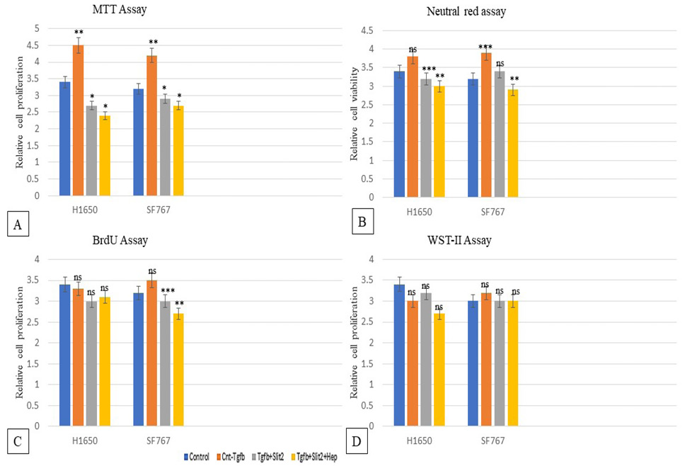
Cell functional assays in H1650 and Sf767 cells. MTT assay (A) showed decreased metabolic activity in presence of Slit2 and heparin. Neutral red assay (B) showed decreased cell viability with Slit2 and heparin treatment. BrdU assay (C) and WST-II assay (D) showed slight down-regulation in cell proliferation with Slit2 and heparin.
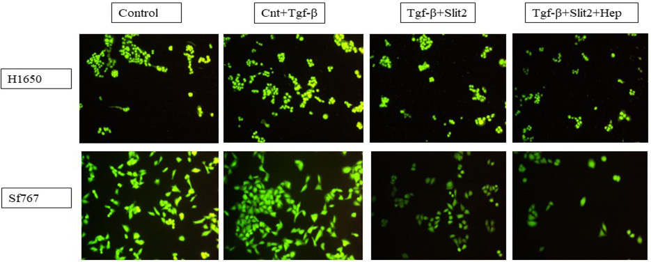
FDA stained viable/live H1650 cells and SF767 cells. The number of viable cells decreased with Slit2 addition and with heparin even in presence of TGF-β.
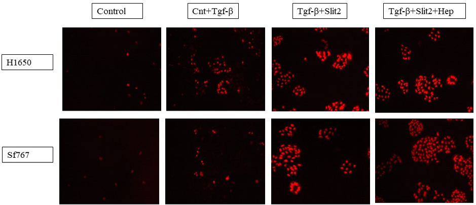
PI stained dead/non-viable H1650 and SF767 cells. The number of non-viable PI-stained cells increased after addition of Slit2 along with heparin.
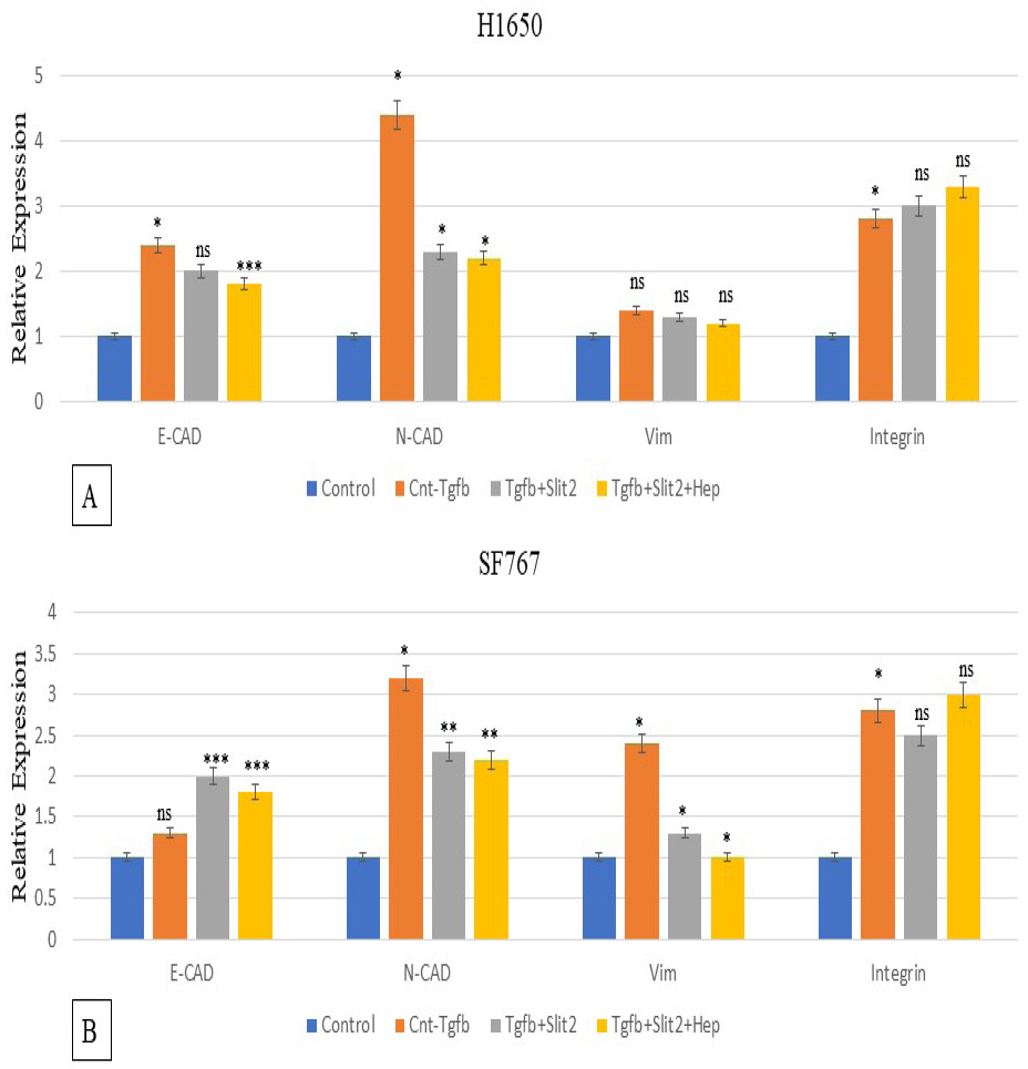
Expression analysis of cell adhesion genes in H1650 (A) and SF767 cells (B). There was significant down-regulation in expression of vimentin and N-Cad in presence of Slit2 that further amplifies in presence of heparin along with Slit2. Integrin beta 1 expression remained unchanged in H1650 and Sf767 cells.
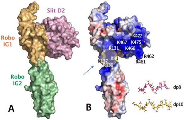
A model of human Slit2 (Domain 2) and Robo1 (IG1) complex. (A) shows the surface representation. (B) shows the electrostatic representation. The position of heparin binding to Slit2 and Robo is shown. The location of heparin oligosacharides in Robo1 is indicated by arrow. Figure taken from Fakuhara et al. (2008).
References
- Chang, J., Lan, T., Li, C., Ji, X., Zheng, L., Gou, H., Ou, Y., Wu, T., Qi, C. and Zhang, Q., 2015. Activation of Slit2-Robo1 signaling promotes liver fibrosis. J. Hepatol., 63: 1413-1420. https://doi.org/10.1016/j.jhep.2015.07.033[CrossRef]
- Chen, J., Liu, J., Gao, S., Qiu, Y., Wang, Y., Zhang, Y., Gao, L., Qi, G., Wu, Y. and Lash, G.E., 2021. Role of Slit2 upregulation in recurrent miscarriage through regulation of stromal decidualization. Placenta, 103: 1-9. https://doi.org/10.1016/j.placenta.2020.10.008[CrossRef]
- Dallol, A., Da Silva, N.F., Viacava, P., Minna, J.D., Bieche, I., Maher, E.R. and Latif, F., 2002. SLIT2, a human homologue of the Drosophila Slit2 gene, has tumor suppressor activity and is frequently inactivated in lung and breast cancers. Cancer Res., 62: 5874-5880.
- Dudás, J., Ladányi, A., Ingruber, J., Steinbichler, T.B. and Riechelmann, H., 2020. Epithelial to mesenchymal transition: a mechanism that fuels cancer radio/chemoresistance. Cells, 9: 428. https://doi.org/10.3390/cells9020428[CrossRef]
- Figiel, S., Vasseur, C., Bruyere, F., Rozet, F., Maheo, K. and Fromont, G., 2017. Clinical significance of epithelial-mesenchymal transition markers in prostate cancer. Hum. Pathol., 61: 26-32. https://doi.org/10.1016/j.humpath.2016.10.013[CrossRef]
- Fukuhara, N., Howitt, J.A., Hussain, S.A. and Hohenester, E., 2008. Structural and functional analysis of slit and heparin binding to immunoglobulin-like domains 1 and 2 of Drosophila Robo. J. biol. Chem., 283: 16226-16234. https://doi.org/10.1074/jbc.M800688200[CrossRef]
- Geraldo, L.H., Xu, Y., Jacob, L., Pibouin-Fragner, L., Rao, R., Maissa, N., Verreault, M., Lemaire, N., Knosp, C. and Lesaffre, C., 2021. SLIT2/ROBO signaling in tumor-associated microglia and macrophages drives glioblastoma immunosuppression and vascular dysmorphia. J. clin. Invest., 131: e141083. https://doi.org/10.1172/JCI141083[CrossRef]
- Hohenester, E., Hussain, S. and Howitt, J., 2006. Interaction of the guidance molecule Slit with cellular receptors. Biochem. Soc. Trans., 34: 418-421. https://doi.org/10.1042/BST0340418[CrossRef]
- Hu, H., 2001. Cell-surface heparan sulfate is involved in the repulsive guidance activities of Slit2 protein. Nat. Neurosci., 4: 695-701. https://doi.org/10.1038/89482[CrossRef]
- Hua, W., Ten Dijke, P., Kostidis, S., Giera, M. and Hornsveld, M., 2020. TGFβ-induced metabolic reprogramming during epithelial-to-mesenchymal transition in cancer. Cell. Mol. Life Sci., 77: 2103-2123. https://doi.org/10.1007/s00018-019-03398-6[CrossRef]
- Huang, Y., Xie, Y., Abel, P.W., Wei, P., Plowman, J., Toews, M.L., Strah, H., Siddique, A., Bailey, K.L. and Tu, Y., 2020. TGF-β1-induced miR-424 promotes pulmonary myofibroblast differentiation by targeting Slit2 protein expression. Biochem. Pharmacol., 180: 114172. https://doi.org/10.1016/j.bcp.2020.114172[CrossRef]
- Hussain, S.A., Piper, M., Fukuhara, N., Strochlic, L., Cho, G., Howitt, J.A., Ahmed, Y., Powell, A.K., Turnbull, J.E. and Holt, C.E., 2006. A molecular mechanism for the heparan sulfate dependence of slit-robo signaling. J. biol. Chem., 281: 39693-39698. https://doi.org/10.1074/jbc.M609384200[CrossRef]
- Jiang, Z., Liang, G., Xiao, Y., Qin, T., Chen, X., Wu, E., Ma, Q. and Wang, Z., 2019. Targeting the SLIT/ROBO pathway in tumor progression: Molecular mechanisms and therapeutic perspectives. Ther. Adv. med. Oncol., 11: 1758835919855238. https://doi.org/10.1177/1758835919855238[CrossRef]
- Kim, Y., Lee, B.B., Kim, D., Um, S.W., Han, J., Shim, Y.M. and Kim, D.H., 2022. Aberrant methylation of SLIT2 gene in plasma cell-free DNA of non-small cell lung cancer patients. Cancers, 14: 296. https://doi.org/10.3390/cancers14020296[CrossRef]
- Morlot, C., Thielens, N.M., Ravelli, R.B., Hemrika, W., Romijn, R.A., Gros, P., Cusack, S. and McCarthy, A.A., 2007. Structural insights into the Slit-Robo complex. Proc. natl. Acad. Sci., 104: 14923-14928. https://doi.org/10.1073/pnas.0705310104[CrossRef]
- Rezniczek, G.A., Grunwald, C., Hilal, Z., Scheich, J., Reifenberger, G., Tannapfel, A. and Tempfer, C.B., 2019. ROBO1 expression in metastasizing breast and ovarian cancer: SLIT2-induced chemotaxis requires heparan sulfates (heparin). Anticancer Res., 39: 1267-1273. https://doi.org/10.21873/anticanres.13237[CrossRef]
- Steigemann, P., Molitor, A., Fellert, S., Jäckle, H. and Vorbrüggen, G., 2004. Heparan sulfate proteoglycan syndecan promotes axonal and myotube guidance by slit/robo signaling. Curr. Biol., 14: 225-230. https://doi.org/10.1016/j.cub.2004.01.006[CrossRef]
- Yiin, J.J., Hu, B., Jarzynka, M.J., Feng, H., Liu, K.W., Wu, J.Y., Ma, H.I. and Cheng, S.Y., 2009. Slit2 inhibits glioma cell invasion in the brain by suppression of Cdc42 activity. Neurooncology, 11: 779-789. https://doi.org/10.1215/15228517-2009-017[CrossRef]
- Zhang, F., Moniz, H.A., Walcott, B., Moremen, K.W., Linhardt, R.J. and Wang, L., 2013. Characterization of the interaction between Robo1 and heparin and other glycosaminoglycans. Biochimie, 95: 2345-2353. https://doi.org/10.1016/j.biochi.2013.08.018[CrossRef]
- Zhang, F., Ronca, F., Linhardt, R.J. and Margolis, R.U., 2004. Structural determinants of heparan sulfate interactions with Slit proteins. Biochem. biophys. Res. Commun., 317: 352-357. https://doi.org/10.1016/j.bbrc.2004.03.059[CrossRef]
Services
Article usage statistics
Loading…