
Lumpy skin disease (LSD) is among the most significant poxvirus diseases that affect livestock, with high morbidity but low mortality. The disease spreads in various climatic zones, including Africa, Asia, and Europe, causing substantial economic losses in the animal industry. This study aimed to investigate the epidemiological state and risk factors for LSD virus (LSDV) in the six governorates of Egypt (Al-Menia, Alwady ALgaded, Al-Bhera, Al-Dakahlia, Al-Menofia, and Al-Sharqia), based on viral isolation and molecular detection. The required data were collected using a pre-structured questionnaire during fieldwork. This study examined 575 cattle from 25 herds during the LSD outbreak from June 2020 to May 2022. Approximately 185 of 575 cows showed typical LSDV clinical signs that varied from mild to serious. Diseased animals had fever, anorexia, and decrease milk yield, as well as superficial lymph nodes expansion, the sudden appearance of several nodules varying in size from 0.5 to 6 cm in diameter, and numbers according to infection severity. The infected calves displayed serious signs of illness, such as extensive skin lesions and respiratory manifestations. The morbidity, mortality, and case fatality rates were 32.5% (187/575), 6.1% (35/575), and 18.7% (35/187), respectively. There was no significant difference in disease occurrence between different age groups, but animals aged <1 year revealed high mortality. The analysis confirmed that the Holstein breed, females, summer season, and communal water and feeding system all have a major impact on LSD occurrence in cattle. To control the spread of LSD infection, introducing of new animals into the herd must be monitored and vaccination by Neethling strain is recommended. Conventional PCR rely on the EEV Glycoprotein gene is more accurate, sensitive, and time-efficient for LSDV diagnosis than virus isolation on CAM of embryo chicken eggs. As 62 of 81 (76.5%) from skin nodule biopsies and nasal swabs samples showed characteristic Pock lesions when inoculated on CAM of embryo chicken eggs while 39 of 40 (97.5%) of the samples tested positive for LSDV depending on partial amplification of the EEV glycoprotein gene (958-bp) in conventional PCR.
Research Article · Volume 56, Issue 5, 13 August 2024 · Open Access
Epidemiological Study of Lumpy Skin Disease Outbreaks in Egypt Based on Viral Isolation and Molecular Detection
· · ·
Affiliations & Notes ⌄
- 1Department of Animal Medicine, Division of Infectious Diseases, Faculty of Veterinary Medicine, Zagazig University, Zagazig 44511, Egypt. 2Tissue Culture Unit, Virology Department, Animal Health Research Institute, Dokki, El-Giza, Egypt. * Corresponding author: hendelshikh@zu.edu.eg
- 2hendelsheikh91@yahoo.com
Article Info ⌄
- Journal:
- Pakistan Journal of Zoology
- Published:
- 13 August 2024

From the article
- Thus, this study aimed to investigate the epidemiological state and risk factors for LSDV in the six governorates of Egypt (Al-Menia, Alwady ALgaded, Al-Bhera, Al-Dakahlia, Al-Menofia, and Al-Sharqia) based on viral isolation and molecular detection.
- Approximately 185 cows out of 575 displayed typical LSDV clinical signs ranging from mild to serious.
- Lumpy skin disease is an endemic disease that has spread throughout Egypt’s governorates, causing substantial economic losses in the animal husbandry industry.
- To control the spread of LSD infection, introducing of new animals into the herd must be monitored and vaccination by Neethling strain is recommended.
Selected from the article's own text.
Abstract
INTRODUCTION
The livestock industry in Egypt consists of approximately 9.86 million head of cows and buffaloes . Ruminants are crucial to food production. They provide approximately 51% of all protein (67 percent from milk and 33 percent from meat) . Lumpy skin disease (LSD) is among the most significant poxvirus diseases affecting cattle, with high morbidity (up to 50%) and a low mortality rate of less than 10% . Infecting all ages and breeds of cattle . In 1929, LSD was discovered for its first time in Zambia and was identified as pseudo-urticaria . In Egypt, it initially emerged on the 7th of May, 1988 in the province of Suez then Ismalia on the 30th of October, 1988 . The disease spreads in a variety of ecological and climatic zones, including Africa, Asia, the Middle East, and Europe . The prototype strain of LSD is Neethling strain . LSD virus (LSDV), along with sheep pox and goat pox viruses comprise the genus Capripoxvirus, which relates to the subfamily Chordopoxvirinae of the family Poxviridae . Fever, nodules on the skin, mucous membranes, loss of weight, swollen lymph nodes and edema are all manifestations of lumpy skin disease in cattle (OIE, 2017). LSD is diagnosed through inspection of distinctive feature clinical signs, virus isolation, as well as molecular methods (OIE, 2017). For LSDV isolation, the best way is to use chicken embryos. LSDV was isolated from samples taken from naturally infected cattle in Egypt by inoculating specific pathogen free (SPF) embryo chicken eggs (ECEs) on the chorioallantois membrane (CAM). After the first passage, characteristic pock lesions were noticed, which became obvious after the third passage (Salem et al., 2021). The preferred test for detecting and identifying the LSDV causal agent is polymerase chain reaction targeting EEV glycoprotein gene as it is a useful target for genetic differentiation amongst Capripoxvirus members . LSD control is primarily based on vaccination of animals, whether heterologous with sheep pox or homologous with Neethling LSDV, in conjunction with other tools such as appropriate hygienic measures, regulations for effective quarantine to newly introduced animals, removal of carcasses, and the restriction of animal movement both within the country and across land borders with neighboring countries . This study aimed to investigate the epidemiological state and assess some risk factors for LSDV such as breed, sex, ages, seasons, feeding, watering system, introduction of new animals to the herd and state of vaccination in six governorates of Egypt (Al-Menia, Alwady ALgaded, Al-Bhera, Al-Dakahlia, Al-Menofia, and Al-Sharqia), based on trials for isolation of LSD virus from suspected cows on CAM of ECEs with further identification by conventional PCR.
MATERIALS AND METHODS
Field examination
Several LSD outbreaks were observed in six governorates (Al-Menia, Alwady ALgaded, Al-Bhera, Al-Dakahlia, Al-Menofia, and Al-Sharqia) in Egypt from June 2020 to May 2022. Approximately 185 of 575 cows from 25 herds exhibited skin nodules that led to a suspicion of LSD infection when clinically investigated according to Radostits et al. (2007). Some animals reared on communal feeding and water system and others reared on separate as mentioned on Table I. The most of animals were fed adlibitum corn silage and total mixed ration with citrus silage.
Data collection
During fieldwork, the required data was collected using a pre-structured questionnaire. The full case history and information on the animals and herds were documented, including breed, sex, ages, seasons, feeding, watering system, introduction of new animals to the herd and state of vaccination. The animals were split into 3 age groups: Less than one year, one to three years, and more than three years. The majority of animal’s owners not aware about LSD vaccination.
Samples
Eighty-one samples (72 biopsies of skin nodules and 9 nasal swabs) were collected from diseased cows for viral isolation (Table IV). Representative forty samples (32 skin nodules and 8 nasal swabs) from all 81 samples were examined by conventional PCR by using partial EEV glycoprotein gene (Table IV). The specimens were taken in an aseptic manner and delivered to the Virology Department of the Animal Health Research Institute in Dokki, El-Giza, in a cool box. For subsequent processing, the specimens were preserved at -80°C.
Sample preparation
According to Abdallah et al. (2018), skin nodule biopsy samples were sliced and ground with sterile sand before being transferred to sterile tubes containing 10 ml sterile phosphate-buffered saline (PBS) containing antibiotic and centrifuged at 5000 rpm for 10 min. Take the supernatant and preserve it at -80°C until needed. Nasal swabs were collected on saline containing 10% pen-strop-amphotericin B and centrifuged for 10 min at 2000 rpm. Take the supernatant and keep it at -80°C until the laboratory testing.
Isolation of LSDV on SPF of ECEs
Embryo chicken eggs 10-12 days old were injected with suspect prepared samples on CAM, then sealed, rotated, and incubated at 37°C with regular shaking, ventilation, and regulated humidity in egg incubator (70%). For the first 5-7 days after incubation, the incubated eggs were checked every day. The CAM were collected and washed three times in PBS and checked visually for pock lesions and other pathological features. When no lesion was found, two subsequent passes were performed, and the sample was only judged negative if no pock lesion was found after three passages .
Conventional PCR
Viral DNA was extracted from prepared samples as directed by the manufacturer (QIAamp DNA mini kit instructions; Metabion, Germany). The LSDV partial EEV glycoprotein gene was amplified 931-958 bp (958 bp for LSD and 931 bp for SPPV, GTPV) using conventional PCR. Partial EEV glycoprotein F- 5′-ATGGGAATAGTATCTGTTGTATACG-3′ and
Table I. Morbidity, mortality, case fatality rates and odds ratio of LSD for each investigated risk factor in 6 governorates.
| Predictors | Factor levels | Total No. of animals | No. of diseased | No. of death | Morbidity rate (%) | Mortality rate (%) | Case fatality rate (%) |
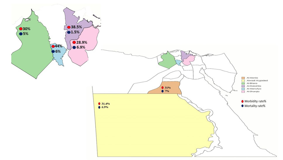
| Al-Menia | 100 | 31 | 7 | 31 | 7 | 22.6 | |
| Alwady ALgaded | 175 | 55 | 12 | 31.4 | 6.9 | 21.8 | |
| Al-Bhera | 40 | 12 | 2 | 30 | 5 | 25 | |
| Al-Dakahlia | 65 | 25 | 1 | 38.5 | 1.5 | 4 | |
| Al-Menofia | 50 | 22 | 3 | 44 | 6 | 13.6 | |
| Al-Sharqia | 145 | 42 | 10 | 28.9 | 6.9 | 23.8 | |
| Total | 575 | 187 | 35 | 32.5 | 6.1 | 18.7 | |
| Breed | Balady | 79 | 12 | 3 | 15.2 | 3.8 | 25 |
| Holstein | 289 | 104 | 18 | 36 | 6.2 | 17.3 | |
| Mixed | 207 | 71 | 14 | 34.3 | 6.8 | 19.7 | |
| Sex | Female | 416 | 145 | 29 | 34.9 | 7 | 20 |
| Male | 159 | 42 | 6 | 26.4 | 3.8 | 14.3 | |
| Age | > 3y | 200 | 58 | 11 | 29 | 5.5 | 19 |
| 1-3 y | 253 | 85 | 10 | 33.6 | 4 | 11.8 | |
| 0-1 y | 122 | 44 | 14 | 36.1 | 11.5 | 31.8 | |
| Season | Autumn | 189 | 68 | 6 | 36 | 3.2 | 8.8 |
| Spring | 74 | 24 | 3 | 32.4 | 4.1 | 12.5 | |
| Summer | 226 | 86 | 21 | 38.1 | 9.3 | 24.4 | |
| Winter | 86 | 9 | 5 | 10.5 | 5.8 | 55.6 | |
| Feeding and watering system | Communal system | 371 | 138 | 28 | 37.2 | 7.5 | 20.3 |
| separate | 204 | 49 | 7 | 24 | 3.4 | 14.3 | |
| Introducing new cattle | Yes | 305 | 132 | 27 | 43.3 | 8.9 | 20.5 |
| no | 270 | 55 | 8 | 20.4 | 3 | 14.5 | |
| Vaccination | Vaccinated | 244 | 29 | 2 | 11.9 | 8 | 6.9 |
| unknown | 177 | 95 | 9 | 53.7 | 5.1 | 9.5 | |
| unvaccinated | 154 | 63 | 24 | 40.9 | 15.6 | 38.1 |
EEV glycoprotein R-5′- CGAACCCCTATTTACTTGAGAA-3′ . Temperature and time conditions of the primer during PCR are shown in Supplementary Table I according to Emerald Amp GT PCR mastermix (Takara) kit.
Statistical analysis
The statistical analysis was performed with the SPSS program (SPSS 25 for Windows, SPSS Inc., Chicago, IL, USA). Data presented as count and percent of mortality and morbidity rates. The odds ratio (OR) was calculated for each risk factor. In all the analyses, confidence levels at 95% were calculated, and a value of P<0.05 was accepted as statistically significant. Univariate logistic regression analysis was used to compute the strength of contribution of the risk factors to LSD occurrence. Predictors with p < 0.25 were used in final multinomial logistic regression. The model was built with stepwise forward selection. The association of vaccination type with the clinical case of LSD was evaluated using the Chi-square (χ2) test.
RESULTS
Clinical findings
This study describes the occurrence of LSDV outbreaks in cows in six governorates in Egypt (Al-Menia, Alwady ALgaded, Al-Bhera, Al-Dakahlia, Al-Menofia, and Al-Sharqia) from June 2020 to May 2022. In total, 185 of the 575 examined cows from 25 herds, 185 displayed typical LSD clinical signs ranging from mild progressed to serious.
As in the case of a mild infection, diseased animals suffered from transient fever (39°C), depression, anorexia and the sudden appearance of a small number of nodules that disappeared quickly within 2 weeks (Fig. 1). While in moderate infection, diseased animals developed fever (Biphasic) and significant decrease in milk yield in dairy cattle. Expansion of superficial lymph nodes, particularly the prescapular and prefemoral. The sudden appearance of several nodules varying in size from (0.5 to 6 cm) in diameter and numbers that may continue as solid lumps or become wet, necrotic, and sloughed divided by a thin circle of hemorrhage makes a hole of full skin thickness and the distinctive feature lesion of “inverted cone-shaped area” of necrosis, defined as sit fast (Fig. 2). Serious form of infection is similar to the moderate form, but there are more nodules, and a wide area of skin is sloughed away, creating deep ulcers. Nodules on the nasal mucosa and along the respiratory system cause severe dyspnea and respiratory distress, Legs and brisket edema. This is a common finding in calves (Fig. 3).
Epidemiological finding
Multinomial logistic regression (MLR)
The MLR revealed that all predictors showed a significant effect on LSD (p < 0.001), except for the governorate and age group. Animals with the Balady breed were significantly less likely to be infected with LSD than Holstein breed animals (OR = 0.30; p = 0.0003). Females were 1.6 times more susceptible to LSD infection than males (OR = 1.6; p < 0.0001). The season is a very important predictor in the LSD model. Clinical cases in summer were 5 times more than in winter (OR = 5.13; p < 0.001. Monitoring the introduction of new animals decrease the susceptibility of animals to LSDV infection (OR = 0.3; p < 0.0001) and decrease death rate (OR = 0.21; p < 0.001). Animals, which reported with unknown state of vaccination and/or unknown type of vaccination, were 10 times more likely than vaccinated ones to have LSD or clinical signs (OR = 9.55; P < 0.0001), while unvaccinated animals were 7 times more likely to have LSD than vaccinated (OR = 7.12; p < 0.0001). Mortality in unvaccinated animals was 39 times more likely than in vaccinated animals (OR = 38.73; p < 0.0001) (Table II).
Multivariable multinomial logistic regression
Governorates, breed, season, feeding and watering systems, the introduction of new animals, and vaccination were significant risk factors for LSD in final multivariable multinomial logistic regression (Table III).
The two-dimensional plot of multiple correspondence analyses (MCA)
The two-dimensional plot of MCA revealed a strong relationship between vaccinated and healthy animals, in the upper left quarter of the plot. Additionally, the quarter of healthy animals includes Balady breed and separate feeding and watering system but farther apart from the other categories in the quarter. Conversely, the lower right quarter includes the diseased cases which are highly related to the summer season and the communal system of rearing. The dead animal cases are more related to the un-vaccination condition, and other categories or variables in the data did not have a strong relationship with the deaths of LSD (Fig. 5).
Table II. Multinomial logistic regression results for LSD, ORs and 95% confidence intervals showing correlates of diseased and dead animals versus healthy ones.
| Predictors | Factor levels | Diseased | Dead | P-value | ||||
| OR | CI | P-value | OR | CI | P-value | |||
| Governorate | Al-Menia | 1.107 | 0.63-1.95 | > 0.05 | 1.050 | 0.38-2.91 | > 0.05 | > 0.05 |
| Al-wady Algaded | 1.128 | 0.69-1.84 | > 0.05 | 1.033 | 0.43-2.50 | > 0.05 | ||
| Al-Bhera | 1.022 | 0.47-2.22 | > 0.05 | .715 | 0.15-3.47 | > 0.05 | ||
| AL-Dakahlia | 1.419 | 0.76-2.64 | > 0.05 | .238 | 0.03-1.93 | > 0.05 | ||
| AL-Menofia | 1.949 | 0.99-3.84 | > 0.05 | 1.116 | 0.29-4.36 | > 0.05 | ||
| AL-Sharqia | ||||||||
| Breed | Balady | 0.30 | 0.15- 0.58 | 0.0003 | 0.43 | 0.12 – 1.51 | > 0.05 | 0.002 |
| Mixed | 0.92 | 0.63 – 1.35 | > 0.05 | 1.05 | 0.51 – 2.21 | >0.05 | ||
| Holestin | Ref. | |||||||
| Sex | Female | 1.60 | 1.06 -2.41 | < 0.0001 | 2.23 | 0.89- 5.52 | > 0.05 | 0.02 |
| Male | Ref. | |||||||
| Age | 0-1 y | 1.55 | 0.95-2.54 | > 0.05 | 2.6 | 1.12 -6.06 | .03 | 0.021 |
| 1-3 y | 1.21 | 0.81-1.82 | > 0.05 | 0.75 | 0.31-1.83 | > 0.05 | ||
| > 3y | Ref. | |||||||
| Season | Autumn | 4.19 | 2.03-8.68 | < 0.0001 | 0.74 | 0.22-2.52 | >0.05 | < 0.0001 |
| Spring | 3.63 | 1.59-8.27 | 0.002 | 0.91 | 0.21-3.97 | >0.05 | ||
| Summer | 5.13 | 2.50-10.52 | <0.0001 | 2.51 | 0.91-6.94 | > 0.05 | ||
| Winter | Ref. | |||||||
| Feeding and watering system | Communal system | 2.06 | 1.39-3.04 | < 0.0001 | 2.9 | 1.23-6.82 | 0.01 | < 0.001 |
| Separate | Ref. | |||||||
| Introducing new cattle | No | 0.30 | 0.21-0.44 | < 0.0001 | 0.21 | 0.09-0.48 | < 0.001 | < 0.001 |
| Yes | Ref. | |||||||
| Vaccination | Unknown | 9.55 | 5.84-15.66 | < 0.0001 | 13.13 | 2.77-61.18 | 0.001 | < 0.001 |
| Unvaccinated | 7.12 | 4.24-11.96 | < 0.0001 | 38.73 | 8.92-166.21 | < 0.0001 | ||
| Vaccinated | Reference category (Ref) | |||||||
Table III. Multivariable logistic regression showing significant predictors with forward stepwise selection.
| Predictors | Factor levels | Diseased | Dead | P-value | ||||
| OR | CI | P-value | OR | CI | P-value | |||
| Governrates | Al-Menia | 2.665 | 1.19-5.98 | .018 | 1.783 | 0.46-6.89 | > 0.05 | 0.001 |
| Alwady ALgaded | 0.607 | 0.29-1.27 | .185 | .607 | 0.16-2.28 | > 0.05 | ||
| Al-Bhera | .199 | 0.07-0.56 | .002 | .313 | 0.05-1.95 | > 0.05 | ||
| Al-Dakahlia | .812 | 0.37-1.78 | .602 | .177 | 0.02-1.66 | > 0.05 | ||
| Al-Menofia | 1.416 | 0.60-3.34 | .427 | 1.060 | 0.21-5.27 | > 0.05 | ||
| Al-Sharqia | Ref. | |||||||
| Breed | Balady | .213 | 0.09-0.53 | 0.001 | .340 | 0.08-1.51 | > 0.05 | 0.005 |
| Mixed | .624 | 0.36-1.09 | > 0.05 | 1.360 | 0.46-4.05 | > 0.05 | ||
| Holestin | Ref. | |||||||
| Season | Autumn | 4.105 | 1.49-11.29 | 0.006 | .391 | 0.07-2.32 | 0.30 | 0.001 |
| Spring | 1.515 | 0.48-4.83 | > 0.05 | .095 | 0.01-0.83 | 0.03 | ||
| Summer | 1.529 | 0.57-4.13 | > 0.05 | .407 | 0.08-1.96 | > 0.05 | ||
| Winter | Ref. | |||||||
| Feedingand watering system | Communal system | 3.05 | 1.80-5.14 | < 0.001 | 2.29 | 0.82-6.47 | > 0.05 | 0.00008 |
| separate | Ref. | |||||||
| Vaccination | Unknown | 17.14 | 9.26-31.7 | < 0.0001 | 23.24 | 4.38-123.30 | < 0.0001 | < 0.000001 |
| unvaccinated | 8.5 | 4.48-16.14 | < 0.0001 | 54.71 | 11.11-269.4 | < 0.0001 | ||
| vaccinated | Ref. | |||||||
Chi-square test
Chi-square test showed that clinical signs of LSD and type of vaccination significantly related P< 0.05. Neethling vaccine did not reveal mortality rate of LSD, and lower number of diseased animals compared by sheep pox vaccine. Unvaccinated and animals with unknown condition of vaccination recorded the highest diseases and deaths of LSD (Supplementary Table II; Fig. 6).
Isolation of samples on CAM of SPF of ECEs
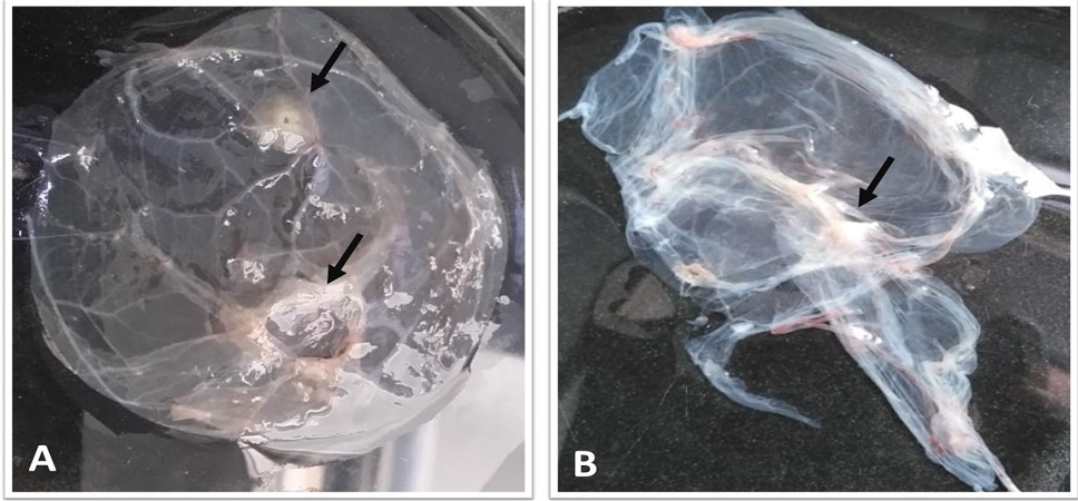
The results revealed that 62 out of 81 (76.5%) [59/72 (81.9%) skin nodule biopsies and 3/9 (33.3%)] nasal swabs samples were positive (Table IV). The CAMs of positive samples were hemorrhagic and congested at the first passage on the seventh day of inoculation, whereas at the second passage on the seventh day of inoculation, pock lesion was detected in the form of an extending white line (such a line could be owing to virus cell to cell infection) and became more prominent at the third passage (Fig. 7).
Virus detection in clinical samples using conventional PCR
The LSDV was detected using conventional PCR reactions depending on partial amplification of the EEV glycoprotein gene (958-bp). The findings indicated that 39 of 40 (97.5%) samples tested positive for LSDV (32/32 [100%] skin nodule biopsies and 7/8 [87.5%] nasal swab samples) (Table IV and Supplementary Fig. 1).
DISCUSSION
LSD is classified as an international concern animal disease due to the severity of its losses, ability to spread to other countries, and great influence on trade and food security . LSD was first detected in Egypt in 1988 . Over the last ten years, PCR have been used to diagnose the disease in various locations through most of Egypt . Therefore, LSDV is regarded being one of the most serious viral diseases of cattle, causing massive financial losses in Egypt due to lower feed consumption, milk yield, weight conversion, and traction power, in addition to abortion, fertility problems, damaged skin, treatment, and vaccination fees at the herd level . Thus, this study aimed to investigate the epidemiological state and risk factors for LSDV in the six governorates of Egypt (Al-Menia, Alwady ALgaded, Al-Bhera, Al-Dakahlia, Al-Menofia, and Al-Sharqia) based on viral isolation and molecular detection. Several outbreaks of LSDV were observed between June 2020 and May 2022. All animals were subjected to a general clinical examination. Approximately 185 cows out of 575 displayed typical LSDV clinical signs ranging from mild to serious. Sick animals suffered from fever, lack of appetite, and a massive reduction in milk output in dairy cows. Sudden development of many lumps ranging in size from (0.5 to 6 cm) and numbers depending on the severity of infection. In mild infection, nodules may disappear quickly, whereas in moderate and serious cases, nodules may persist as hard lumps and becoming wet, necrotic, and removed from the body, creating sit fast. These signs and symptoms match those described by Abdallah et al. (2018). In this research, serious form of infection is similar to the moderate form, but there are more nodules, and a wide area of skin is sloughed away, creating deep ulcers.
Table IV. Virus isolation and conventional PCR positive sample percentage.
| Governorate | No. of examined animals | No. Of positive samples isolated in ECE | No. of positive samples examined by conventional PCR | |||||||||
| Animals without clinical signs | Suspected diseased animals | Dead animals | Total | Skin nodules | Nasal swabs | Total | Skin nodules | Nasal swab | Total | |||
| Al-Menia | 62 | 31 | 7 | 100 | 12/14 (85.7%) | 0 | 12/14 (85.7%) | 6/6 (100%) | 0 | 6/6 (100%) | ||
| Alwady Algaded | 108 | 55 | 12 | 175 | 12/18 (66.7%) | 0 | 15/25 (60%) | 9/9 (100%) | 0 | 9/9 (100%) | ||
| Al-Bhera | 26 | 12 | 2 | 40 | 5/7 (71.4%) | 0 | 5/7 (71.4%) | 4/4 (100%) | 0 | 4/4 (100%) | ||
| Al-Dakahlia | 39 | 25 | 1 | 65 | 9/10 (90%) | 1/2 (50%) | 10/12 (83.3%) | 4/4 (100%) | 2/2 (100%) | 6/6 (100%) | ||
| Al-Menofia | 25 | 22 | 3 | 50 | 10/10 (100%) | 0 | 10/10 (100%) | 3/3 (100%) | 0 | 3/3 (100%) | ||
| Al-Sharqia | 93 | 42 | 10 | 145 | 11/13 (84.6%) | 2/7 (28.6%) | 13/20 (65%) | 6/6 (100%) | 5/6 (83.3%) | 11/12 (92.3%) | ||
| Total | 355 | 187 | 35 | 575 | 59/72 (81.9%) | 3/9 (33.3%) | 62/81 (76.5%) | 32/32 (100%) | 7/8 (87.5%) | 39/40 (97.5%) | ||
Nodules on the nasal mucosa and along the respiratory system cause severe dyspnea and respiratory distress especially in calves. These symptoms are consistent with those described according to Constable et al. (2017). Depending on clinical signs of LSD, the current study found that the morbidity, mortality, and case fatality rates were 32.5%, 6.1%, and 18.7%, respectively. Comparison with previous study in Egypt Salib and Osman (2011), recorded higher morbidity rate of LSD among examined Egyptian cattle reach to 100% but lower mortality and case fatality rates were 1.8%, and 1.8%, respectively. The current study attempted to investigate the probability of common breeds in Egypt to LSDV. The results revealed that the Holstein breeds are more susceptible to LSD infection 3 times more than Balady. These observations were similar to past research by Selim et al. (2021). In this investigation, there was significant difference in LSD occurrence between females and males as female more likely to infection than male by 2 times (OR=1.5). The current findings concur with those of Ayelet et al. (2013). Females have a higher morbidity rate because lactation and pregnancy periods provoke physiological stress and reduced immunity . Our findings revealed that all estimated age groups were susceptible to LSDV infection with no significant differences, but the mortality rate was high in animals less than one year. On the contrary, Sameea et al. (2017) found that LSD occurs more frequently in older cattle than in young cattle. The current study concluded that the prevalence of LSD varied across four seasons. The summer season has a higher rate of infection by 6 times (OR= 5.13) than winter season. The seasonal variation of LSD presented in this research could be attributed to the virus’s ability to live in extreme conditions and enhance vector activity during the summer time . The present study revealed that cattle raised in a communal feeding system or using common water supply were more likely to be infected with LSDV by twice more than cattle raised in a separate system (OR= 2.06). The probable reason is mixing animals in same pasture and watering source raises the risk of infection and disease transmission . This is most likely due to an increased possibility for mechanical virus transmission by Stomoxys spp. and mosquitoes . Our findings highlight the significance of diagnostic tests and quarantine monitoring periods for newly introduced animals Selim et al. (2021) where the implementation of new animals has the potential to increase the prevalence of LSD. This is supported by research from Ethiopia and Egypt .
LSD control is primarily based on vaccination of animals, whether heterologous with sheep pox or homologous with Neethling LSD vaccines. According to our findings, the Neethling strain vaccine resulted in no LSD mortalities and a lower morbidity than sheep pox vaccine. Our findings are corroborated by Ben-Gera et al. (2015), who found that the LSDV vaccine was more effective than the RM65 SPPV (10X) vaccine. Contrary to our research, Hailu et al. (2014) found no correlation between vaccination and LSD incidence.
The current study reported the isolation of LSDV from diseased cow skin lesions and nasal swaps on the CAM of ECEs. The findings revealed that 59 of 72 (81.9%) skin biopsies and 3 of 9 (33.3%) nasal swab samples showed characteristic pin-point pock lesions when inoculated on CAM of SPF embryo chicken eggs. Understanding of Capri poxvirus pathogenesis implies that skin lesions are the most effective samples for virus isolation and virus can be isolated for about 35 days after the skin nodules develop . The CAMs of positive samples seem to be hemorrhagic and congested during the first passage of inoculation, while pock lesions were observed during the second passage and became more prominent during the third passage. This finding agree with those demonstrated by El-Kenawy and El-Tholoth (2011). There are multiple diseases that cause symptoms similar LSDV, such as pseudo lumpy skin disease, dermodicosis, oncocercasiosis, and insect bite allergies . So, it is crucial to gain a definitive diagnosis by using molecular technique. In this study, LSDV was detected and identified via using conventional PCR reactions depending on partial amplification of the EEV Glycoprotein gene (958-bp). Skin biopsies were found to be positive for LSDV in 32 out of 32 (100%) and nasal swabs in 7 out of 8 (87.5%) samples. Our findings were validated by Hodhod et al. (2020) mentioned that, PCR is a speedy diagnostic technique with good specificity and sensitivity when compared to virus isolation.
CONCLUSION
Lumpy skin disease is an endemic disease that has spread throughout Egypt’s governorates, causing substantial economic losses in the animal husbandry industry. The analysis confirmed that the Holstein breed, female animals, summer season and communal water and feeding system all have a major impact on the occurrence of LSD in cattle. To control the spread of LSD infection, introducing of new animals into the herd must be monitored and vaccination by Neethling strain is recommended.
Conventional PCR rely on using EEV glycoprotein gene are more accurate, sensitive, and time-efficient for LSDV diagnosis than virus isolation on CAM of embryo chicken eggs.
Supplementary material
There is supplementary material associated with this article. Access the material online at: https://dx.doi.org/10.17582/journal.pjz/20220802170851
Statement of conflict of interest
The authors have declared no conflict of interest.
Figures
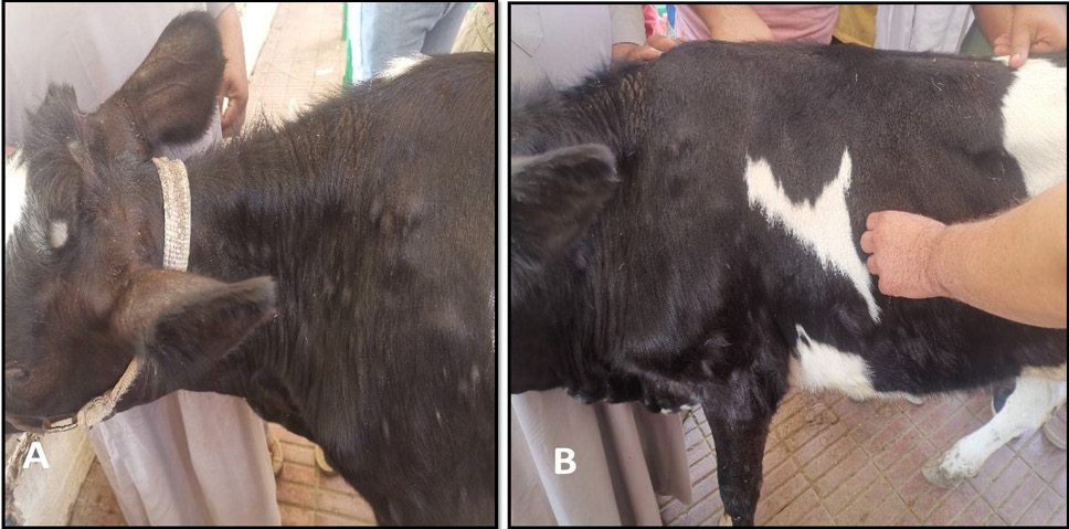
Mild form of LSDV revealed few numbers of closed nodules as shown in A and B.
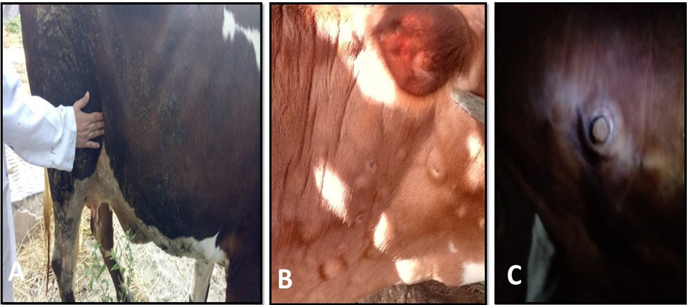
Moderate form of LSDV. (A) Swollen prefemoral lymph node. (B, C) distinctive feature lesions of LSDV sit fast.
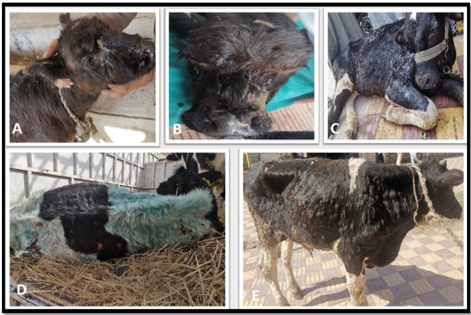
Severe form of LSDV. (A) Diseased calf showed, extreme dyspnea, nasal, ocular discharge, and respiratory manifestations. (B) Nodules present on the muzzle, as well as within the nasal and buccal mucosa. (C) Edema in legs and brisket and infected calf unable to stand. (D, E) The cutaneous lesion coalesced and large area of skin is sloughed creating deep ulcers.
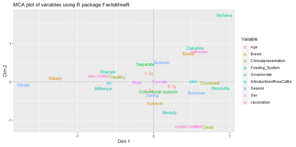
Two-dimensional plot of multiple correspondence analyses representing relationship between predictor’s categories.
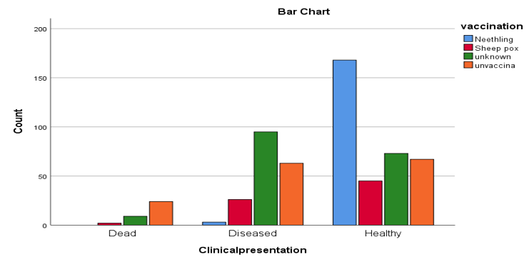
Relationship between vaccination type and clinical case of LSDV: Neethling strain vaccine did not show any deaths of LSD, and the lower number of diseased animals compared by sheep pox vaccine. Unvaccinated and animals with unknown condition of vaccination recorded the highest diseases and deaths of LSD.
References
- Abdallah, F.M., El Damaty, H.M. and Kotb, G.F., 2018. Sporadic cases of lumpy skin disease among cattle in sharkia province, Egypt: Genetic characterization of lumpy skin disease virus isolates and pathological findings. Vet. World, 11: 1150. https://doi.org/10.14202/vetworld.2018.1150-1158[CrossRef]
- Abutarbush, S.M., Ababneh, M.M., Al Zoubi, I.G., Al Sheyab, O.M., Al Zoubi, M.G., Alekish, M.O. and Al Gharabat, R.J., 2015. Lumpy skin disease in Jordan: Disease emergence, clinical signs, complications and preliminary-associated economic losses. Transbound. Emerg. Dis., 62: 549-554.
- Alexander, R.A., Plowright, W. and Haig, D.A., 1957. Cytopathogenic agents associated with lumpy skin disease of cattle. Bull. Epizoot. Dis. Afr., 5: 489-492.
- Ayelet, G., Abate, Y., Sisay, T., Nigussie, H., Gelaye, E., Jemberie, S. and Asmare, K., 2013. Lumpy skin disease: Preliminary vaccine efficacy assessment and overview on outbreak impact in dairy cattle at Debre Zeit, Central Ethiopia. Antivir. Res., 98: 261-265. https://doi.org/10.1016/j.antiviral.2013.02.008[CrossRef]
- Badhy, S.C., Chowdhury, M.G.A., Settypalli, T.B.K., Cattoli, G., Lamien, C.E., Fakir, M.A.U., Akter, S., Osmani, M.G., Talukdar, F. and Begum, N., 2021. Molecular characterization of lumpy skin disease virus (LSDV) emerged in Bangladesh reveals unique genetic features compared to contemporary field strains. BMC Vet. Res., 17: 1-11. https://doi.org/10.1186/s12917-021-02751-x[CrossRef]
- Beillard, M.J. and Omar, S., 2020. Livestock and products annual 2020. Gain.
- Ben-Gera, J., Klement, E., Khinich, E., Stram, Y. and Shpigel, N.Y., 2015. Comparison of the efficacy of neethling lumpy skin disease virus and x 10rm65 sheep-pox live attenuated vaccines for the prevention of lumpy skin disease the results of a randomized controlled field study. Vaccine, 33: 4837-4842. https://doi.org/10.1016/j.vaccine.2015.07.071[CrossRef]
- Chakraborty, S., Kumar, N., Dhama, K., Verma, A.K., Tiwari, R., Kumar, A., Kapoor, S. and Singh, S.V., 2014. Foot-and-mouth disease, an economically important disease of animals. Adv. Anim. Vet. Sci., 2: 1-18. https://doi.org/10.14737/journal.aavs/2014/2.2s.1.18[Full text][CrossRef]
- Constable, P.D., Hinchcliff, K.W., Done, S.H. and Grünberg, W., 2017. Veterinary medicine: A textbook of the diseases of cattle, horses, sheep, pigs and goats. Elsevier Health Sciences.
- El-Kenawy, A.A. and El-Tholoth, M.S., 2011. Lumpy skin disease virus identification in different tissues of naturally infected cattle and chorioallantoic membrane of emberyonated chicken eggs using immunofluorescence, immunoperoxidase techniques and polymerase chain reaction. Int. J. Virol., 7: 158-166. https://doi.org/10.3923/ijv.2011.158.166[CrossRef]
- Fauquet, C.M., Mayo, M.A., Maniloff, J., Desselberger, U. and Ball, L.A., 2005. 8th report of the international committee on taxonomy of viruses. Virus taxonomy. https://doi.org/10.1016/B978-0-12-249951-7.50004-3[CrossRef]
- Gari, G., Waret-Szkuta, A., Grosbois, V., Jacquiet, P. and Roger, F., 2010. Risk factors associated with observed clinical lumpy skin disease in Ethiopia. Epidemiol. Infect., 138: 1657-1666. https://doi.org/10.1017/S0950268810000506[CrossRef]
- Hailu, B., Tolosa, T., Gari, G., Teklue, T. and Beyene, B., 2014. Estimated prevalence and risk factors associated with clinical lumpy skin disease in North-Eastern Ethiopia. Prev. Vet. Med., 115: 64-68. https://doi.org/10.1016/j.prevetmed.2014.03.013[CrossRef]
- Hodhod, A., Elgendy, E., Abd El-Moniem, M.I. and Ibrahim, M.S., 2020. Isolation and molecular characterization of lumpy skin disease virus in Egypt during 2017-2018. BMC Vet. Res., 18: 302.
- House, J.A., Wilson, T.M., Nakashly, S.E., Karim, I.A., Ismail, I., Danaf, N.E., Moussa, A.M. and Ayoub, N.N., 1990. The isolation of lumpy skin disease virus and bovine herpes virus-from cattle in Egypt. J. Vet. Diagn., 2: 111-115. https://doi.org/10.1177/104063879000200205[CrossRef]
- Ince, O.B. and Türk, T., 2019. Analyzing risk factors for lumpy skin disease by a geographic information system (gis) in Turkey. J. Hell. Vet., 70: 1797-1804. https://doi.org/10.12681/jhvms.22222[CrossRef]
- Issimov, A., Kutumbetov, L., Orynbayev, M.B., Khairullin, B., Myrzakhmetova, B., Sultankulova, K. and White, P.J., 2020. Mechanical transmission of lumpy skin disease virus by Stomoxys spp. (Stomoxys calsitrans, Stomoxys sitiens, Stomoxys indica), Diptera: Muscidae. Animals, 10: 477. https://doi.org/10.3390/ani10030477[CrossRef]
- MacDonald, R.A.S., 1931. Pseudo-urticaria of cattle. Department of animal health. Government of Northern Rhodesia. pp. 20-21. https://doi.org/10.2307/209366[CrossRef]
- Menasherow, S., Erster, O., Rubinstein-Giuni, M., Kovtunenko, A., Eyngor, E., Gelman, B., Khinich, E., and Stram, Y., 2016. A high-resolution melting (HRM) assay for the differentiation between israeli field and neethling vaccine lumpy skin disease viruses. J. Virol. Methods, 232: 12-15. https://doi.org/10.1016/j.jviromet.2016.02.008[CrossRef]
- Ocaido, M., Otim, C.P. and Kakaire, D., 2009. Impact of major diseases and vectors in smallholder cattle production systems in different agro-ecological zones and farming systems in uganda. Crops, 56: 51-54.
- OIE, 2017. Manual of diagnostic tests and vaccines for terrestrial animals. Sheep pox and goat pox. Office International des Epizooties, OIE World Organisation for Animal Health.
- Radostits, O.M., Gay, C.C., Hinchcliff, K.W. and Constable, P.D., 2007. A textbook of the diseases of cattle, horses, sheep, pigs and goats. Vet. Med., 10: 2045-2050.
- Rossiter, P.B. and Al Hammadi, N., 2009. Living with transboundary animal diseases (tads). Trop. Anim. Hlth. Prod., 41: 999-1004. https://doi.org/10.1007/s11250-008-9266-7[CrossRef]
- Salem, H., A., Abouelyazeed, E., A., Ali, H.M., Khattab, O.M. and Moussa, S., A., 2021. Molecular characterization of lumpy skin disease virus in cattle (2017). Egypt. J. Anim. Hlth., 1: 45-52. Available from https://doi.org/10.21608/ejchem.2017.1586.1124[CrossRef]
- Salib, F.A. and Osman, A.H., 2011. Incidence of lumpy skin disease among Egyptian cattle in Giza Governorate, Egypt. Vet. World, 4: 162-167.
- Sameea, Y., P., Mardani, K., Dalir-Naghadeh, B. and Jalilzadeh-Amin, G., 2017. Epidemiological study of lumpy skin disease outbreaks in north-western iran. Transbound. Emerg. Dis., 64: 1782-1789. https://doi.org/10.1111/tbed.12565[CrossRef]
- Selim, A., Manaa, E. and Khater, H., 2021. Seroprevalence and risk factors for lumpy skin disease in cattle in northern Egypt. Trop. Anim. Hlth Prod., 53: 1-8. https://doi.org/10.1007/s11250-021-02786-0[CrossRef]
- Şevik, M., Avci, O., Doğan, M. and İnce, Ö.B., 2016. Serum biochemistry of lumpy skin disease virus-infected cattle. Biomed. Res. Int., https://doi.org/10.1155/2016/6257984[CrossRef]
- Sharawi, S.S.A. and Abd El-Rahim, I.H.A., 2011. The utility of polymerase chain reaction for diagnosis of lumpy skin disease in cattle and water buffaloes in Egypt. Rev. Sci. Tech. OIE, 30: 821. https://doi.org/10.20506/rst.30.3.2075[CrossRef]
- Tasioudi, K.E., Antoniou, S.E., Iliadou, P., Sachpatzidis, A., Plevraki, E., Agianniotaki, E.I., Fouki, C., Mangana-Vougiouka, O., Chondrokouki, E. and Dile, C., 2016. Emergence of lumpy skin disease in Greece, 2015. Transbound. Emerg. Dis., 63: 260-265. https://doi.org/10.1111/tbed.12497[CrossRef]
- Tuppurainen, E.S.M., Stoltsz, W.H., Troskie, M., Wallace, D.B., Oura, C.A.L., Mellor, P.S., Coetzer, J.A.W. and Venter, E.H., 2011. A potential role for ixodid (hard) tick vectors in the transmission of lumpy skin disease virus in cattle. Transbound. Emerg. Dis., 58: 93-104. https://doi.org/10.1111/j.1865-1682.2010.01184.x[CrossRef]
- Van Rooyen, P.J., Munz, E.K. and Weiss, K.E., 1969. The optimal conditions for the multiplication of neethling-type lumpy skin disease virus in embryonated eggs. Onderstepoort. J. Vet. Res., 36:165-174.
Supplementary material
Services
Article usage statistics
Loading…