
Oviductus ranae (OR) is an animal-based traditional medicine. To explore its mechanism of antidepressant effect, healthy Institute of Cancer Research (ICR) male mice were divided into control, chronic unpredictable mild stress (CUMS), fluoxetine (3 mg/kg), OR800 (OR 800 mg/kg) and OR400 (OR 400 mg/kg) dose groups. After the last administration, the content of corticotropin-releasing hormone (CRH), adrenocorticotrophic hormone (ACTH) and corticosterone (CORT) in serum were detected. Hematoxylin and Eosin (H&E) staining was used to observe the hippocampal histomorphological changes. The protein levels of glucocorticoid receptor (GR), mineralocorticoid receptor (MR), brain-derived neurotrophic factor (BDNF), trosine kinase B (TrkB) and cyclic adenosine monophosphatec response element binding protein (CREB) in hippocampus were detected by western blotting. The results showed that, OR could significantly reduce the contents of CRH, ACTH and CORT in serum and improve the damage of hippocampal structure, and increase the expression levels of GR, MR, BDNF, TrkB and CREB in hippocampus. In conclusion, Oviductus ranae can reduce hypothalamic-pituitary-adrenal (HPA) axis hyperfunction induced by CUMS, increase the expression level of BDNF related signal pathway proteins, improve hippocampal tissue damage, and thus play an antidepressant role by regulating the negative feedback of HPA.
Short Communication · Volume 55, Issue 3, 20 April 2023 · Open Access
Protective Effects of Oviductus Ranae Mediated HPA Axis Regulation on Depressive Model of Mice
· · · · · ·
Affiliations & Notes ⌄
- 1School of Pharmaceutical Sciences, Changchun University of Chinese Medicine, Changchun, 130117, China 2Jilin Ginseng Academy, Changchun University of Chinese Medicine, Changchun, 130117, China * Corresponding author: linzhe1228@163.com, lvgf@ccucm.edu.cn
Article Info ⌄
- Journal:
- Pakistan Journal of Zoology
- Published:
- 20 April 2023

From the article
- The results showed that the contents of CREB, BDNF, TrkB in CUMS group decreased significantly, however, their contents in OR800 and OR400 groups increased significantly after administration.
- H&E staining showed that the mice in CUMS group had obvious structural damages such as cell atrophy, reduced density and more disordered arrangement, while that in OR800 and OR400 groups showed a gradually normal in cell structures, an increase in the density and significantly reduced damage, compared with CUMS group.
- Its mechanism is to restore the excessive activation of the HPA axis caused by stress and reduce glucocorticoids, alleviate the decline in BNDF caused by glucocorticoids, and promote the expression of BNDF by up-regulating CREB.
- Moreover, up-regulate the expression level of TrkB further for activating downstream signaling pathways, promote the neuron growth and neuron protection of neurotrophic factors, promote the repair of hippocampal tissue damage.
Selected from the article's own text.
Abstract
Materials and methods
Oviductus ranae was provided by Tonghua Dingshen Pharmaceuticals Company (Tonghua, China). Fluoxetine hydrochloride capsules were purchased from Sinochem Pharmaceutical Industry Co., Ltd. (Suzhou, China). Institute of Cancer Research (ICR) male mice were purchased from Yisi Experimental Animal Technology Co., Ltd (Changchun, China). The other main reagents and equipment are listed in Supplementary Table SI.
Fifty male ICR mice were housed for 7 days under standard conditions at temperature 24 ± 1°C, 12 h:12 h light: dark cycles, with normal diet and drinking water. The mice were randomly divided into 5 groups (10 mice/group): control (equal volume of distilled water), CUMS (fluoxetine 3 mg/kg), OR800 ( 800 mg/kg) and OR400 (400 mg/kg) dose groups, and administered intragastrically.
The CUMS model was established and evaluated accordingly ; . The control group was normally fed, while the other groups received 28 days of chronic unpredictable stressors. The details including: electric foot-shock (24 V for 5 min), fasting and water deprivation (24 h), tilting the cage at 45° (24 h), force swimming in ice water at 4°C (3 min), shaking the cage horizontally (10 min), hot environment at 45°C (5 min), noising stimulation (10 min) and tail-clamping (5 min). To prevent mice from adapting, 1 to 2 different stressors listed above were given a day.
Mice in each group were anesthetized, their blood samples were taken from eyeballs and centrifuged 3000 r/min at 4°C for 10 min. The upper serum was collected and placed in a 2 mL EP tube, and stored at -80 °C. ELISA was used to detect the contents of CRH, ACTH and CORT in the serum .
After the mice were sacrificed, the brain tissue was collected, and the hippocampal tissues were quickly separated on ice and put into 4% paraformaldehyde fixative. After dehydration, paraffin embedding and sectioning, the hippocampal tissue was sliced into paraffin sections and the changes of tissue structure were observed by H&E staining under a microscope .
Hippocampal tissue samples were placed in tube, lysed in lysate buffer, homogenized on ice, centrifuged to obtain the supernatant and stored at –80°C for later use. The concentration of protein in hippocampus were measured by bicinchoninic acid (BCA) assay. Proteins were resolved on SDS-PAGE by electrophoresis and are transferred to PVDF membrane (Mishra et al., 2017). After incubation with specific antibody at 4°C overnight, the membrane was exposed to secondary antibody for 1 h, washed with TBST for 3 times. Then, chemiluminescence and gel imaging analyses were performed. The ratio of target protein to internal reference GAPDH was used as the relative protein expression.
All data were presented as the mean of three samples with standard deviation. One-way analysis of variance (ANOVA) and Tukey’s range test were used to determine differences between groups, p < 0.05 was considered as statistically significant.
Results
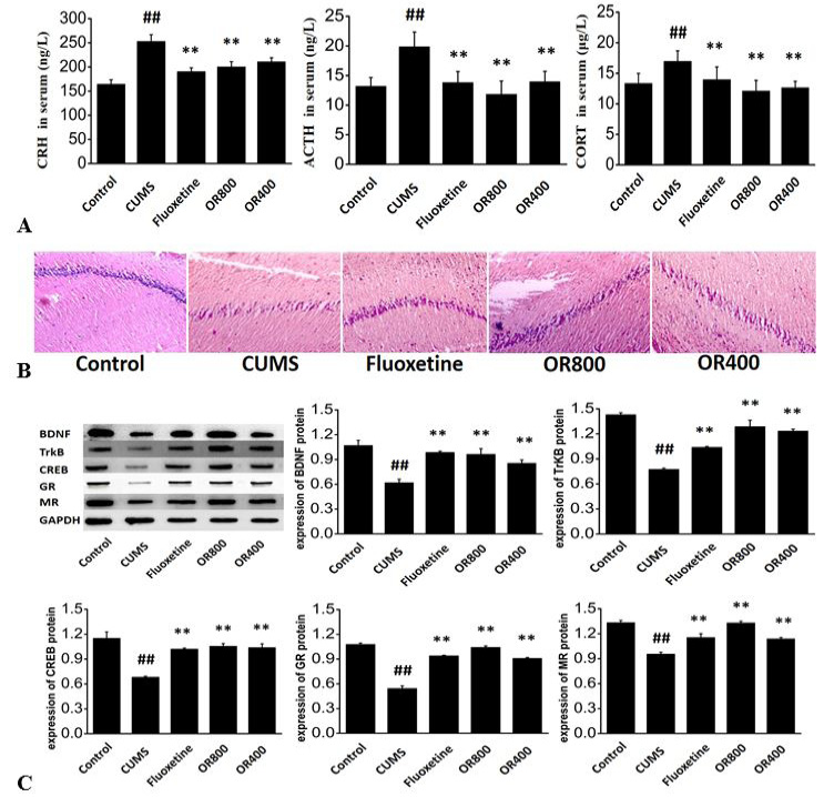
The contents of CRH, ACTH and CORT in serum are shown in Figure 1A. In CUMS group, the contents of CRH, ACTH, and CORT were increased significantly compared with that of the control group (p <0.01). Compared with the CUMS group, the contents of serum CRH, ACTH and CORT in fluoxetine group, OR800 and OR400 groups were significantly reduced (p <0.01). Long-term stress can cause HPA axis hyperfunction and increase the contents of CRH, ACTH and CORT.
The pathological changes in hippocampal tissues were shown in Figure 1B. The hippocampal CA1 cells in control group were normal, plump, dense, arranged neatly and no obvious atrophy, while that of the CUMS group showed cells atrophy, decreased density, disordered arrangement, large gaps in the middle, and obvious degree of damage. Compared with CUMS group, the structure of cells in the OR800 and OR400 groups became normal gradually, density increased, the space between the cells decreased, the overall arrangement was more orderly, and the damage was significantly alleviated.
The expression of BDNF, TrkB, CREB, GR and MR proteins in hippocampal tissues were shown in Figure 1C. Compared with control group, the expression of BDNF, TrkB, CREB, GR, and MR proteins in CUMS group decreased significantly (p<0.01). While the fluoxetine group and OR800 and OR400 groups were significantly up-regulated (p<0.01), compared with that of CUMS group. The results showed that OR could enhance the inhibition of HPA axis by increasing the cortical hormone receptor, reduce the damage and repair the hippocampal tissue by increasing the neurotrophic factors and receptors.
Discussion
When exposing under long-term chronic pressure, the HPA axis of the body will be activated. CRH secreted by hypothalamus can stimulate the pituitary gland to secrete ACTH, then stimulated adrenal glands to release a large amount of glucocorticoids, resulting in the increase of glucocorticoids in body. Hippocampus plays an important role in the condition of HPA axis. Glucocorticoids in body will first bind to MR in hippocampus at a low level; only when at a high level, it will bind to GR. Meanwhile, hippocampus regulation inhibits the excessive secretion of HPA axis, maintains it to a steady state, and normalize glucocorticoid levels in vivo . The results in this study indicated that can increase the contents of hippocampal GR and MR receptors, enhance the hippocampus’ negative feedback regulation of the HPA axis, inhibit the over-activation of the HPA axis, reduce the body’s glucocorticoids and relieve the hippocampal tissue damage caused by excessive glucocorticoids.
BDNF exists in hippocampus and cortex, which participates in the growth and development of neurons, and also protects the physiological functions of neurons from being damaged. The increase of glucocorticoids leads to the low expression of BNDF and damages the nervous system from being repaired, resulting in depression . As a receptor with high affinity with BDNF, TrkB can activate downstream signaling pathways, such as mitogen-activated protein kinase (MAPK), phosphoinositide 3 kinase (PI3K), etc., and play biological effects after binding to BDNF . Phosphorylated CREB can combine with CRE of downstream BDNF sequence, play biological functions and promote the expression of BDNF . The results showed that the contents of CREB, BDNF, TrkB in CUMS group decreased significantly, however, their contents in OR800 and OR400 groups increased significantly after administration. H&E staining showed that the mice in CUMS group had obvious structural damages such as cell atrophy, reduced density and more disordered arrangement, while that in OR800 and OR400 groups showed a gradually normal in cell structures, an increase in the density and significantly reduced damage, compared with CUMS group.
Studies showed that oleic acid and linolenic acid were two fatty acids in OR. It was found that linolenic acid could reduce the incidence of female depression , oleic acid can play an antidepressant role as an inhibitor of autoinducer-2 (AL-2) . These fatty acids may be the material basis for the antidepressant effect of OR, which need to be further studied.
Conclusion
Oviductus ranae has an antidepressant effect. Its mechanism is to restore the excessive activation of the HPA axis caused by stress and reduce glucocorticoids, alleviate the decline in BNDF caused by glucocorticoids, and promote the expression of BNDF by up-regulating CREB. Moreover, up-regulate the expression level of TrkB further for activating downstream signaling pathways, promote the neuron growth and neuron protection of neurotrophic factors, promote the repair of hippocampal tissue damage.
Acknowledgment
This work was supported by the Scientific Research Planning Project of Education Department of Jilin Province (JJKH20200901KJ), Administration of Traditional Chinese Medicine of Jilin Province (2020165), Health Commission of Jilin Province (2019Q027).
Ethics statement
This study was approved by the Ethics Committee of Changchun University of Traditional Chinese Medicine (Approval No: 2020107).
Supplementary material
There is supplementary material associated with this article. Access the material online at: https://dx.doi.org/10.17582/journal.pjz/20210513050530
Statement of conflict of interest
The authors have declared no conflict of interest.
Figures
References
- Chen, J., Wang, Z.Z., Zhang, S., Zuo, W. and Chen, N.H., 2016. Life Sci., 152: 76-81. https://doi.org/10.1016/j.lfs.2016.03.022[CrossRef]
- Ge, L., Liu, L., Liu, H., Liu, S., Xue, H., Wang, X., Yuan, L., Wang, Z. and Liu, D., 2015. Eur. J. Pharmacol., 768: 49-57. https://doi.org/10.1016/j.ejphar.2015.10.026[CrossRef]
- Guo, H., Gan, Y., Liu, M., Wang, S., Ni, S., Zhou, Y., Xiao, Y., Wang, Z. and Wang, Y., 2019. Foods, 8: 322. https://doi.org/10.3390/foods8080322[CrossRef]
- Katz, D.A., Locke, C., Greco, N., Liu, W. and Tracy, K.A., 2017. Brain Behav., 7: e00628. https://doi.org/10.1002/brb3.628[CrossRef]
- Keller, J., Gomez, R., Williams, G., Lembke, A., Lazzeroni, L., Murphy Jr, G.M. and Schatzberg, A.F., 2017. Mol. Psychiat., 22: 527-536. https://doi.org/10.1038/mp.2016.120[CrossRef]
- Kikuchi, T., Suzuki, T., Uchida, H., Watanabe, K. and Mimura, M., 2013. Psychiat. Res., 210: 127-133. https://doi.org/10.1016/j.psychres.2013.05.007[CrossRef]
- Li, Y.J., Li, Y.J., Yang, L.D., Zhang, K., Zheng, K.Y., Wei, X.M., Yang, Q., Niu, W.M., Zhao, M.G. and Wu, Y.M., 2018. Behav. Brain Res., 348: 184-191. https://doi.org/10.1016/j.bbr.2018.04.025[CrossRef]
- Liu, S., Xu, S., Wang, Z., Guo, Y., Pan, W. and Shen, Z., 2018. Med. Sci. Monit., 24: 7646-7653. https://doi.org/10.12659/MSM.908422[CrossRef]
- Lu, Y., Xu, X., Jiang, T., Jin, L., Zhao, X.D., Cheng, J.H., Jin, X.J., Ma, J., Piao, H.N. and Piao, L.X., 2019. Int. Immunopharmacol., 67: 119-128. https://doi.org/10.1016/j.intimp.2018.12.011[CrossRef]
- Lucas, M., Mirzaei, F., O’Reilly, E.J., Pan, A., Willett, W.C., Kawachi, I., Koenen, K., and Ascherio, A., 2011. Am. J. clin. Nutr., 93: 1337-1343. https://doi.org/10.3945/ajcn.111.011817[CrossRef]
- Medina-Rodriguez, E.M., Madorma D., O’Connor, G., Mason, B.L., Han D., Deo, S.K., Oppenheimer, M., Nemeroff, C.B., Trivedi, M.H., Daunert, S. and Beurel, E., 2020. Am. J. Psychiat., 177: 974-990. https://doi.org/10.1176/appi.ajp.2020.19090960[CrossRef]
- Meyer, U., van Kampen, M., Isovich, E., Flugge, G. and Fuchs, E., 2001. Hippocampus, 11: 329-336. https://doi.org/10.1002/hipo.1047[CrossRef]
- Mishra, M., Tiwari, S. and Gomes, A.V., 2017. Expert Rev. Proteom., 14: 1037-1053. https://doi.org/10.1080/14789450.2017.1388167[CrossRef]
- Naoi, M., Maruyama, W. and Shamoto-Nagai, M., 2018. J. Neural Transm., 125: 53-66. https://doi.org/10.1007/s00702-017-1709-8[CrossRef]
- Pei, G., Xu, L., Huang, W. and Yin, J., 2020. Int. Immunopharmacol., 78: 106076. https://doi.org/10.1016/j.intimp.2019.106076[CrossRef]
- Peng, Y., Zhang, C., Su, Y., Wang, Z. and Jiang, Y., 2018. Electrophoresis, 40: 1245-1250. https://doi.org/10.1002/elps.201800381[CrossRef]
- Phillips, C., 2017. Neural Plast., 2017: 7260130. https://doi.org/10.1155/2017/7260130[CrossRef]
- Roversi, K., de David Antoniazzi, C.T., Milanesi, L.H., Rosa, H.Z., Kronbauer, M., Rossato, D.R., Duarte, T., Duarte, M.M. and Burger, M.E., 2019. Mol. Neurobiol., 56: 6239-6250. https://doi.org/10.1007/s12035-019-1522-5[CrossRef]
- Serra, M.P., Poddighe, L., Boi, M., Sanna, F., Piludu, M.A., Sanna, F., Corda, M.G., Giorgi, O. and Quartu, M., 2018. Int. J. mol. Sci., 19: 3745. https://doi.org/10.3390/ijms19123745[CrossRef]
- Shelton, R.C., Claiborne, J., Sidoryk-Wegrzynowicz, M., Reddy, R., Aschner, M., Lewis, D.A. and Mirnics, K., 2011. Mol. Psychiat., 16: 751-762. https://doi.org/10.1038/mp.2010.52[CrossRef]
- Shirayama, Y., Fujita, Y., Oda, Y., Iwata, M., Muneoka, K. and Hashimoto, K., 2020. Behav. Brain Res., 390: 112670. https://doi.org/10.1016/j.bbr.2020.112670[CrossRef]
- Song, Y., Zhong, M. and Cai, F.C., 2018. Eur. Rev. Med. Pharmacol. Sci., 22: 250-261. https://doi.org/10.26355/eurrev_201801_14126[CrossRef]
- Su, W.J., Zhang, Y., Chen, Y., Gong, H., Lian, Y.J., Peng, W., Liu, Y.Z., Wang, Y.X., You, Z.L., Feng, S.J., Zong, Y., Lu, G.C. and Jiang, C.L., 2017. Behav. Brain Res., 322: 1-8. https://doi.org/10.1016/j.bbr.2017.01.018[CrossRef]
- Suzumura, A., Kaneko, H., Funahashi, Y., Takayama, K., Nagaya, M., Ito, S., Okuno, T., Hirakata, T., Nonobe, N., Kataoka, K., Shimizu, H., Namba, R., Yamada, K., Ye, F., Ozawa, Y., Yokomizo, T. and Terasaki, H., 2020. Diabetes, 69: 724-735. https://doi.org/10.2337/db19-0550[CrossRef]
- Uddin, M.F., Alweis, R., Shah, S.R., Lateef, N., Shahnawaz, W., Ochani, R.K., Dharani, A.M. and Shah, S.A., 2017. J. clin. Diagn. Res., 11: OE05-OE07. https://doi.org/10.7860/JCDR/2017/29473.10696[CrossRef]
- Xu, Q., Dou, C., Liu, X., Yang, L., Ni, C., Wang, J., Guo, Y., Yang, W., Tong, X. and Huang, D., 2018. Biomed. Pharmacother., 107: 1692-1704. https://doi.org/10.1016/j.biopha.2018.07.071[CrossRef]
- Zhao, X., Cao, F., Liu, Q., Li, X., Xu, G., Liu, G., Zhang, Y., Yang, X., Yi, S., Xu, F., Fan, K. and Ma, J., 2019. Behav. Brain Res., 364: 494-502. https://doi.org/10.1016/j.bbr.2017.05.064[CrossRef]
Supplementary material
Services
Article usage statistics
Loading…