
Cadmium (Cd), a toxic heavy metal, is a potential carcinogen, mutagen and teratogen through bioaccumulation in tissues. Aim of present study was to analyze various developmental abnormalities by a sub-lethal dose of cadmium chloride and protective role of garlic (Allium sativum), to minimize the intensity of these toxicities. For this purpose, fertilized eggs of Gallus domesticus were randomly divided into four groups of forty eggs each. Control group was intact and untreated. Eggs of one group were injected with a sub-lethal dose of cadmium chloride (1.5 µg/egg) in albumin on 7th day of incubation. In another group, eggs were treated with cadmium chloride and fresh garlic juice (0.2 µg / egg) after a short interval of 10 minutes. Antidote group was injected with fresh garlic juice (0.2 µg / egg). Eggs of each group were incubated at 37 ± 0.5 °Cand at 50-60% relative humidity till hatching. Natural hatching was obtained as 92.5, 32.5, 72.5, and 87.5% in control, dose, dose+antidote, and anitodote groups respectively with delayed hatching in some cases in dose and dose+antidote groups. Chicks and dead embryos were studied for morphological and morphometric analysis. Liver tissues were dissected out from each group for histological analysis. Cadmium chloride induced significant developmental abnormalities such as reduced body weight and crown rump length, exencephaly, ablepharia, crossed beak, gastroschisis, crooked toes, non-alcoholic fatty liver and pyknosis. Fresh garlic juice has successfully attenuated these toxicities and increased the body weight, crown rump length. Results of present study determine that garlic is a potential antidote to ameliorate the cadmium induced teratogenic and hepatotoxic defects in developing Gallus domesticus.
Research Article · Volume 33, Issue 1, 10 May 2018 · Open Access
Protection Against Cadmium-Induced Abnormalities and Hepatotoxicity in ovo by Allium sativum
· ·
Affiliations & Notes ⌄
- 1University of Education, Division of Science & Technology, College Road Township, Lahore
- 2Department of Zoology, University of the Punjab, Lahore, Pakistan. , *Corresponding author Dr. Shagufta Andleeb, drshagufta@ue.edu.pk, s_andleeb@yahoo.com
Article Info ⌄
- Journal:
- Punjab University Journal of Zoology
- Published:
- 10 May 2018
From the article
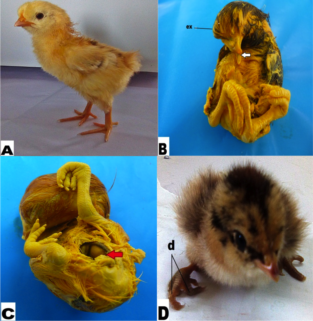
- A: Control group (untreated); B & C: Dose group treated with 1.5µg/0.05ml/egg of CdCl2; D: Dose+antidote group treated with 1.5µg/0.05ml/egg of CdCl2 and 0.2 µg/0.05ml/egg of garlic juice.
- Results of present study authenticate the potent ameliorative nature of garlic against cadmium and suggest its use in any way at regular basis to withstand the harmfulness of unseen toxins like cadmium through food and water.
Selected from the article's own text.
Abstract
Introduction
Heavy metals including cadmium owing to human activities have become common environmental pollutants and bio accumulated in human through food chain . Cadmium is known as group 1 carcinogen . Prenatal exposure in mice has resulted into deleterious changes on the behavioral activities, neurotransmitters, oxidative stress, and brain neurons morphology , and altered thymocyte development in mice . It is found to be renotoxic , cytotoxic and embyotoxic ; ; ; . It affects semen quality in human and alters feeding pattern and urine volume in Wistar rat . Interestingly, cadmium has been found to exert sex specific effects on birth size, DNA methylation ; birth length and fetal growth . It is reported as gametotoxic , tumor angiogen , neurotoxic , and osteotoxic . Prenatal exposure and placental crossing over induces hepatotoxicity and DNA methylation ; ; . Neurulation is found to be most vulnerable state leading to neural tube defects in mice (Robinson et al., 2011).
Garlic is well known for its protective role against cancer and so many other diseases for its therapeutic and prophylactic effects owing to biologically active antioxidant substances like alliinase, allicin, alliin, and S-allylcysteine ; . Administration of garlic juice during pregnancy and lactation is known to protect from apoptosis in rat offspring’s eye retina . Aged garlic extract (AGE), even can reduce side effects of anticancer drugs .Keeping in view the potential ameliorative role of garlic, the present study is designed to find its protective activity in developing chicks against cadmium.
Materials and Methods
Chemical used and dose preparation
Cadmium chloride (SIGMA-ALDRICH), was used as source of cadmium in the study. A sublethal dose of cadmium (Dżugan et al., 2011) was prepared in sterilized 0.7% avian saline in such a way that each 0.05ml contained 1.5 µg of cadmium.
Preparation of antidote
Fresh garlic, Purple Glazer (Allium sativum) was used as antidote. Fresh garlic bulbs were purchased from local market in order to prepare fresh garlic juice. For this purpose, garlic cloves (2g) were peeled, washed, chopped and ground in pastel, mixed with distilled water and filtered afterwards and filtrate was used to prepare the desired concentration of 0.2 µg/0.05ml.
Experimental animal
Embryos of Gallus domesticus (domestic fowl, Comb Leghorn) were used as experimental animal. Fresh fertilized eggs (n=160) were purchased from a local hatchery at Pasrur road, Narowal. Eggs were cleaned, labelled and incubated horizontally at standard conditions (incubation days: D0-D21, temperature: 37 ± 0.5 °C, relative humidity: 50-60%) in a rolling egg incubator (24̋ × 30̋ × 17̋). Humidity was maintained by keeping the water filled tray inside the incubator which was replaced after every 24 hours and its level was maintained to provide eggs with equal percentage of humidity throughout the incubation period. Eggs were observed through the candler on 4th day (D4) of incubation to check the embryonic development to remove the unfertilized eggs.
Experimental grouping and drug administration
On 7th day (D7) of incubation, all the fertilized eggs were randomly divided into four groups of 40 eggs each as follows:
Control group: untreated
Dose group: CdCl21.5 µg /0.05 ml/egg
Dose + Antidote group: CdCl2 1.5µg/0.05ml/egg + Garlic juice 0.2 µg/0.05 ml/ egg (after a short interval of 10 minutes)
Antidote group: Garlic juice 0.2 µg/0.05ml/egg
Dose administration was carried out through a hole in the egg shell into albumen at blunt end using a sterile needle in sterilized environment of laminar air flow. Following injections, holes were sealed with molten paraffin wax immediately to avoid contamination. The remaining incubation period was continued safely until hatching.
Hatching of chicks
Fertilized eggs of each group were allowed to hatch naturally on 21st day of incubation. Immediately after hatching chicks were kept separately in an environmentally controlled room at 27-30 °C with a photocycle of 14 hours light and 10 hours dark for two days. Chicks were constantly supplied with pearl millet and fresh tap water for drinking during this time period.
Morphological analysis and macrophotography
Un-hatched embryos, hatched chicks of all groups were studied morphologically for developmental abnormalities of skull, beak, eyes, limbs, tail, vertebral column and abdomen.
Morphometric analysis
Body weight and crown rump length of each embryo was recorded for each group.
Histological preparation
Liver tissues were dissected out and subsequently chopped into small pieces with sharp cutter and preserved in Bouin’s fluid for fixation for 48 hours and processed for histological analysis using paraffin wax and hematoxylin-eosin staining technique.
Microphotography and histological study
Histological sections of liver were observed for various anomalies using microscope SWIFT (M4000-D) and microphotographed with the help of digital camera BESTSCOPE (BUC2-500C).
Statistical analysis
Data were analyzed using one-way analysis of variance (ANOVA) Tukey test using GraphPad Prism (Version 5.01) to find out the significant difference (p< 0.05) among various groups.
A: Control group (untreated); B & C: Dose group treated with 1.5µg/0.05ml/egg of CdCl2; D: Dose+antidote group treated with 1.5µg/0.05ml/egg of CdCl2 and 0.2 µg/0.05ml/egg of garlic juice. Note: (ex: exencephaly; Arrow white: crossed beak; Arrow red: gastroschisis; d: crooked toes).
Results
Morphological analysis
Chick hatchlings as well as embryos of control group were quite healthy and uniform in appearance and in other anatomical details. The body was well differentiated into head, neck and trunk regions. They had completely developed morphological features, including head crown, beak, eyes, fore limbs, hind limbs and digits. In dose group, 32.5% of embryos hatched naturally at day 21.These embryos showed minor morphological abnormalities, retarded growth and weight loss as compared to control group. Among rest in this group, 48.14% dead embryos were recovered on 23rd day. Embryos recovered on D23 showed various anomalies. Exencephaly, ablepharia, crossed beak, gastroschisis, and crooked toes was observed in 7.14, 21.43, 57.1 and 35.71 of embryos respectively in dose group, while crooking of toes was also observed in 21.43% of chicks in dose +antidote group (Table 1 and Figure 1 A-D). Hence, overall percentage of resorbed embryos in dose group was 35% and malformed embryos was 42.5%.
In dose+antidote group, 72.5% embryos hatched naturally at 21st day of incubation. A total of 10% delayed hatching was recorded in dose and dose+antidote group on 23rd day of incubation. All the hatchlings were subjected to morphological studies (Table 1). With the exception of some malformed hatchlings, all were quite healthy. They had completely developed morphological features, including head crown, beak, eyes, fore limbs and hind limbs. In this experimental group, 15% were malformed while 22.5% embryos were resorbed during incubation. A total of 87.5% naturally hatched chicks of antidote group were similar to control group with only 12.5% embryonic resorption (Table 1).
Morphometric analysis
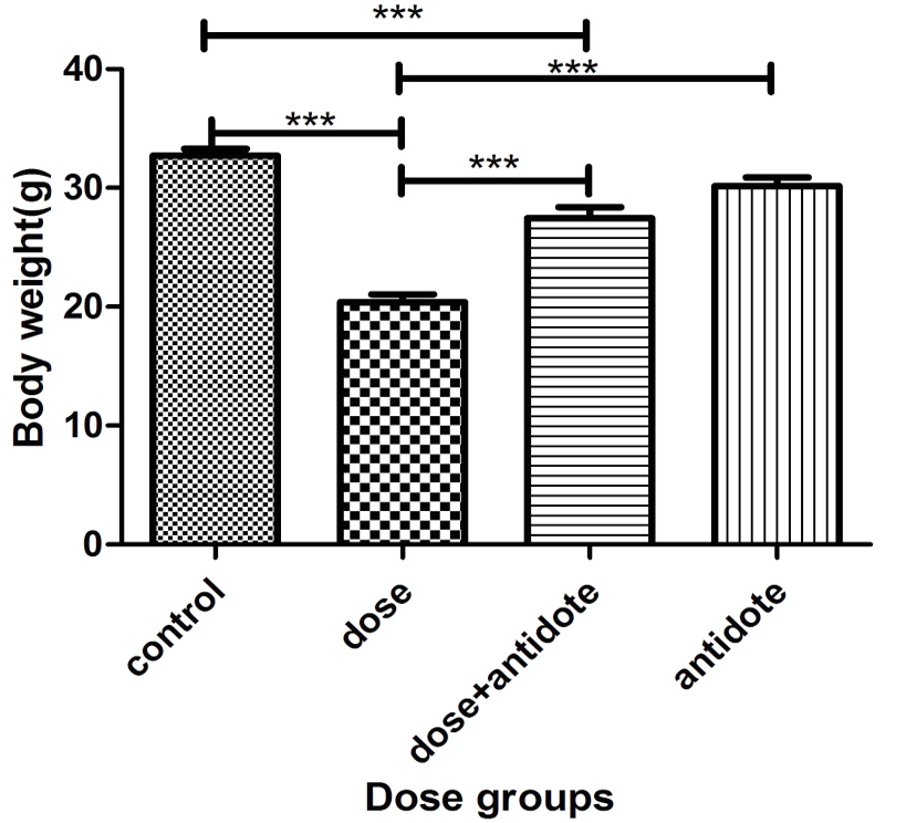
Significant difference in birth weight was observed among all groups except control and antidote as well as dose+antidote and antidote group (Figure 2).
Histological analysis
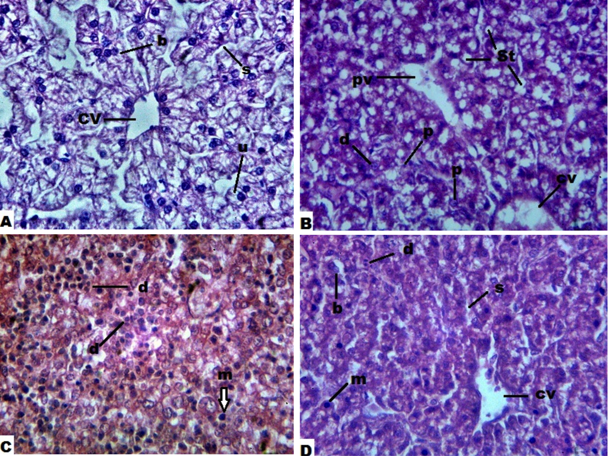
Histological analysis of liver of chicks from control group appeared with intact and normal association of sinusoids and hepatic cords. A cross section of liver of chick embryo treated with 1.5µg/0.05 ml/egg cadmium chloride showed various abnormalities including steatosis leading to pyknosis. Section through liver of chicks treated with dose plus antidote shows increased number of dividing cells indicating recovery from toxicity of cadmium. Antidote group showed normal association of sinusoids and hepatocytes similar to control group (Figure 3A-D).
Control: untreated ; Dose group: treated with 1.5µg/0.05ml/egg of CdCl2; Dose+antidote group: treated with 1.5µg/0.05ml/egg of CdCl2 and 0.2 µg/0.05ml/egg of garlic juice; antidote group: treated with 0.2 µg/0.05ml/egg of garlic juice (*** indicate significant difference at p<0.05).
Discussion
Cadmium is notorious for its toxicity in adults as well as in prenatal exposure . In this study it has been found to reduce body weight (Figure 2)
Table I: Ameliorative effects of fresh garlic juice against cadmium induced teratogenicities in chick embryos against control.
| Group | Total No. of eggs | % of natural hatching | % of deaths | % of malfor-mation | % of resor-ption | CR length mm Mean ± SEM | Major abnormalities% | ||||
| Exenc-ephaly | Ablep-haria | Cro-ssed beak | Gastro-schisis | Croo-ked toes | |||||||
| Control (untreated) | 40 | 92.5 | - | - | 7.5 | 101.20±2.311a | - | - | - | - | - |
| Dose (1.5µg/0.05 ml/egg) | 40 | 32.5 | 48.14 | 42.5 | 35 | 78.80±8.157b | 7.14 | 21.43 | 57.1 | 35.71 | 35.71 |
| Dose (1.5µg/0.05 ml/egg) +Antidote (0.2µg/0.05 ml/egg) | 40 | 72.5 | 18.18 | 15 | 22.5 | 96.20±4.042a | - | - | - | - | 21.43 |
| Antidote (0.2µg/0.05 ml/egg) | 40 | 87.5 | - | - | 12.5 | 110.20±4.420a | - | - | - | - | - |
Values not sharing common letters are significantly different from each other
A: Control group (untreated); B & C: Dose group treated with 1.5µg/0.05ml/egg of CdCl2; D: Dose+antidote group treated with 1.5µg/0.05ml/egg of CdCl2 and 0.2 µg/0.05ml/egg of garlic juice. Note: b: bi-nucleated hepatocyte; s: sinusoid; cv: central vein; u,m: uni-nucleated hepatocyte; pv: portal vein; st: steatosis; d: dividing hepatocyte; p: pyknosis
and successful natural hatching, induce simultaneously the malformations, resorption and death as compared to control. Among deformities, crossed beak was found to be the most prevalent. Gastroschisis, the most lethal one was second major anomaly obtained. Crooked toes and ablepharia were third abundant malformation, while exencephaly was encountered with least incidences. Such craniofacial and skeletal malformations and retarded growth in form of exencephaly, ablephary have also been recorded in mice and in vitro studies ; Arguelles-Velazquez et al., 2013). Inhalation of cadmium even is found toxic and causes cataractogenesis in chronic smokers . Crooking of toes may result from decreased bone mineral density, as it is known to cause bone brittleness for such reason in rat . In chick embryos, such anomalies have dose relationship . Among 11 differentially expressed genes (DEGs) in chicken, which are linked with beak deformity in addition to biosynthesis of unsaturated fatty acids and glycerolipid metabolism , over-expression of LOC426217 in the beak is thought to be the actual responsible (Bai et al., 2016). Cadmium is found to induce delayed hatching in this study as also reported by Dzugan and Lis (2016).
Histological analysis in current study revealed non- alcoholic fatty liver (steatosis) leading to pyknotic cells with shrunk nuclei. Such results are also evidenced in human ; that may be due to altered gene expression in human hepatocellular carcinoma (HepG2) cells , or decrease in hepatic enzymatic and non-enzymatic antioxidants reduced glutathione, catalase, superoxide dismutase .
Mechanism of toxicity of cadmium , like other metals is oxidative stress, DNA damage, ER stress lipoperoxidation, mitochondrial fragmentation, and cell death ; ; ; ; ; ; ; ; through either MAPK pathways , or by decreasing expression and activity of SIRT3 protein and promotes the acetylation of superoxide dismutase 2 ; .
In the group, co-treated with garlic along with cadmium, increased body weight, least malformations and recovery of fatty liver with increased number of normal and dividing cells, possibly due to its anti-Cd properties, radical scavenging assay, ferric reducing ability power assay, chelating activities, superoxide, and hydroxyl scavenging assay ; , complexation of Cd to glutathione (GSH) and metallothionein (MT), prevention of endoplasmic reticulum(ER) stress, mitophagy and metabolic stress, as well as expression of chaperones . Aged garlic extract (AGE) contains S-allylcysteine (SAC) activates Nrf2 factor and inhibits prooxidant enzymes, and chelating effects (Cola-N-Gonzalez et al., 2012). Thiacremonone, another constituent of garlic has potent anti-inflammatory and anti-arthritic properties through the inhibition of NF-κB .
Cadmium toxicity is suggested to overcome by high water intake, chemical antidotes , however, dietary strategies including plants is likely to be more cheaper to encounter such unseen hazards , as the use of fresh garlic along with high-fat diet keeps safe from its hepatotoxicity . Results of present study authenticate the potent ameliorative nature of garlic against cadmium and suggest its use in any way at regular basis to withstand the harmfulness of unseen toxins like cadmium through food and water.
Figures
A macrophotograph of chicks of different dose groups.
A bar graph showing body weight of chicks of different dose groups in ovo exposure to cadmium at 7th day of incubation.
Microphotographs through histological sections of liver of chicks of different dose groups in ovo exposure to cadmium at 7th day of incubation (H&E 400X).
References
- Allam, A.A., Maodaa, S.N., Abo-Eleneen, R., and Ajarem, J., 2016. Protective effect of parsley juice (Petroselinum crispum, Apiaceae) against cadmium deleterious changes in the developed albino mice newborns (Mus musculus) brain. Oxid. Med. Cell Longev., 2016: 264-6840. https://doi.org/10.1155/2016/2646840[CrossRef]
- Arguelles-Velazquez, N., Alvarez-Gonzalez, I., Madrigal-Bujaidar, E., and Chamorro-Cevallos, G., 2013. Amelioration of cadmium-produced teratogenicity and genotoxicity in mice given Arthrospira maxima (Spirulina) treatment. Evid.Based.Complement. Alternat. Med., 2013: 604535. https://doi.org/10.1155/2013/604535[CrossRef]
- Bai, H., Sun, Y., Zhu, J., Liu, N., Li, D., Xue, F., Li, Y. and Chen, J., 2016. Study on LOC426217 as a candidate gene for beak deformity in chicken. BMC. Genet., 17: 44. https://doi.org/10.1186/s12863-016-0353-x[CrossRef]
- Bai, H., Zhu, J., Sun, Y., Liu, R., Liu, N., Li, D., Wen, J., and Chen, J. 2014. Identification of genes related to beak deformity of chickens using digital gene expression profiling. PLoS One., 9(9): e107050. https://doi.org/10.1371/journal.pone.0107050[CrossRef]
- Ban, J.O., Oh, J.H., Kim, T.M., Kim, D.J., Jeong, H.S., Han, S.B. and Hong, J.T., 2009. Anti-inflammatory and arthritic effects of thiacremonone, a novel sulfurcompound isolated from garlic via inhibition of NF-κB. Arthritis Res.Ther., 11: R145. https://doi.org/10.1186/ar2819[CrossRef]
- Bhattacharyya, M.H., 2009. Cadmium Osteotoxicity in Experimental Animals: Mechanisms and Relationship to Human Exposures. Toxicol. Appl. Pharmacol., 238: 258-265. https://doi.org/10.1016/j.taap.2009.05.015[CrossRef]
- Boonpeng, S., Siripongvutikorn, S., Sae-Wong, C. and Sutthirak, P., 2014. The antioxidant and anti-cadmium toxicity properties of garlic extracts. Food Sci. Nutr., 2: 792-801. https://doi.org/10.1002/fsn3.164[CrossRef]
- Cartularo, L., Laulicht, F., Sun, H., Kluz, T., Freedman, J.H. and Costa, M., 2015. Gene expression and pathway analysis of human hepatocellular carcinoma cells treated with cadmium. Toxicol. Appl. Pharmacol., 288: 399-408. https://doi.org/10.1016/j.taap.2015.08.011[CrossRef]
- Castillo, P., Ibanez, F., Guajardo, A., Llanos, M.N. and Ronco, A.M., 2012. Impact of cadmium exposure during pregnancy on hepatic glucocorticoid receptor methylation and expression in rat fetus. PLoS One., 7: e44139. https://doi.org/10.1371/journal.pone.0044139[CrossRef]
- Chen, C., Zhang, S., Liu, Z., Tian, Y. and Sun, Q., 2015. Cadmium toxicity induces ER stress and apoptosis via impairing energy homoeostasis in cardiomyocytes. Biosci. Rep., 35: e00214.
- Chen, X., Li, J., Cheng, Z., Xu, Y., Wang, X., Li, X., Xu, D., Kapron, C.M. and Liu, J. 2016. Low dose cadmium inhibits proliferation of human renal mesangial cells via activation of the JNK pathway. Int. J. Environ. Res. Public Health, 13: 990.
- Cola-N-Gonzalez, A.L., Santana, R.A., Silva-Islas, C.A., Chanez-Cardenas, M.E., SantamaraA, A. and Maldonado, P.D., 2012. The antioxidant mechanisms underlying the aged garlic extract- and s-allylcysteine-induced protection. Oxid. Med. Cellul. Longev., 2012: 907162.
- Cruz, C., Correa-Rotter, R., Sanchez-Gonzalez, D.J., Hernandez-Pando, R., Maldonado, P.D., Martinez-Martinez, C.M., Medina-Campos, O.N., Tapia, E., Aguilar, D., Chirino, Y.I. and Pedraza-Chaverri, J., 2007. Renoprotective and antihypertensive effects of S-allylcysteine in 5/6 nephrectomized rats. Am. J. Physiol. Renal Physiol., 293: 1691-1698. https://doi.org/10.1152/ajprenal.00235.2007[CrossRef]
- Dzugan, M., Lis, M., Droba, M. and Neidzioika, J.W., 2011. Effect of cadmium injected in ovo on hatching results and the activity of plasms hydrolytic enzymes in newly hatched chicks. Acta Vet. Hung., 59: 337-347. https://doi.org/10.1556/AVet.2011.020[CrossRef]
- Dzugan, M. and Lis, M., 2016. Cadmium-induced changes in hatchability and in the activity of aminotransaminases and selected lysosomal hydrolases in the blood plasma of Muscovy ducklings (Cairina moschata). Acta Vet. Hungarica, 64: 239-249. https://doi.org/10.1556/004.2016.024[CrossRef]
- Go, Y.M., Sutliff, R.L., Chandler, J.D., Khalidur, R., Kang, B.Y., Anania, F.A., Orr, M., Hao, L., Fowler, B.A. and Jones, D.P. 2015. Low-dose cadmium causes metabolic and genetic dysregulation associated with fatty liver disease in mice. Toxicol. Sci., 147: 524-534. https://doi.org/10.1093/toxsci/kfv149[CrossRef]
- Guo, P., Pi, H., Xu, S., Zhang, L., Li, Y., Li, M., Cao, Z., Tian, L., Xie, J., Li, R., He, M., Lu, Y., Liu, C., Duan, W., Yu, Z. and Zhou, Z., 2014. melatonin improves mitochondrial function by promoting mt1/sirt1/pgc-1 alpha-dependent mitochondrial biogenesis in cadmium-induced hepatotoxicity in vitro. Toxicol. Sci., 142: 182-195. https://doi.org/10.1093/toxsci/kfu164[CrossRef]
- Gupta, V.K., Singh, S., Agrawal, A., Siddiqi, N.J. and Sharma, B., 2015. Phytochemicals mediated remediation of neurotoxicity induced by heavy metals. Biochem. Res. Int., 2015: 534769. https://doi.org/10.1155/2015/534769[CrossRef]
- Ha, T.T., Burwell, S.T., Goodwin, M.L., Noeker, J.A. and Heggland, S.J., 2016. Pleiotropic roles of Ca+2/calmodulin-dependent pathways in regulating cadmium-induced toxicity in human osteoblast-like cell lines. Toxicol. Lett., 260: 18-27. https://doi.org/10.1016/j.toxlet.2016.08.020[CrossRef]
- Hanson, M.L., Brundage, K.M., Schafer, R., Tou, J.C. and Barnett, J.B., 2010. Prenatal cadmium exposure dysregulates sonic hedgehog and Wnt/β-catenin signaling in the thymus resulting in altered thymocyte development1. Toxicol. Appl. Pharmacol., 242: 136-145.
- Hyder, O., Chung, M., Cosgrove, D., Herman, J.M., Li, Z., Firoozmand, A., Gurakar, A., Koteish, A. and Pawlik, T.M., 2013. Cadmium exposure and liver disease among US adults. J. Gastrointest. Surg., 17: 1265-1273.
- Imafidon, C.E., Akomolafe, R.O., Sanusi, A.A., Ogundipe, O.J., Olukiran, O.S. and Ayowole, O.A., 2015. Polyphenol-rich extract of Vernonia amygdalina (Del.) leaves ameliorated cadmium-induced alterations in feeding pattern and urine volume of male Wistar rats. J. Intercult. Ethnopharmacol., 4: 284-292. https://doi.org/10.5455/jice.20151107021034[CrossRef]
- Jacobo-Estrada, T., Santoyo-Sanchez, M.P., Thevenod, F. and Barbier, O., 2017. Cadmium handling, toxicity and molecular targets involved during pregnancy: Lessons from experimental models. Int. J. Mol. Sci., 18: 1590. https://doi.org/10.3390/ijms18071590[CrossRef]
- Jamakala, O. and Rani, U.A. 2015. Amelioration effect of zinc and iron supplementation on selected oxidative stress enzymes in liver and kidney of cadmium-treated male albino rat. Toxicol. Int., 22: 1-9. https://doi.org/10.4103/0971-6580.172289[CrossRef]
- Jan, A.T., Azam, M., Siddiqui, K., Ali, A., Choi, I. and Haq, Q.M.R., 2015. Heavy metals and human health: mechanistic insight into toxicity and counter defense system of antioxidants. Int. J. Mol. Sci., 16: 29592-29630.
- Khordad, E., Fazel, A. and Ebrahimzadeh, B.A., 2013. The effect of ascorbic acid and garlic administration on lead-induced apoptosis in rat offspring’s eye retina. Iran Biomed. J., 17: 206-213.
- Kim, H.S., Kim, Y.J. and Seo, Y.R. 2015. An overview of carcinogenic heavy metal: Molecular toxicity mechanism and prevention. J. Cancer Prev., 20: 232-240.
- Kippler, M., Engstra, M.K., Mlakar, S.J., Bottai, M., Ahmed, S., Hossain, M.B., Raqib, R., Vahter, M. and Broberg, K., 2013. Sex-specific effects of early life cadmium exposure on DNA methylation and implications for birth weight. Epigenetics., 8: 494-503.
- Kippler, M., Tofail, F., Gardner, R., Rahman, A., Hamadani, J. D., Bottai, M. and Vahter, M., 2012. Maternal Cadmium Exposure during Pregnancy and Size at Birth: A Prospective Cohort Study. Environ. Health Perspect., 120(2): 284-289. https://doi.org/10.1289/ehp.1103711[CrossRef]
- Majewski, M., 2014. Allium sativum: facts and myths regarding human health. Rocz. Panstw. Zakl. Hig., 65: 1-8.
- Meeker, J.D., Rossano, M.G., Protas, B., Diamond, M.P., Puscheck, E., Daly, D., Paneth, N. and Wirth, J.J. 2008. Cadmium, lead, and other metals in relation to semen quality: human evidence for molybdenum as a male reproductive toxicant. Environ. Health Perspect., 116(11): 1473-1479. https://doi.org/10.1289/ehp.11490[CrossRef]
- Memon, S. and Pratten, M., 2013. Effects of Multivitamins and Known Teratogens on Chick Cardiomyocytes Micromass Culture Assay. Iran J. Basic Med. Sci., 16: 996-1003.
- Nair, A.R., Degheselle, O., Smeets, K., Van, K.E. and Cuypers, A., 2013. Cadmium-Induced Pathologies: Where Is the Oxidative Balance Lost (or Not)? Int. J. Mol. Sci., 14: 6116-6143. https://doi.org/10.3390/ijms14036116[CrossRef]
- Nasr, A.Y. 2014. Protective effect of aged garlic extract against the oxidative stress induced by cisplatin on blood cells parameters and hepatic antioxidant enzymes in rats. Toxicol. Rep., 1: 682-691. https://doi.org/10.1016/j.toxrep.2014.09.003[CrossRef]
- Oyinloye, B.E., Adenowo, A.F., Osunsanmi, F.O., Ogunyinka, B.I., Nwozo, S.O. and Kappo, A.P. 2016. Aqueous extract of Monodora myristica ameliorates cadmium-induced hepatotoxicity in male rats. Springerplus., 5: 641. https://doi.org/10.1186/s40064-016-2228-z[CrossRef]
- Paniagua-Castro, N., Escalona-Cardoso, G., Madrigal-Bujaidar, E., Martinez-Galero, E. and Chamorro-Cevallos, G., 2008. Protection against cadmium-induced teratogenicity in vitro by glycine. Toxicol. In Vitro, 22(1): 75-79. https://doi.org/10.1016/j.tiv.2007.08.005[CrossRef]
- Pawlak, K., Dżugan, M., Wojtysiak, D., Lis, M. and Niedziółka, J., 2013. Effect of in ovo injection of cadmium on chicken embryo heart. African J. Agr. Res., 8: 1534–1539. https://doi.org/10.5897/AJAR12.2116[CrossRef]
- Pi, H., Xu, S., Reiter, R. J., Guo, P., Zhang, L., Li, Y., Li, M., Cao, Z., Tian, L., Xie, J., Zhang, R., He, M., Lu, Y., Liu, C., Duan, W., Yu, Z. and Zhou, Z., 2015. SIRT3-SOD2-mROS-dependent autophagy in cadmium-induced hepatotoxicity and salvage by melatonin. Autophagy., 11:1037-1051. https://doi.org/10.1080/15548627.2015.1052208[CrossRef]
- Poljsak, B. and Fink, R., 2014. The Protective Role of Antioxidants in the Defence against ROS/RNS-Mediated Environmental Pollution. Oxid. Med. Cell Longev., 2014: 671539.
- Qamar, A., Usmani, A., Waqar, H., Siddiqui, A. and Kumar, H., 2016. Ameliorating effect of Allium Sativum on high-fat diet induced fatty liver in albino rats. Pak. J. Med. Sci., 32: 403-407.
- Rafati, R.M., Rafati, R.M., Kazemi, S. and Moghadamnia, A., 2017. Cadmium toxicity and treatment: An update. Caspian. J. Intern. Med., 8: 135-145.
- Ramakrishnan, S., Sulochana, K.N., Selvaraj, T., Abdul, R.A., Lakshmi, M. and Arunagiri, K., 1995. Smoking of beedies and cataract: cadmium and vitamin C in the lens and blood. Br. J. Ophthalmol., 79: 202-206. https://doi.org/10.1136/bjo.79.3.202[CrossRef]
- Robinson, J.F., Yu, X., Hong, S., Zhou, C., Kim, N., Demasi, D. and Faustman, E.M. 2010. Embryonic toxicokinetic and dynamic differences underlying strain sensitivity to cadmium during neurulation. Reprod. Toxicol., 29: 279-285. https://doi.org/10.1016/j.reprotox.2009.12.004[CrossRef]
- Robinson, J.F., Yu, X., Moreira, E.G., Hong, S. and Faustman, E.M., 2011. Arsenic- and Cadmium-induced toxicogenomic response in mouse embryos undergoing neurulation. Toxicol. Appl. Pharmacol., 250: 117-129. https://doi.org/10.1016/j.taap.2010.09.018[CrossRef]
- Rodriguez-Fragoso, P., Reyes-Esparza, J., Leon-Buitimea, A. and Rodriguez-Fragoso, L., 2012. Synthesis, characterization and toxicological evaluation of maltodextrin capped cadmium sulfide nanoparticles in human cell lines and chicken embryos. J. Nanobiotechnol., 10: 47. https://doi.org/10.1186/1477-3155-10-47[CrossRef]
- Romano, M.E., Enquobahrie, D.A., Simpson, C., Checkoway, H. and Williams, M.A., 2016. Maternal body burden of cadmium and offspring size at birth. Environ. Res., 147: 461-468. https://doi.org/10.1016/j.envres.2016.02.029[CrossRef]
- Ruiter, S., Sippel, J., Bouwmeester, M.C., Lommelaars, T., Beekhof, P., Hodemaekers, H.M., Bakker, F., Van Den Brandhof, E.J., Pennings, J.L.A. and Van Der Ven, L.T.M., 2016. Programmed effects in neurobehavior and antioxidative physiology in zebrafish embryonically exposed to cadmium: Observations and hypothesized adverse outcome pathway framework. Int. J. Mol. Sci., 17: 1830. https://doi.org/10.3390/ijms17111830[CrossRef]
- Sandbichler, A.M. and Hãckner, M., 2016. Cadmium protection strategies for a hidden trade-off? Int. J. Mol. Sci., 17: 139. https://doi.org/10.3390/ijms17010139[CrossRef]
- Sanders, A.P., Smeester, L., Rojas, D., Debussycher, T., Wu, M.C., Wright, F.A., Zhou, Y.H., Laine, J.E., Rager, J.E., Swamy, G.K., Ashley-Koch, A., Lynn, M.M. and Fry, R.C., 2014. Cadmium exposure and the epigenome: Exposure-associated patterns of DNA methylation in leukocytes from mother-baby pairs. Epigenetics., 9: 212-221. https://doi.org/10.4161/epi.26798[CrossRef]
- Setiawan, V.W., Yu, G.P., Lu, Q.Y., Lu, M.L., Yu, S.Z., Mu, L., Zhang, J.G., Kurtz, R.C., Cai, L., Hsieh, C.C. and Zhang, Z.F., 2005. Allium vegetables and stomach cancer risk in China. Asian Pac. J. Cancer Prev., 6: 387-395.
- Tualla, I.P.B. and Bitacura, J.G., 2016. Effects of cadmium and zinc on the gamete viability, fertilization, and embryonic development of Tripneustes gratilla (Linnaeus). Scientifica.(Cairo.), 2016: 8175213. https://doi.org/10.1155/2016/8175213[CrossRef]
- Ugwuja, E.I., Erejuwa, O.O. and Ugwu, N.C., 2016. Spices mixture containing garlic, ginger and nutmeg has protective effects on the kidneys and liver of cadmium exposed rats. Adv. Pharm. Bull., 6: 271-274.
- Veeriah, V., Saran, U., Swaminathan, A., Balaguru, U.M., Thangaraj, P., Nagarajan, S., Rajendran, V.K. and Chatterjee, S., 2015. Cadmium-induced embryopathy: nitric oxide rescues teratogenic effects of cadmium. Toxicol. Sci., 144: 90-104. https://doi.org/10.1093/toxsci/kfu258[CrossRef]
- Vilahur, N., Vahter, M. and Broberg, K., 2015. The Epigenetic Effects of Prenatal Cadmium Exposure. Curr.Environ.Health Rep., 2(2): 195-203. https://doi.org/10.1007/s40572-015-0049-9[CrossRef]
- Wei, Q., Juanjuan, B., Longlong, T., Zhan, L., Peng, L. and Wangsuo, W., 2014. The effect of multiwalled carbon nanotubes on hepatotoxicity of Cd2+ in accumulated cadmium-metallothione in mice. Biomed. Res. Int., 2014: 463161. https://doi.org/10.1155/2014/463161[CrossRef]
- Wei, T., Jia, J., Wada, Y., Kapron, C.M. and Liu, J., 2017. Dose dependent effects of cadmium on tumor angiogenesis. Oncotarget., 8: 44944-44959.
- Witeska, M., Sarnowski, P., Ugowska, K. and Kowal, E., 2014. The effects of cadmium and copper on embryonic and larval development of ide Leuciscus idus L. Fish. Physiol. Biochem., 40: 151-163. https://doi.org/10.1007/s10695-013-9832-4[CrossRef]
- Xu, S., Pi, H., Chen, Y., Zhang, N., Guo, P., Lu, Y., He, M., Xie, J., Zhong, M., Zhang, Y., Yu, Z. and Zhou, Z., 2013. Cadmium induced Drp1-dependent mitochondrial fragmentation by disturbing calcium homeostasis in its hepatotoxicity. Cell Death. Dis., 4: e540. https://doi.org/10.1038/cddis.2013.7[CrossRef]
- Xu, Z., Jin, X., Pan, T., Liu, T., Wan, N. and Li, S., 2017. Antagonistic effects of selenium on cadmium-induced apoptosis by restoring the mitochondrial dynamic equilibrium and energy metabolism in chicken spleens. Oncotarget., 8: 52629-52641.
- Yiran, Z., Chenyang, J., Jiajing, W., Yan, Y., Jianhong, G., Jianchun, B., Xuezhong, L. and Zongping, L., 2013. Oxidative stress and mitogen-activated protein kinase pathways involved in cadmium-induced BRL 3A cell apoptosis. Oxid. Med. Cell Longev., 2013: 516051. https://doi.org/10.1155/2013/516051[CrossRef]
- Zhai, Q., Narbad, A. and Chen, W., 2015. Dietary strategies for the treatment of cadmium and lead toxicity. Nutrients., 7: 552-571. https://doi.org/10.3390/nu7010552[CrossRef]
- Zhao, L., Ru, Y., Liu, M., Tang, J., Zheng, J., Wu, B., Gu, Y. and Shi, H., 2017. Reproductive effects of cadmium on sperm function and early embryonic development in vitro. PLoS.One., 12(11): e018672. https://doi.org/10.1371/journal.pone.0186727[CrossRef]
Services
Article usage statistics
Loading…