
Acute kidney injury (AKI) is a life-threatening condition which may result from extensive injury to the kidney due to various causative agents. Renal insult can result from ischemia, drugs, renal obstruction, inflammation, or extensive muscle injury. The current study focused only on Rhabdomyolysis-induced AKI. We make an attempt to uncover the preventive efficacy of cinnamic acid (CA) and CA-conjugated gold nanoparticles (CA-AuNPs) against rhabdomyolysis-induced AKI induced in mice. CA (50 mg/Kg) and CA-AuNPs (30 mg/Kg) were given to the animals for four days. On the fourth day and after 24 h of water deprivation, 50% glycerol intra-muscular was administered. Blood urea and creatinine were tested. Kidney histopathology was done and damaged kidney areas were measured. Immunohistochemistry for actin and cyclooxygenase-2 (COX-2) were also carried out. Real time RT-PCR studies were performed to check mRNA expressions of nuclear factor-κB (NFκB) p50, inducible nitric oxide synthase (iNOS), kidney injury molecule-1 (Kim-1) and hemeoxygenase-1 (HO-1). CA and CA-AuNPs improved blood urea and creatinine levels when compared with the glycerol induced AKI group. The compounds kept the actin intact, decreased COX-2 protein expression, down-regulated the expressions of NFkB p50 and iNOS, and up-regulated Kim-1 and HO-1. The tested compounds (CA and CA-AuNPs) prevented the kidney from injury in the rhabdomyolysis-induced AKI animal models. However, almost complete protection is observed in CA-AuNPs treated animals at a relatively lower dose.
Research Article · Volume 56, Issue 2, 21 January 2024 · Open Access
Prevention of Rhabdomyolysis-Induced Acute Kidney Injury via Attenuation of Oxidative Injury and Inflammation by Cinnamic Acid Coated Gold Nanoparticles in Mice
· · · · · ·
Affiliations & Notes ⌄
- 1Department of Molecular Medicine, Ziauddin University, Karachi, Pakistan. 2Dr. Panjwani Center for Molecular Medicine and Drug Research, International Center for Chemical and Biological Sciences, University of Karachi, Karachi-75270, Pakistan. 3H.E.J. Research Institute of Chemistry, International Center for Chemical and Biological Sciences, University of Karachi, Karachi-75270, Pakistan. 4Institute of Biological Sciences, Faculty of Science, University of Malaya, 50603 Kuala Lumpur, Malaysia. 5
Article Info ⌄
- Journal:
- Pakistan Journal of Zoology
- Published:
- 21 January 2024

From the article
- This anti-oxidant activity was also presented in our study with the evidence of less injury to the kidney from glycerol-induced AKI.
- Thus, we can conclude that CA and CA-Au-NPs have anti-inflammatory actions with protective activity against rhabdomyolysis-induced AKI.
- CA prevents rhabdomyolysis-induced AKI shown by significantly lower blood urea and creatinine levels.
- The kidney protective mechanisms of these compounds could involve downregulation of the COX-2 protein, and thus, reducing the effect of oxidant injury and inflammation.
Selected from the article's own text.
Abstract
Introduction
Acute kidney injury (AKI) is defined as complete or partial detrition in renal functions which may repercuss from injury caused by any drug, inflammation, hindrance in renal flow or ischemia . Rhabdomyolysis is an important pathological process which may account for approximately 50% of cases of AKI ; . Rhabdomyolysis can be defined as destruction of skeletal muscle leading to an increase in the levels of myoglobin and/or hemoglobin, and its deposition in the kidneys, resulting in severe kidney damage. This damage to skeletal muscles can also result from infections such as malaria, crush injury, drug intoxication, seizures etc. . This type of kidney failure was the major reason for death of the injured soldiers in both the 1st and 2nd World Wars.
Even with the recent advances in health and medicine, currently there are no specific therapeutics available for AKI other than supportive treatment or renal transplant . Although limited, the tubular epithelial cells do have the ability to regenerate and resume normal physiological function if the noxious stimulus is removed ; . However, renal tissues can be irreversibly damaged if the stimulus is severe or long-term. Irreversible kidney damage accounts for 50–70% of death rate which has not declined in last ten decades . Preventive therapeutics are therefore an important aspect to explore for AKI which affect in lowering the mortality rate of AKI.
There are many established animal models for AKI one of which is rhabdomyolysis-induced AKI. Glycerol administration at a dose of 10ml/kg body weight intramuscularly can cause rhabdomyolysis due to oxidative injury to the renal tubular cells ; Ustundag et al., 2005; ; ; . Intramuscular (IM) glycerol along with damage to myoglobin can also cause wreckage in red blood cells leading to release of hemoglobin in circulation, ultimately playing an additional role in the sequalae of kidney injury . Furthermore, inflammatory mediators are also activated and released in the blood. These mediators recruit leukocytes at the injury site leading to production of reactive oxygen species eventually causing necrosis of renal tissues . The process of inflammation is reported to be initiated by activation of NF-κB which instigates the inflammatory process via expression of various pro-inflammatory genes. Moreover, iNOS and COX-2 have been described as major activators of the inflammatory pathway . Henceforth, renal protective players also play an important part in AKI where Kim-1 and HO-1 expression plays a vital part in the protection of kidney morphology. This can be assessed by increment in expression of these in response to any injury to kidney cells . Keeping in mind the physiological response of renal cells to any insult, it can be elucidated that increment in renal protective genes and inhibition of inflammatory genes with scavenging of free radicals may halt or delay the entire process of kidney injury.
Cinnamic acid (CA) is found abundantly in Cinnamon cassia as one of its active compounds and a major constituent. The plant is used in flavours, as a synthetic azure and in pharmaceutical companies as well. Recent studies showed that CA also possesses antidiabetic activity by enhancing insulin secretion and sensitivity . In this study the selection of this compound was based on its anti-oxidant and anti-inflammatory activities.
Delivering a drug to the site of injury is a key component in treating many diseases. Because nano-particle can easily pass through cell membranes, they can provide us with improved drug bioavailability at specific sites while at the same time reducing cross-toxicity due to low dose requirements . Therefore, in this study we also investigated gold conjugated nanoparticles of CA.
Materials and Methods
Synthesis of CA-AuNPs
To synthesize the CA-conjugated AuNPs, CA (1 mM solution) was mixed with HAuCl4.3H2O (1 mM solution) and stirred. After 30 minutes, 0.4 mL of 40 mM NaBH4 was mixed, resulting in a complete reduction of gold ions to form the CA-conjugated AuNPs. The CA-conjugated AuNPs were obtained after freeze drying. Their UV–Visible spectra were taken by Thermo Scientific Evolution 300 UV-Visible spectrophotometer. The shape and size of the CA-conjugated AuNPs was also determined by using Atomic Force Microscope (AFM, Agilent 5500) operating in tapping mode.
Animals
International guidelines were followed for all animal procedures and institutional ethical committee approval was obtained for proper animal use (Protocol No.: 2016-0002). Animal house facility at International Center for Chemical and Biological Sciences, University of Karachi, Karachi was used to breed and maintain male albino mice. Experimental mice were kept with free access to rodent chow and water in conventional cages water at 22-23°C with a 12-h light-dark cycle. All animals were acclimatized prior to the start of experiments.
Treatment regime
Animals were segregated into four groups with six mice in each. Animals in test groups were pretreated with CA (50 mg/Kg) and CA-AuNPs (30 mg/Kg) for 4 days. On the fourth day, all the mice were water deprived for 24-h followed by IM injection of glycerol. 24 h later, all animals were sacrificed humanly under anesthesia (60 mg/Kg pentobarbital sodium) for further studies.
Biochemical investigations
After sacrificing the animals, blood was drawn through cardiac puncture. Serum was isolated and urea-creatinine levels were measured by Reflotron® Dry Chemistry Analyzer (Roche, Switzerland) to assess renal functions in all four animal groups.
Histopathology and immunohistochemistry
Kidney tissues were fixed in Bouin’s fixative for four h followed by dehydration in graded isopropyl alcohol. The tissues were then paraffin embedded. Six micrometer sections were cut followed by hematoxylin and eosin (H and E) and periodic acid Shiff’s (PAS) staining. For immunohistochemistry of actin and COX-2, we followed our previous protocol .
Real time PCR analysis
Mouse kidney tissues were homogenized with TRIzol reagent (Life Technologies, USA) and mRNA was isolated following standard protocol. The cDNA was synthesized using the RevertAid first strand cDNA synthesis kit (Fermentas, USA). Real-time PCR studies were carried out using the Agilent Technologies (USA) for mice iNOS, NFkB p50, Kim-1 and HO-1. The primer sequences and Real Time RT-PCR protocol was followed according to our previous study .
Statistical analysis
Statistical analysis of data was performed using SPSS version 20. All values were expressed as mean ± SEM. The data obtained from results were compared among or within groups by t-test. Significant difference was considered if the P-value was found to be of less than 0.05.
Results
Characterization of CA-AuNPs
Synthesized CA-conjugated AuNPs were characterized by UV-Visible spectroscopy. The AuNPs showed characteristics surface Plasmon resonance at around 520-550 nm. The UV-Visible spectrum of CA-AuNPs (Fig. 1A) showed an absorption maximum at 527 nm indicating the formation of CA conjugated AuNPs. Size determination and morphological studies of the synthesized CA-AuNPs were carried out using AFM technique. The AFM results showed that the CA conjugated AuNPs were sphere-shaped having average size ranges from 10-30 nm (Fig. 1B).
Cast deposition and tubular necrosis is prevented by CA and CA-AuNPs
Figure 2A and E shows normal mice kidney cortex and medulla. In animals treated with 50% glycerol at a dose of 10 mL/kg body weight, an increase in proximal convoluted tubular necrosis was observed (Fig. 2B), and the casts were found to be deposited in the loop of Henle (LH). Furthermore, damage to the LH cells was also seen (Fig. 2F). However, the animals pre-treated with CA at a dose of 50 mg/kg body weight demonstrated remarkable much less in tubular damage and protein cast depositions (Fig. 2C, G). The CA-AuNPs pre-treated (30 mg/kg) animals revealed almost complete protection against damage as shown in Figure 2D, H. The extent of tubular necrosis was also measured by the software NIS-Elements AR 3.2 (Nikon, Japan) and data showed a significant decrease (p <0.001) in tubular necrosis in both treatment groups (Fig. 3). However, it is seen that there is a notable reduction in the damage treated by CA-AuNPs at a lower dose as compared to CA alone.
CA and CA-AuNPs decreases serum urea and creatinine levels
CA and CA-AuNPs prevent damage to PCT and LH brush borders
PAS stained sections of the kidney (Fig. 5) shows normal pattern of PCT and LH brush borders is shown in Figure 5A and E. Remarkable damage was seen in the brush borders of the AKI group (Fig. 5B and F). CA treatment decreased the damage to the brush border induced by glycerol (Fig. 5C and G). Conversely, almost thorough protection of the brush borders was seen in the animals treated with CA-AuNPs (Fig. 5D and H).
CA and CA-AuNPs maintain actin cytoskeletal integrity
The test compound and its nanoformulation were also investigated for actin cytoskeleton immunohistochemistry (Fig. 6A-D). Figure 6A shows normal actin cytoskeleton pattern while a massive destruction of actin was observed in the animals treated with glycerol (Fig. 6B). CA was observed to reduction in actin disruption (Fig. 6C). However, the treatment of CA-AuNPs almost entirely prevented the actin cytoskeleton (Fig. 7D).
CA and CA-2-AuNPs reduce the expression of COX-2
Figure 6E shows no immunohistochemical staining of COX-2 in sections of the normal group. As expected, increased expression was seen in the AKI group (Fig. 6F). As compared to AKI group, animals in CA (Fig. 6G) and CA-AuNPs (Fig. 6H) treated groups exhibited marked decrease in the expression of COX-2.
CA and CA-AuNPs cause upregulation HO-1 and Kim-1, down-regulation of the mRNA expressions of iNOS and NFκB p50
mRNA expressions of iNOS, NFκB p50, HO-1 and Kim-1 are shown in Figure 7. A significant increase in iNOS (p <0.001) and NFκB p50 (p <0.001) mRNA expression was observed in AKI control animals. In comparison to the control group, a significant decrease of iNOS (p <0.001) and NFκB p50 (p <0.001) was seen in CA and CA-AuNPs treated groups (Fig. 7A and B) despite the glycerol injection. The HO-1 mRNA level was not significantly higher in AKI group compared to the normal controls. However, the level of HO-1 was significantly higher (p <0.05) in the animals treated with CA and CA-AuNPs as compared to the glycerol treated group (Fig. 7C). The Kim-1 expression was significantly higher (p <0.05) in AKI treated group as compared to the normal controls, whereas the level of Kim-1 in CA and CA-AuNPs treated groups were noted to be significantly higher (p <0.05) than in the AKI group.
Discussion
Natural or synthetic products that have been proposed to be active against AKI have been discovered using the rhabdomyolysis- induced AKI which establishes the fact of this model being widely accepted. One of the major factors for this is the adjacency with the human pathogenesis of AKI. Inflammatory cascades that are cause of necrosis in the renal cells, are consequence of oxidative injury caused to the proximal convoluted tubules by glycerol. In our study, we not only found necrotic cells but also observed the presence of condensed nuclei, thus showing the process of apoptosis in the cells. Therefore, this model was used to study the protective effects of CA and CA-AuNP on mice.
Potent anti-oxidant activity of CA has been reported previously in diabetic models of rat . Anti-oxidant property was the prime reason for selection of this compound as oxidative injury is a major factor in rhabdomyolysis-induced AKI. This anti-oxidant activity was also presented in our study with the evidence of less injury to the kidney from glycerol-induced AKI. The test compound also prevented damage to tubular brush borders concomitantly reducing serum urea and creatinine levels in comparison to AKI group. CA preserved the architecture of actin cytoskeleton present in the extracellular matrix and brush borders. Protein expression for COX-2, an initiator of inflammatory response was also inhibited by CA and CA-AuNPs. This inhibition of the inflammatory cascade further enhances the protective effect of CA and CA-AuNPs to the kidney cells.
Bioavailability has always been a major factor for exhibition of clinical activity for any moiety for a probable drug candidate. Hence, drug delivery is an important concern in clinical research practice. Many drugs have inefficient bioavailability to their specific organ or site of activity. Furthermore, the toxicities of some drugs are very high, specifically towards the liver and kidney. To address these important issues, gold nanoparticles conjugated with CA were used in this study. The gold nanoparticles appear to be safe as no apparent cytotoxicity in animals treated with gold nanoparticles alone were observed (data not shown). CA-AuNPs showed protective activity on AKI better than CA alone. Next, we also elucidated the molecular pathways of protection by CA and its gold nanoparticles and investigated the role of iNOS, NF-κB, HO-1 and Kim-1 following CA treatment in animals with AKI.
We know that nitric oxide species promote production of other inflammatory cytokines . In accordance to this, we also observed a significant rise in iNOS mRNA expression in the glycerol treated animals. Conversely, it was down regulated in CA and CA-AuNPs treated animals. This implicates that CA and CA-AuNPs treatment negatively modulates the high oxidative environment in AKI group.
NF-κB regulates transcriptions of various genes by complex mechanisms . It also contributes in the production of cytokines and survival of cells ; . NF-κB is mainly responsible for inflammatory feedback mechanisms through various pathways affecting the duration and degree of the inflammation ; . Findings in this study are in accordance with these reports as we also observed raised NF-κB expression in the AKI group in comparison to normal controls. Conversely, the NF-κB expression levels were noticeably decreased in CA and CA-AuNPs treated animals.
Heme oxygenase (HO) enzyme catalyzes the degradation of heme and produces iron, biliverdin and carbon monoxide . HO-1 is induced in stress such as during oxidative burst. Protective role of HO-1 is reported during oxidative stress. It also plays an anti-inflammatory role by up-regulating interleukin-1R and interleukin-10 antagonist expression . The cellular preventative effects of HO-1 are exerted by inhibition of a variety of immune system mediated inflammatory pathways. In this study, we observed significantly increased levels of HO-1 in CA and CA-Au-NPs treated animals as compared to normal and glycerol treated animals. This explains the protective role of the compound through HO-1 modulation.
Kidney injury molecule-1 (Kim-1) is a new kidney injury marker ; . The Kim-1 expression is increased in inflammation. In accordance to these reports, we also observed increased Kim-1 expression in glycerol treated animals in comparison to normal controls. All treatment groups revealed much higher mRNA expression of Kim-1. Consequently, it may have a crucial role in the inhibition of inflammation. Thus, we can conclude that CA and CA-Au-NPs have anti-inflammatory actions with protective activity against rhabdomyolysis-induced AKI.
Conclusion
CA prevents rhabdomyolysis-induced AKI shown by significantly lower blood urea and creatinine levels. The nano-formulations of CA (CA-AuNPs) can shield kidney architecture such as kidney tubular brush borders and actin cytoskeleton very well at a much less dose compared to CA alone. The kidney protective mechanisms of these compounds could involve downregulation of the COX-2 protein, and thus, reducing the effect of oxidant injury and inflammation. Moreover, they inhibit mRNA expressions of NF-κB, and iNOS and escalate HO-1 and Kim-1 expressions.
Acknowledgement
The authors are grateful to Dr. Panjwani Center for Molecular Medicine and Drug Research, ICCBS, University of Karachi, for providing the facility and funds to conduct the research work.
Ethical statement
The International Center for Chemical and Biological Sciences Institutional Animal Care and Use Committee (IACUC) approved the use of animals on the ASP referenced above for one year period (04/08/2014 through 13/08/2015). The animals were taken care of for the entire study as per the guidelines of the committee for the purpose of control and supervision of of experimental animals.
Statement of conflict of interest
The authors have declared no conflict of interest.
Figures
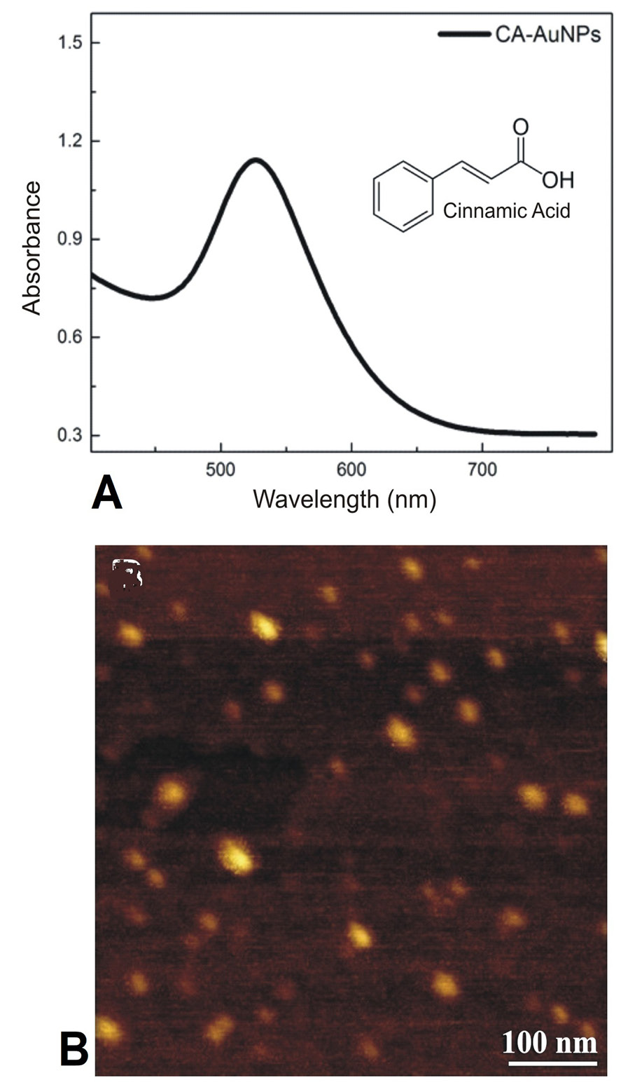
(A) UV-visible spectrum and (B) AFM analysis of CA coated AuNPs.
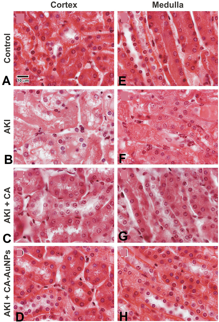
H and E stained microscopic images demonstrating kidney cortex and medulla. Images A and E are showing normal kidney architectures. Images B and F are of glycerol treated AKI sections showing damaged proximal tubules and cast deposition in tubules. Images C and g are CA treated group presenting notable decline in tubular cast deposition and damage. While images D and H are CA-AuNPs treated kidney sections displaying more or less complete protection. (Magnification; 600x).
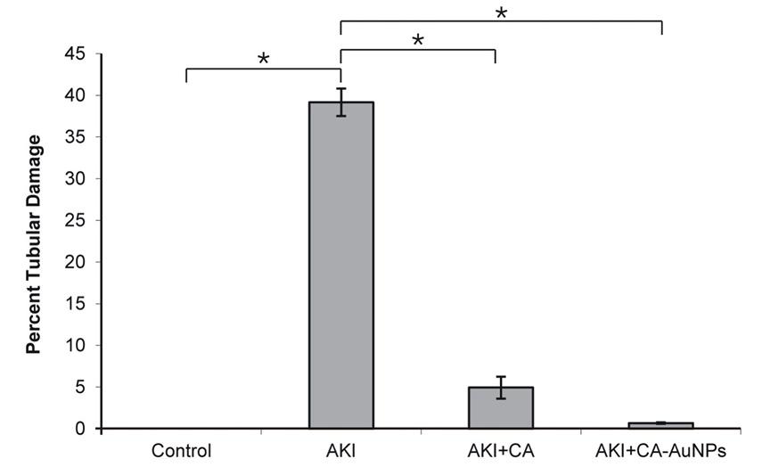
Calculation of Damaged areas in proximal convoluted tubules in different animal groups. The graph demonstrating no proximal tubular damage in normal control. Whereas, there is a marked increase in damaged area in glycerol treated AKI group compared to the normal control (*p <0.001). A significant decrease in the damaged areas was observed in animals treated with CA as compared with the glycerol treated AKI group (*p <0.001) and nearly complete protection can be seen in CA-AuNPs treated groups at a relatively low dose (*p <0.001).
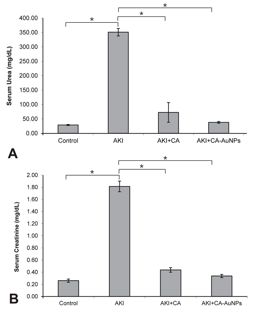
Serum urea and creatinine levels. Serum urea and creatinine levels were significantly elevated in the glycerol treated AKI group as compared to the normal control (*p <0.001). The levels were significantly decreased in CA and CA-AuNPs treated animals as compared to the glycerol treated AKI group (*p <0.001).
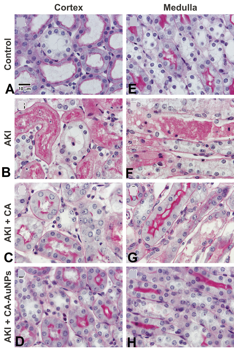
PAS stained microscopic images demonstrating brush borders of kidney tubules in the cortex and medulla. Images A and E shows normal tubular brush borders. Images B and F are of glycerol treated AKI group showing damaged brush borders of loop of Henle and proximal tubules. Images C and G are taken from CA treated animals, a remarkable reduction in the damage of brush borders can be observed while images D and H displays protection with CA-AuNPs. (Magnification; 600x).
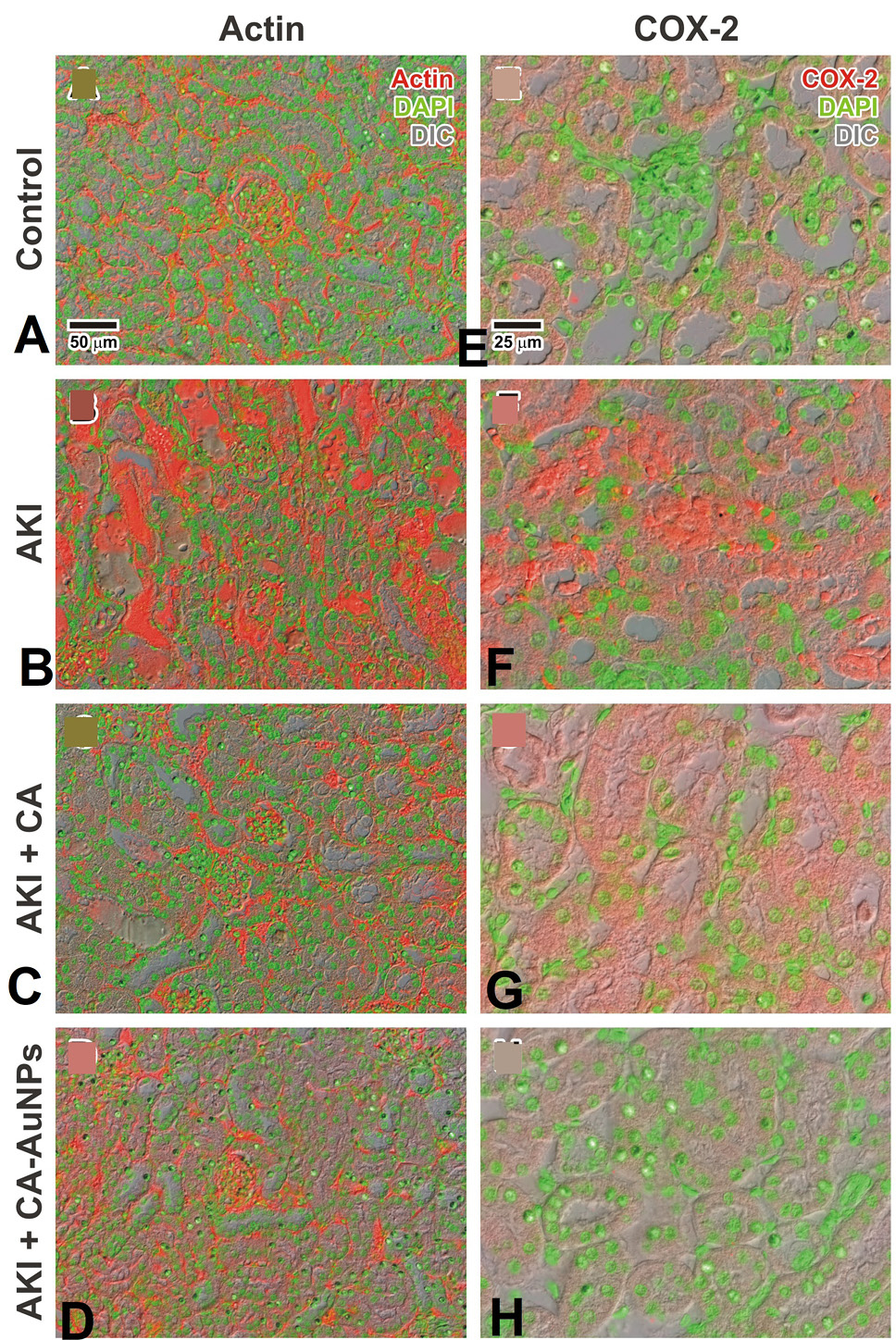
CA and CA-AuNPs prevent damage in actin cytoskeleton and reduces expression of COX-2. Normal distribution of actic can be seen in figure A while figure B shows disrupted actin cytoskeleton. The protection of actin cytoskeleton can be observed in figure C and D which were treated with CA and CA-AuNPs respectively. (Magnification; 200x). There is no expression of COX-2 in normal control (E) whereas higher expression level can be observed in glycerol treated AKI group (F). A decrease expression of COX-2 can be noticed in CA (G) and CA-AuNPs (H) treated groups. Red is actin and COX-2, green is DAPI and gray is DIC image. (Magnification; 600x).
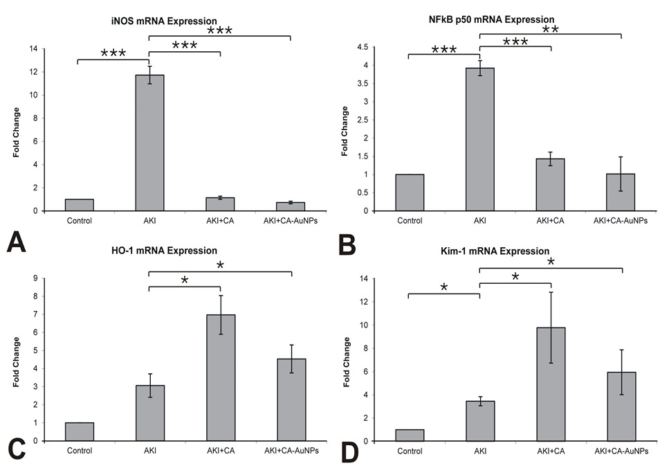
Effects of CA and CA-AuNPs on iNOS, NF-κB, HO-1 and Kim-1 mRNA expressions. CA and CA-AuNPs attenuates mRNA expression of iNOS (A) and NF-κB (B) while both increases the mRNA expression of HO-1 (c) and Kim-1 (d) (***p < 0.001, **p < 0.01, * p < 0.05).
References
- Baliga, R., Ueda, N., Walker, P.D. and Shah, S.V., 1997. Oxidant mechanisms in toxic acute renal failure. Am. J. Kidney Dis., 29: 465-477. https://doi.org/10.1016/S0272-6386(97)90212-2[CrossRef]
- Bosch, X., Poch, E. and Grau, J.M., 2009. Rhabdomyolysis and acute kidney injury. N. Eng. J. Med., 361: 62-72. https://doi.org/10.1056/NEJMra0801327[CrossRef]
- Chander, V., Singh, D. and Chopra, K., 2003. Catechin, a natural antioxidant protects against rhabdomyolysis-induced myoglobinuric acute renal failure. Pharmacol. Res., 48: 503-509. https://doi.org/10.1016/S1043-6618(03)00207-X[CrossRef]
- Cil, O., Ertunc, M., Gucer, K.S., Ozaltin, F., Iskit, A.B. and Onur, R., 2012. Endothelial dysfunction and increased responses to renal nerve stimulation in rat kidneys during rhabdomyolysis-induced acute renal failure: role of hydroxyl radical. Ren. Fail., 34: 211-220. https://doi.org/10.3109/0886022X.2011.643389[CrossRef]
- Colledge, N.R., Walker, B.R. and Ralston, S.H., 2010. Davidson’s principles and practice of medicine. Elsevier Limited.
- Feather, A., Randall, D. and Waterhouse, M., 2020. Kumar and Clark’s clinical medicine (E-Book). Elsevier Health Sciences.
- Fishman, A.I., Alexander, B., Eshghi, M., Choudhury, M. and Konno, S., 2012. Nephrotoxin-induced renal cell injury involving biochemical alterations and its prevention with antioxidant. J. clin. med. Res., 4: 95. https://doi.org/10.4021/jocmr833w[CrossRef]
- Gilmore, T.D., 2006. Introduction to NF-κ B: Players, pathways, perspectives. Oncogene, 25: 6680-6684. https://doi.org/10.1038/sj.onc.1209954[CrossRef]
- Hafizur, R.M., Hameed, A., Shukrana, M., Raza, S.A., Chishti, S., Kabir, N. and Siddiqui, R.A., 2015. Cinnamic acid exerts anti-diabetic activity by improving glucose tolerance in vivo and by stimulating insulin secretion in vitro. Phytomedicine, 22: 297-300. https://doi.org/10.1016/j.phymed.2015.01.003[CrossRef]
- Hayden, M.S. and Ghosh, S., 2008. Shared principles in NF-κB signaling. Cell, 132: 344-362. https://doi.org/10.1016/j.cell.2008.01.020[CrossRef]
- Kikuchi, G., Yoshida, T. and Noguchi, M., 2005. Heme oxygenase and heme degradation. Biochem. biophys. Res. Commun., 338: 558-567. https://doi.org/10.1016/j.bbrc.2005.08.020[CrossRef]
- Kim, J.H., Lee, S.S., Jung, M.H., Yeo, H.D., Kim, H.J., Yang, J.I., Roh, G.S., Chang, S.H. and Park, D.J., 2010. N-acetylcysteine attenuates glycerol-induced acute kidney injury by regulating MAPKs and Bcl-2 family proteins. Nephrol. Dial. Transplantat., 25: 1435-1443. https://doi.org/10.1093/ndt/gfp659[CrossRef]
- Kumar, A., Abbas, A.K. and Jon, C., 2015. Aster: Robbins and Cotran pathologic basis of disease. Professional Edition.
- Lawrence, T., 2009. The nuclear factor NF-κB pathway in inflammation. Cold Spring Harbor Persp. Biol., 1: a001651. https://doi.org/10.1101/cshperspect.a001651[CrossRef]
- Lim, A.I., Tang, S.C., Lai, K.N. and Leung, J.C., 2013. Kidney injury molecule-1: More than just an injury marker of tubular epithelial cells? J. Cell. Physiol., 228: 917-924. https://doi.org/10.1002/jcp.24267[CrossRef]
- Martis, E.A., Badve, R.R. and Degwekar, M.D., 2012. Nanotechnology based devices and applications in medicine: An overview. Chron. Young Scient., 3: 68-73. https://doi.org/10.4103/2229-5186.94320[CrossRef]
- Mungrue, I.N., Husain, M. and Stewart, D.J., 2004. The role of NOS in heart failure: Lessons from murine genetic models. In: The role of nitric oxide in heart failure. pp. 113-128. https://doi.org/10.1007/1-4020-7960-5_10[CrossRef]
- Park, C.H., Tanaka, T., Cho, E.J., Park, J.C., Shibahara, N. and Yokozawa, T., 2012. Glycerol-induced renal damage improved by 7-O-galloyl-D-sedoheptulose treatment through attenuating oxidative stress. Biol. Pharma. Bull., 35: 34-41. https://doi.org/10.1248/bpb.35.34[CrossRef]
- Perkins, N.D., 2007. Integrating cell-signalling pathways with NF-κB and IKK function. Nat. Rev. Mol. Cell Biol., 8: 49-62. https://doi.org/10.1038/nrm2083[CrossRef]
- Piantadosi, C.A., Withers, C.M., Bartz, R.R., MacGarvey, N.C., Fu, P., Sweeney, T.E., Welty-Wolf, K.E. and Suliman, H.B., 2011. Heme oxygenase-1 couples activation of mitochondrial biogenesis to anti-inflammatory cytokine expression. J. biol. Chem., 286: 16374-16385. https://doi.org/10.1074/jbc.M110.207738[CrossRef]
- Rahimi, R., Nikfar, S., Larijani, B. and Abdollahi, M., 2005. A review on the role of antioxidants in the management of diabetes and its complications. Biomed. Pharmacother., 59: 365-373. https://doi.org/10.1016/j.biopha.2005.07.002[CrossRef]
- Sana, A.B., Sanchea-Nino, M.D., Ramos, A.M., 2010. NF-κB in renal inflammation. J. Am. Soc. Nephrol., 21: 1254-1262. https://doi.org/10.1681/ASN.2010020218[CrossRef]
- Siddiqui, R.A., Simjee, S.U., Kabir, N., Ateeq, M., Shah, M.R. and Hussain, S.S., 2019. N-(2-hydroxyphenyl) acetamide and its gold nanoparticle conjugation prevent glycerol-induced acute kidney injury by attenuating inflammation and oxidative injury in mice. Mol. Cell. Biochem., 450: 43-52. https://doi.org/10.1007/s11010-018-3371-3[CrossRef]
- Ustundag, S., Yalcın, O., Sen, S., Cukur, Z., Ciftci, S. and Demirkan, B., 2008. Experimental myoglobinuric acute renal failure: The effect of vitamin C. Ren. Fail., 30: 727-735. https://doi.org/10.1080/08860220802212965[CrossRef]
- Yang, L., Brooks, C.R., Xiao, S., Sabbisetti, V., Yeung, M.Y., Hsiao, L.L., Ichimura, T., Kuchroo, V. and Bonventre, J.V., 2015. KIM-1 mediated phagocytosis reduces acute injury to the kidney. J. clin. Invest., 125: 1620-1636. https://doi.org/10.1172/JCI75417[CrossRef]
Services
Article usage statistics
Loading…