
Donor screening and testing are two mainstays in blood processing centres. Torque teno virus (TTV) is highly prevalent among the general population throughout the world. TTV was discovered in 1997 in the serum of a Japanese patient. TTV is a non-enveloped small virus of 3.8 kb containing a circular single-stranded negative-sense DNA genome. TTV represents the first circovirus-like virus found in humans. The study was conducted to assess the molecular epidemiology of TTV in healthy blood donors and find its relationship with hepatitis B and hepatitis C seropositivity. The study was conducted at the Department of Pathology and Transfusion Medicine, Shaheed Zulfiqar Ali Medical University, Islamabad, from November 2016 to June 2017. Total 282 samples were selected after routine blood screening of Hepatitis B and C, of which 75 were HCV positive (asymptomatic), 75 were HBV positive (asymptomatic) and 132 were from healthy (without HCV and HBV) voluntary blood donors. Selected samples were tested for TTV presence by ELISA and PCR assays. TTV DNA was detected in 55.2% of the healthy donor’s samples. ELISA showed 78/282 positive samples from which 15/75 were HCV positive, 27/75 for HBV and 36/132 TTV positive resulted from healthy donor samples. PCR showed 78/282 positive samples from which 18/75 were positive for HCV, 30/75 for HBV and 30/132 positive for TTV. Collective percentage of TTV prevalence in HCV, HBV and healthy blood donors by ELISA and PCR were 22%, 38%, and 25%, respectively. To conclude, TTV is found with other transfusion transmitted infections, hence, preventive measures should be taken for better health of general population as blood donors represent healthy pool of society. TTV prevalence knowledge in Pakistan will help to develop strategy to control virus transmission in healthy population.
Short Communication · Volume 53, Issue 3, 4 April 2021 · Open Access
Prevalence of Torque Teno Virus in Blood Donors and its Implication on Blood Safety in Pakistan
· · · · · · · · ·
Affiliations & Notes ⌄
- 1Department of Biological Sciences, International Islamic University, Islamabad 2Department of Pathology and Transfusion Medicine, Shaheed Zulfiqar Ali Bhutto Medical University, Islamabad 3Safe Blood Transfusion Programme, Ministry of National Health Services, Government of Pakistan 4Institute of Basic Medical Sciences, Khyber Medical University, Peshawar 5Department of Zoology, University of AJK, Muzaffarabad, AJK , * Corresponding author: usman.waheed07@gmail.com
Article Info ⌄
- Journal:
- Pakistan Journal of Zoology
- Published:
- 4 April 2021

From the article
- Collectively ELISA showed 78/282 positive samples from which 15/75 were from HCV, 27/75 from HBV and 36/132 positive results from healthy donors samples.
- TTV can be transmitted by blood and blood products similar to other TTIs.
- TTV prevalence has been reported globally and its prevalence information in Pakistan will help to formulate a strategy for virus control in the healthy population.
- The study provides sufficient evidence on the significance of TTV for blood safety purposes.
Selected from the article's own text.
Abstract
Materials and methods
The Ethical Review Board (ERB) of Shaheed Zulfiqar Ali Bhutto Medical University (SZABMU) endorsed the study. Samples were collected from Department of Transfusion Medicine, SZABMU. The donors belonged to different geographical areas of Pakistan.
A total of 9,000 samples were collected from November 2016 to June 2017 from healthy blood donors. Out of these, 75 HBV positive (asymptomatic), 75 HCV positive (asymptomatic) and 132 healthy donor samples (non-reactive for HBV, HCV) were included in the study. The sample tubes were centrifuged at 14,000 rpm for 5 min to separate serum/plasma from whole blood. The resulting supernatant was transferred to 1.5ml eppendorf that was used for further serological testing of HBV, HCV and TTV.
For HBV and HCV screening, the samples were tested on Abbott’s Architect analyzer through chemiluminescence immunoassay (CLIA) as per manufacturer’s instructions. Hepatitis B was confirmed by testing for HBsAg while in the case of HCV, testing was performed for anti-HCV antibodies.
All collected samples were tested for ALT level with Chemwell analyzer, (Awareness Technologies, USA). For the qualitative detection of TTV antigen, automated ELISA kits (Abbexaabx053944, Abbexa Ltd., Cambridge Science Park, Cambridge, CB4 0FN, UK) were used, according to manufacturer’s instruction.
For PCR amplification, DNA isolation was carried out by using Sambrook and Russel (2001) DNA isolation protocol from serum samples. The isolated DNA was run on 1% agarose gel. Primers shown below and PCR protocol was taken from Ninomiya et al. (2008).
Forward primers:5- ACA TGT ACC GAA TGG CTG AGT TT–3
Reverse primers:3- CCC CTT GAC TCG TCG GTG TGT AA–5
Results
The average of ALT level found in this study was 35 IU/L. Samples were run on 96 well ELISA kits. Collectively ELISA showed 78/282 positive samples from which 15/75 were from HCV, 27/75 from HBV and 36/132 positive results from healthy donors samples. The percentage of TTV infection detected on ELISA among HCV, HBV and healthy donor’s samples was 20%, 36% and 27.27%, respectively as shown in the Table I.
Samples were then run on PCR thermocycler. Collectively PCR showed 78/282 positive samples of which 18/75 were from HCV, 30/75 from HBV and 30/132 positive results from healthy donor’s samples. After PCR samples were checked on 2% agarose gel.
The percentage of TTV infection detected by PCR by counting gel bands on agarose gel electrophoresis among HCV, HBV and healthy donors samples was 24%, 40% and 22.7%, respectively (Table II).
p-value was calculated using SPSS version 20 by 1 sample t-test. P-value of the results found equal to 1.00. The results showed both ELISA and PCR of equal significance. Comparison of both the test (ELISA and PCR) is shown in Figure 1.
Table I. Frequency of TTV positive results on ELISA.
| No | Sample type | Sample size | ELISA result (+ive) | ELISA results (-ive) | %age of +ive results |
| 1 | HCV (asymptomatic) | 75 | 15 | 60 | 20% |
| 2 | HBV (asymptomatic) | 75 | 27 | 48 | 36% |
| 3 | Healthy (without HCV & HBV) | 132 | 36 | 96 | 27.27% |
Table II. PCR test results for TTV on selected samples.
| No | Sample type | Sample size | PCR result +ve | PCR result -ve | %age of +ve results |
| 1 | HCV (asymptomatic) | 75 | 18 | 57 | 24% |
| 2 | HBV (asymptomatic) | 75 | 30 | 45 | 40% |
| 3 | Healthy (without HCV & HBV) | 132 | 30 | 102 | 22.7% |
Discussion
Screening for transfusion-transmissible infections (TTIs) is a critical part of the process of ensuring that transfusion is as safe as possible. Unsafe blood transfusion is very costly both from a human and an economic point of view. These TTIs include HBV, HCV, HIV, syphilis and malaria. In addition, dengue and HEV are yet to be documented as important TTIs.
TTV is capable of transmission through blood and blood products . Frequency of TTV has been reported as high as 90% in Hong Kong and the lowest reported was 29.7% from Belgium blood donors . Other studies have reported different percentages, e.g. in Brazil the prevalence of TTV was 60%, and 62% in northern and southern region of Brazil, respectively . In France, 66% prevalence of TTV was reported in 2006 . Chattopadhyay and fellows in 2005 and Magua and group in 2015 reported TTV prevalence of Indian population as 43% and 72%, respectively ; . In comparison to all these reports, the TTV prevalence in our studied population was found to be the lowest, i.e. 25%. The results, therefore, highlights the prevalent nature TTV globally but less in Pakistani population.
A study from UAE on TTV prevalence reported 97.9% and 95.7% TTV prevalence in patients with HBV and HCV which are greater than our findings of 38% and 22% . Similarly, in India 87.6% and 77% TTV positive viraemia was reported in HBV and HCV which is much higher than our findings .
In the present study, most of the TTV samples had normal level of ALT, i.e. 35 IU/L. Hence, it was observed that the level of ALT does not have any effect on pathogenicity of TTV. The same findings were reported in Saudi blood donors .
Conclusion
TTV can be transmitted by blood and blood products similar to other TTIs. Therefore, safe blood supply is an uphill task for the national blood transfusion system. TTV prevalence has been reported globally and its prevalence information in Pakistan will help to formulate a strategy for virus control in the healthy population. The study provides sufficient evidence on the significance of TTV for blood safety purposes. Further studies are required to consider inclusion of TTV in routine mandatory screening of blood in blood centers.
Acknowledgements
The authors are grateful to Mr. Shahan Irshad from Sindh Medical Stores for technical support in the conduction of this study.
Statement of conflict of interest
The authors have declared no conflict of interests.
Figures
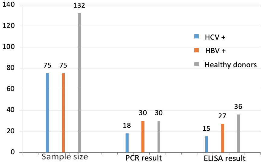
TTV result comparison of ELISA and PCR.
References
- Alfaresi, M.S., Elnazer, A.M., Alzaabi, A.S., Elkoush, A.A. and Islam, A.A., 2006. Saudi med. J., 27: 58-62.
- Ali, S., van Pelt, J.F.,Verslype, C., Nevens, F., Fevery, J. and Yap, S.H., 2004. Acta Gastroenterol. Belg., 67: 161-165.
- Al-Mozaini, M.A., Al-Ahdal, M.N., Kessie, G., Dela-Cruz, D.M., Rezeig, M.A. and Al-Shammary, F.J., 2006. Annal. Saudi Med., 26: 444–449. https://doi.org/10.5144/0256-4947.2006.444[CrossRef]
- Biagini, P., Gallian, P., Cantaloube, J.F., Attoui, H., deMicco, P. and de Lamballerie, X., 2006. J. med. Virol.,78: 298-304. https://doi.org/10.1002/jmv.20539[CrossRef]
- Blood donation screening of transfusion transmissible infections, 2013. Government of Pakistan. Available at http://sbtp.gov.pk/operational-documents-repository/country-strategy-for-ttis-screening-july-2013/ Last visited: 24-8-2017
- Bostan, N., Amen, N. and Bokhari, H., 2013. J. Vacc. Vaccinat., https://doi.org/10.4172/2157-7560.S1-004[CrossRef]
- Chattopadhyay, S., Das, B.C., Gupta, R.K. and Kar, P., 2005. Indian J. med, Res., 122: 29–33.
- de Oliveira, K.B., 2015. Rev. Bras. Hematol. Hemoter., 37: 357-358. https://doi.org/10.1016/j.bjhh.2015.07.009[CrossRef]
- Desai, M.M., Pal, R.B. and Banker, D.D., 2005. J. clin. Gastroenterol., 139: 422–429. https://doi.org/10.1097/01.mcg.0000159219.93160.bc[CrossRef]
- El-Taher, S.M., Fouad, N.A., Fouad, M.A., Mahedy, A.W. and Elnazi, A.K., 2015. Saudi J. Kidney Dis. Transpl., 26: 1215-1222. https://doi.org/10.4103/1319-2442.168643[CrossRef]
- Ishimura, M., Ohga, S., Ichiyama, M., Kusuhara, K., Takada, H., Hara, T., Takahashi, M. and Okamoto, H., 2010. Eur. J. Pediatr., 169: 899-902. https://doi.org/10.1007/s00431-009-1116-8[CrossRef]
- Karimi, G., Gharehbaghian, A., Tafti, M.F. and Vafaiyan, V., 2013. Transfus. Med. Hemother., 40: 210–217. https://doi.org/10.1159/000351540[CrossRef]
- Magua, SK., Kalghatgi, AT. and Bhagat, MR., 2015. Med. J. Armed Forces India,71: 340-344. https://doi.org/10.1016/j.mjafi.2015.06.023[CrossRef]
- Mankotia, D.S. and Irshad, M., 2014. Virol. J., 11: 96. https://doi.org/10.1186/1743-422X-11-96[CrossRef]
- Manzin, A., Mallus, F., Macera, L., Maggi, F. and Blois, S., 2015. J. Infect. Devel. Ctries., 9: 562-570. https://doi.org/10.3855/jidc.6912[CrossRef]
- Niel, C., de Oliveira, J.M., Ross, R.S., Gomes, S.A., Roggendorf, M. and Viazov, S., 1999. J. med. Virol., 57: 259-263. https://doi.org/10.1002/(SICI)1096-9071(199903)57:3<259::AID-JMV8>3.0.CO;2-2[CrossRef]
- Ninomiya, M., Takahashi, M., Nishizawa, T., Shimosegawa, T. and Okamoto, H., 2008. J. clin. Microbiol., 46: 507–514. https://doi.org/10.1128/JCM.01703-07[CrossRef]
- Nishizawa, T., Okamoto, H., Konishi, K., Yoshizawa, H., Miyakawa, Y. and Mayumi, M., 1997. Biochem. biophys. Res. Commun., 241:92–97. https://doi.org/10.1006/bbrc.1997.7765[CrossRef]
- Pinto, W.V., Assis, M.F. and Lemos, J.A., 2007. Cad. Saude Colet., 15: 349-356.
- Sambrook, J. and Russell, D.W., 2001. Molecular cloning. A laboratory manual. Cold Spring Harbor Laboratory Press, Cold Spring Harbor, NY.
- Shaheli, M., Yaghobi, R., Rezaeian, A., Saadi, M.I. and Ramzi, M., 2015. Jundishapur J. Microbiol., 8: e18212. https://doi.org/10.5812/jjm.8(4)2015.18212[CrossRef]
- Spandole, S., Cimponeriu, D., Berca, L.M. and Mihaescu, G., 2015. Arch. Virol., 160: 893-908. https://doi.org/10.1007/s00705-015-2363-9[CrossRef]
- Tokita, H., Murai, S., Kamitsukasa, H., Yagura, M., Harada, H., Takahashi, M. and Okamoto, H., 2002. J. med. Virol., 67: 501–509. https://doi.org/10.1002/jmv.10129[CrossRef]
Services
Article usage statistics
Loading…