Special Issue: Emerging and Re-Emerging Animal Health Challenges in Low and Middle-Income Countries · Volume 13, Issue 1, 7 January 2026 · Open Access
First Molecular Detection and Prevalence of the Toxocara spp. in Pets and Stray Cats
·
Affiliations & Notes ⌄
1Department of Parasitology, Faculty Veterinary medicine, Al-Qasim Green University, Babylon 51013, Iraq. *Correspondence | Safaa M. Kareem, Department of Parasitology, Faculty Veterinary medicine, Al-Qasim Green University, Babylon 51013, Iraq
The present study identified intestinal nematode (Toxocara spp.) in pet and stray cats in Babylon city through microscopic examination with a rate of 24.6% (37/150).
In addition, a recent comparative investigation was conducted to examine the prevalence of intestinal parasites in fecal samples obtained from both domestic and stray cats residing in Baghdad city.
Cats are significant clinical reservoirs and carrier for zoonotic parasites.
In Iraq, Babylon has a high incidence of Toxocara spp. detection.
Selected from the article's own text.
Abstract
Toxocara spp. is a globally prevalent parasitic roundworm that infects cats and dogs. It is a member of the Ascarididae family, which includes one of the most common intestinal parasites. The aim of this study was to investigate the prevalence of Toxocara spp. in a total of 150 individuals from two major animals (75 pets and 75 stray cats). After conducting a comprehensive examination, the clinical sings indicators exhibited by these cats were reported. In order to examine Toxocara spp. eggs under a microscope, we collected litter from each cat. A small sample of faeces was also subjected to molecular characterization of an internal transcribed spacer 2 gene using nested polymerase chain reaction. Our investigations found that infestation rates were about 24.6% (37/150) by microscope method and according to the molecular method a total of 42/150 (28%) (24 in stray and 18 in pet cats) were found positive. Collectively, positive rate based on convention and nested PCR show an elevated prevalence of Toxocara spp. Therefore, it is crucial to develop effective methods for diagnosis, identifying and eliminating Toxocara spp. parasites in cats, while simultaneously prioritizing public education on animal and human health. Recognizing the interconnectedness of animal, environmental, and human health underscores the importance of deworming cats, promoting hygiene, and educating the public to mitigate the risks of this zoonotic condition.
Toxocariasis is a zoonotic disease caused by Toxocara canis and Toxocara cati, which belong to the family Toxocaridae and genus Toxocara. Adult worms live in the small intestine of dogs and cats. Eggs are shed into the environment through feces and become embryonic within 3-6 weeks and infect the host and paratenic host animals including dogs, cats, cattle, sheep, goats, rodents, birds, and man through ingestion of contaminated food . Lactating puppies and kittens appear to get vertically infected via milk during lactation , and tans-placental transmission was reported in dogs .
Human infections are more commonly reported in children, especially those of low age . Infectious larvae can be generated by two capacities to release proteins (MUC-120) that support it to pass through the intestinal wall and then enter the journey and expand many tissues, including the lungs and liver . The disease can be transmitted to humans by contaminated water and food may be transferred by zoonotic agents, with extensive minor linkages to animal secretions . Keeping cats as pets, touching and playing with these animals, by children, free entry of dogs and cats into farmland and public parks, and non-compliance with sanitation in eating non washed vegetables are among the most important risk factors associated with toxocariasis . Ingesting contaminated food and soil with embryonic eggs . The ingested eggs hatch, and the larvae migrate to other tissues and organs, where they remain encapsulated third larval stage . In humans, the migration of larvae produces ocular larval migrans (OLM) or visceral larval migrans (VLM) . Few studies have been carried out in the South and Middle cities of Iraq, and they showed the presence of Toxocara infection in dogs, cats, and humans.
Toxocara species are common ascaridoid nematodes of cats and dogs throughout the world. They are causative agents of toxocariasis, a zoonotic parasitic disease in human with worldwide distribution. The most widespread species of Toxocara in dogs and cats are Toxocara canis and Toxocara cati, respectively . Furthermore, their infection can be detected in cats using clinical elements, and microscopy. Clinical signs of Toxocara spp. infection may include vomiting, diarrhoea, abdominal pain, weight loss, and poor body condition . A molecular study could provide information about the genetic makeup of this parasite in household cats. This might involve amplifying specific genes from Toxocara spp. such as the second internal transcribed spacer (ITS-2) region of ribosomal DNA .
A microscopic study could provide information about the morphological characteristics of T. spp. in feline breeds (pets and stray). This can involve examining fecal samples from infected animal for the presence of T. spp. eggs . As previously reported, T. spp. infection is common in some Iraqi cats. Al-Obaidi, 2012 has found a 40% prevalence in Mosul. In Baghdad, the incidence was 12.9% . Another study reported a 31% in Al-Anbar (15 in Shirazi and 16 in Himalayan) . There is no molecular epidemiological study in household cats in Babylon province. Therefore, the objective of this research was molecular diagnosis of Toxocara spp. in pet and stray cats based on clinical, microscopic and molecular characterizations.
MATERIALS AND METHODS
Study area, sampling, and design
The study was conducted in Babylon city, in the middle province of Iraq. The procedure outlined below was used to collect 150 fecal samples freshly from pet and stray cats from several private veterinary clinics and the streets between October of 2024 to March of 2025 (75 fecal samples from the pet cats and 75 fecal samples from stray cats). There were three age group (from 1 month to 6 months, from 7 to 12 month and more than one year old) for both stray and pet cats. In the 75 samples of stray cats, there were 30 males and 45 females and from 75 samples of pet’s cats, there were 39 males and 36 females. About five grams of fecal samples were placed in sterile plastic container with the date, age, and sex written on it and the rest was frozen for molecular analysis. The samples were then sent to the Parasitology Lab at the Al-Qasim Green University, College of Veterinary Medicine Department of Parasitology for microscopic and molecular analysis.
Microscopic investigation
Microscopic examination of faeces is a widely used to diagnostic the presence of Toxocara spp. in the gastrointestinal tract of animals. The flotation and sedimentation methods are common diagnostic methods used to detect parasite eggs of Toxocara spp. and others parasite. In this method, a small amount of sample is mixed with 40 ml of a saturated a flotation solution of sugar (specific gravity: 1.27) to the sediment, mixed, and centrifuged for 10 min at 1500 rpm. The volume was increased to 45 ml by adding extra sugar solution and then centrifuged for 5 min at 800 rpm. Subsequently, the top 10 ml of supernatant was pipetted and transferred into a clean conical centrifuge tube. The 10 ml transferred supernatants were washed (centrifuged for 5 min at 800 rpm) once with 40 ml of distal water again. After decanting all supernatants, the sediment of each sample was stored at -20° C for further microscopic and molecular evaluation. In terms of microscopic assessment, the observation and identification of Toxocara spp. eggs were performed at magnifications of 100x and 400x based on morphological futures .
DNA extraction and nested PCR
All flotation and sedimented fluids were frozen and thawed three times using liquid nitrogen for three minutes and boiling water for 5 minutes, followed by overnight proteinase K digestion. Following manufacturer’s instructions, genomic DNA was extracted using stool Genomic DNA extraction kit. The primers were lyophilized and dissolved in free ddH2O to give a final concentration of 100 pmol/µl as stock solution and kept a stock at -20 to prepare 10 pmol/µl concentration as work primer suspended. A total of 10 µl of the stock solution in 90 µl of the free ddH2O water was prepared to reach a final volume 100 µl. In the primer ToxCox1, the forward primer 10 picomols/µl (1µl) (5` GATTTTACCTGCTTTTGGTATTATTAG -3`) and reverse primer 10 picomols/µl (1µl)
Table 1: Gene, primer, sequence and PCR product size used to amplify Toxocara spp.
Primer
Sequence
Primer sequence 5'- 3'
Tm (ᵒC)
GC%
Size of product (bp)
ToxCox1
F
GATTTTACCTGCTTTTGGTATTATTAG
6٢.١
55
٤٢٦bp
R
CCAAAGACAGCACCCAAACT
6٥.1
57
(5`-CCAAAGACAGCACCCAAACT-3`) were used to amplify a 426-bp fragment of the mitochondrial cox1 gene . The PCR were accomplished in a final volume of 25μl. The reaction mixture was prepared as follows: 12.5 μl of PCR Master Mix, 10 µl of each primer, 1.5 μl of DNA template, and 9 μl double distal water. The temperature profile was a single cycle of 94° C for 3 min as primary denaturation, followed by 40 cycles of 94° C for 2 min (denaturation), 62° C for 1 min (annealing), 72° C for 2 min (extension), and a final extension of 72° C for 7 min.
The PCR products were electrophoresed using 3 μl of RedSafe nucleic acid staining solution and pouring into the gel cast and left for about 30 minutes to be completely solidified. The gel-plate was transferred into the gel tank and filled with 1X TBE buffer to the point of covering the gel surface. The loading DNA samples (5µl) were mixed with (3µl) DNA loading buffer and loaded in agarose gel wells. The agarose gel electrophoresis was completed at 80V, 65Amp for 1 hour. The DNA was observed by viewed under UV trans illuminator.
Statistical analysis
Statistical analysis was performed using the Statistical Package for Social Science (SPSS) version 27 for window software and Microsoft Excel 2010. Differences between groups were assessed using the chisquare test. All of these statistical analyzes considered the P-value below 0.05 level .
Results
Detection of Toxocara spp. using a microscopic technique
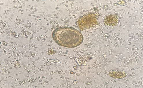
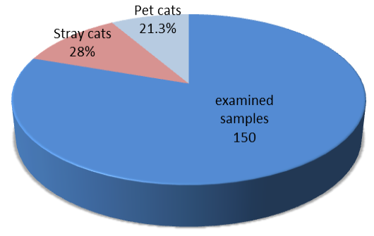
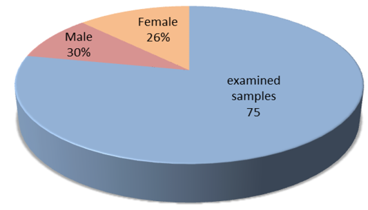
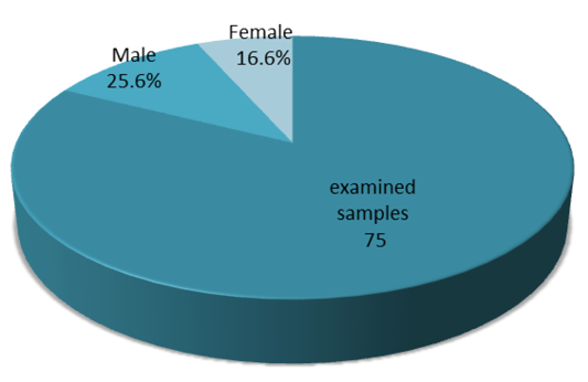
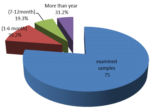
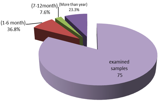
Under 10x and 40× magnification, eggs were dark brown in color, round to ovoid, contains a single-celled embryo and a thick pitted shell (“golf ball” appearance), round to ovoid, and with a thick alveolar capsule granulated content as shown in Figure 1. The total rate of infection in pet and stray cats was 24.6% (37/150) (Figure 2). Regarding the sex of the stray and pet cats, the results of the investigation showed that male have a higher infection rate than female (Figures 3 and 4). According to the present study, the infection rate of Toxocara spp was highest in age 1-6 month which was 39.2% (11/28) and lowest was 19.3% (6/31) in age 7-12 moth in stray cats (Figure 5). While in pet cats, it was 36.8% (7/19) and lowest was 7.6% (2/26) in age 7-12 month in pet cats (Figure 6).
Detection of Toxocara spp. using the PCR technique
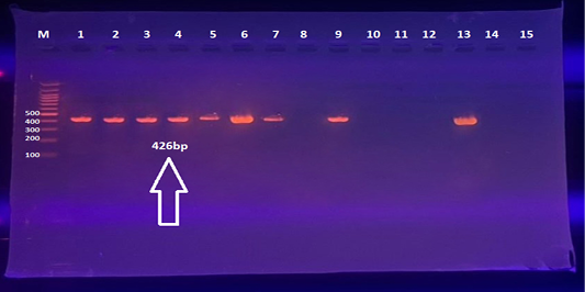
DNA extractions were performed for 150 cats’ faecal sample (75 pet cats and 75 stray cats), from microscopically positive sample (n=37) and negative (n=113) sample for purpose of confirmation. The result showed that PCR amplification of the ToxCox1 was positive for 42/150 sample. The PCR products were as expected, roughly 426 bp in length (Figure 7).
Prevalence of Toxocaras spp. according to breed of pets and stray cats
After confirming the amplification of ITS2 gene by conventional PCR, 28% (42/150) PCR-positive samples, highest infection rate of Toxocaras spp. in stray cats was 32% (24/75), while in pet cats 24% (18/75), with significant differences (P<0.05) as shown in Table 2.
Table 2: Prevalence of Toxocaras spp. according to breed of pets and stray cats.
Host
No. of examined samples
Positive samples
Percentage %
X2
P value
Stray cats
75
24
32%
0.06
0.4
Pet cats
75
18
٢4%
Total
150
42
28%
Prevalence of Toxocaras spp. according to sex of stray cats
According to sex of stray cats, the observation revealed that the highest infection rate of Toxocara spp. was 33.3% (10/30) in male, while in female the infection rate was 31.11% (14/45) which was significantly different (P≤0.05) (Table 3).
Table 3: Prevalence of Toxocaras spp. infection according to sex of stray cats.
Sex
No. of examined samples
Positive samples
Percentage %
X2
P value
Male
30
10
33.3%
0.08
0.02
Female
45
14
31.11%
Total
75
24
Prevalence of Toxocaras spp. according to sex of pet cats
According to sex of pet cats, the observation revealed that the highest infection rate of Toxocara spp. was 28.2% (11/39) in male, while in female the infection rate was 19.4% (7/36) which was significantly different (P≤0.05) (Table 4).
Table 4: Prevalence of Toxocaras spp. infection according to sex of pet cats.
Sex
No. of examined samples
Positive samples
Percentage %
X2
P value
Male
39
11
28.2%
0.48
0.4
Female
36
7
19.4%
Total
75
18
24%
Prevalence of Toxocaras spp. according to age of stray cats
The highest infection rate of Toxocara spp. in stray cats was 46.4% at age group 1-6 moth, while the lowest rate was 19.3% at age group 7-12 moths and a moderate rate of 31.25% (5/16) was observed in the age of more than year with significant differences (P≤0.05) (Table 5).
Table 5: Prevalence of Toxocaras spp. infection according to age of stray cats.
Age
No. of examined samples
Positive samples
Percentage %
X2
P value
(1-6 month)
28
13
46.4%
2.5
0.2
(7-12month)
31
6
19.3%
More than year
16
5
31.25%
Total
75
24
Prevalence of Toxocaras spp. according to age of pet cats
The highest infection rate of Toxocara spp. in pet cats was 36.8.% (7/19) in the age group 1-6 moth, while the lowest was 15.3% (4/26) in the age group of 7-12 moths and moderate rate of 23.3% (7/30) was noticed at the age of more than year with significant differences (P≤0.05) (Table 6).
Table 6: Prevalence of Toxocaras spp. infection according to age of pet cats.
Age
No. of examined samples
Positive samples
Percentage %
X2
P value
(1-6 month)
19
7
36.8%
1.6
0.4
(7-12month)
26
4
15.3%
More than year
30
7
23.3%
Total
75
16
DNA sequencing and phylogenetic tree analysis
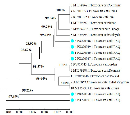
After confirming the amplification by PCR, sequencing was performed to verify the specificity of the PCR amplified bands and to conduct the phylogenetic analysis of the DNA samples. In this study, 8 PCR positive samples from local isolates products were sent to Macrogen Company for sequencing. Based on the analysis, eight PCR products had 99% similarity to the sequences of the COX1 gene of the mitochondrial DNA of T. cati and T. malaysiensis.
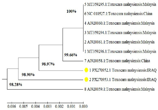
The present sequences were subjected to NCBI’s BLAST analysis. Eight of Toxocara spp. isolates (PX270946, PX270947, PX270948, PX270949, PX270950, PX270951, PX270952, PX270953) were compared with published sequences from various geographical sites to ensure a correct phylogenetic analysis of local T. cati isolates Table 7 and T. malaysiensis isolates Table 8. Finally, the phylogenetic tree was constructed to include T. cati and T. malaysiensis isolates from Iraq with other isolates obtained from other countries based on the above gene sequence (Figures 8 and 9).
Table 7: Comparison of isolate against corresponding Toxocara cati species registered in the NCBI.
The present study identified intestinal nematode (Toxocara spp.) in pet and stray cats in Babylon city through microscopic examination with a rate of 24.6% (37/150). Our results agreed with total infection rate of Toxocara cati in Baghdad region of Iraq. The total infection rate in domestic and stray cats was 23% (19%) in domestic and 27% in stray cats, with a non-significant difference between microscopic method and PCR . Likewise, present study’s results also agreed with a study investigated in the Al-Anbar region of Iraq where they found the infestation rates of 31% according to the microscopy . Rashid et al. (2022) have found in Sulmanya province a higher infection rate. In Babylon, Liravizadeh et al. (2024) have documented 34.75% rate of infection with T. cati in urban and Fahrion et al. (2011) reported 26% rate of T. cati in fecal specimens from housed and stray cats in Al-Diwaniya city. As well as Hajipour (2019) have reported 20% of infection rate in Al-mahaweel reserve/Babylon. Our results disagreed with a study investigated the coprological detection of Toxocariasis in the areas of the Kurdistan region of Iraq. The findings revealed that the prevalence of this parasite among stray cats was 47.62%, which is about four times higher than the infection rate observed in pet cats, standing at 5.5% . In addition, a recent comparative investigation was conducted to examine the prevalence of intestinal parasites in fecal samples obtained from both domestic and stray cats residing in Baghdad city. The study revealed a relatively low infection rate with 1.65% . The overall prevalence rate of Toxocara spp. according to age groups was lower than a study conducted in Iran, which registered a 30% rate in cats over two years of age . The molecular study of cats examined revealed the presence of Toxocara spp. DNA, and the overall infection rate was 28% (42/150), which is relatively compared with the prevalence with another study in Khorramabad city in western Iran, where the infection rate in stray cats was higher than pet cats 20% (19/95) . This is relatively higher compared to another study conducted in Switzerland , where an infection rate of 31.5% was recorded. In Isfahan city, an infection rate of 17.7% was recorded . The results of current study agreed with result of Al-Bayati et al. (2023) who have also recorded the infection rate of 39.58% with Toxocara spp. in the northern of Iraq (Kirkuk) province.
It likely that geographical variables and differences in detection methods may be account for the observed variations in T. cati prevalence among these studies. Based on the findings of De Santis et al (2006), it has been observed that the occurrence of Toxocara spp. infection in feline is higher among stray cats that do not receive veterinary attention in comparison to cats that are owned by individuals. Furthermore, the previous data demonstrated a wide spectrum of prevalence rates, ranging from 5.45% to 67.5% in feral and free-ranging feline, as well as from 1.6% to 30.4% in domesticated cats .
The results are consistent with another study, which recorded an infection rate of 25% with T. cati in Brazil . The current study’s total infection rate was higher than what was previously documented in Mexico, which was recorded at 42.5% by flotation technique, and it was reported that 62.5% of Turkish have T. cati. (Martı-nez-Barbabosa et al., 2003). In Estonia, 48.2% of cats were infected with T. cati; this rate was higher than the studies conducted in the neighboring country, Iran, where 42.6% was documented ; . In Russia, 16.7% of cats were infected with T. cati, whereas other studies found that 52% of cats were infected (Dantas-Torres, 2020; .
Conclusions
Cats are significant clinical reservoirs and carrier for zoonotic parasites. In Iraq, Babylon has a high incidence of Toxocara spp. detection. Compared to conventional methods, PCR is thought to be a more sensitive, and accurate diagnostic procedure that confirms species identity.
Acknowledgments
The authors would like to thank the Al-Qasim Green University, College of Veterinary Medicine, Department of Parasitology, as well as the field technicians who have helped with the study.
Novelty Statement
This study represents the first molecular detection and prevalence analysis of Toxocara spp. in pet and stray cats in Babylon City, Iraq, providing baseline data for further epidemiological studies.
Author’s Contribution
ZAH: Study design, sample collection, laboratory analysis, and manuscript writing. SMK: Data analysis, supervision, and manuscript revision.
Generative AI and AI-assisted technology statement
Artificial intelligence was not used in this research.
Conflict of interest
The authors have declared no conflict of interest.
Figures
Figure 1:.
Eggs of Toxocara spp. “golf ball” appearance isolated from infected cats by floatation method under a microscope at a magnification of 40X.
Figure 2:.
Prevalence of infection rate of Toxocara spp. According to breed of pet and stray cats.
Figure 3:.
Prevalence of Toxocaras spp. infection according to sex of stray cats.
Figure 4:.
Prevalence of Toxocaras spp. infection according to sex of pet cats.
Figure 5:.
Prevalence of Toxocaras spp. infection according to age of stray cats.
Figure 6:.
Prevalence of Toxocaras spp. infection according to age of pet cats.
Figure 7:.
Agarose gel electrophoresis for gene 426bp. Bands were fractionated by electrophoresis on a 1.5% agarose gel, and visualized under UV. Light after staining with red stain. Lane: M (M: 100bp ladder).
Figure 8:.
Dendrogram obtained for T. cati isolates (genotypes) based on the Cox1 gene of the mitochondrial DNA by the maximum likelihood analysis at showing the phylogenetic tree of Iraq isolates with related global strains.
Figure 9:.
Dendrogram obtained for T. malaysiensis isolates (genotypes) based on the Cox1 gene of the mitochondrial DNA by the maximum likelihood analysis at showing the phylogenetic tree of Iraq isolates with related Global strains.
References
Abbas ZTA (2023). Investigation of ecto-endo parasites in some animals in Al-Mahaweel Reserve/Babylon.: Investigation of ecto-endo parasites in some animals in Al-Mahaweel Reserve/Babylon. Wasit J. Pure Sci., 2: 178-191. https://doi.org/10.31185/wjps.156[CrossRef]
Alshawi A, Alhayani O (2024). Molecular detection and prevalence of the Toxocara cati parasite in household cats breeds in the Al-Anbar Province-Iraq. Egypt. J. Vet. Sci., 55(6): 1627-1636. https://doi.org/10.21608/ejvs.2024.263951.1791[CrossRef]
Alani ZK, Kawan MH (2024). Prevalence and molecular analysis of Toxocara cati in Baghdad Province. J. Adv. Vet. Anim. Res., 11(2): 392-397. https://doi.org/10.5455/javar.2024.k788[CrossRef]
Al-Asady SIJ, Al-Hasnawy MH (2024). Investigation of Cryptosporidium species infecting dogs. J. Anim. Health Prod., 12(s1): 174-181. https://doi.org/10.17582/journal.jahp/2024/12.s1.174.181[Full text][CrossRef]
Al-Bayati AM, Jihad LJ, Al-Attar SAA (2023). The effects of Gastro-intestinal Parasites on haemato-biochemical parameters of sheep in Kirkuk province, Iraq. J. Appl. Vet. Sci., 8(4): 62-68.
Al-Obaidi QT (2012). Prevalence of internal Helminthes in stray cats (Felis catus) in Mosul city, Mosul, Iraqi. J. Anim. Vet. Adv., 11(15): 2732–2736. https://doi.org/10.3923/javaa.2012.2732.2736[CrossRef]
Al-Rammahi HM, Kareem SM, Hammadi AK (2014). Prevalence of intestinal helminthes in feral cats in Babylon province/Iraq, urban and rural locations. Mir. Res. Vet. Sci. Anim., 3(2): 44-52.
Al-Taie D, Ahmed A, Al-Khayat F (2022). A Comparative study of some intestinal parasitesin faecal samples of domestic and stray cats in Baghdad, Iraq. Compar. Parasitol., 89(1): 31-35. https://doi.org/10.1654/21-00015[CrossRef]
Azimian H, Shokrani H, Fallahi S (2021). Molecular evaluation of Toxocara species in stray cats using loop-mediated isothermal amplification (lamp) technique as a rapid, sensitive and simple screening assay. Vet. Med. Sci., 7(3): 647. https://doi.org/10.1002/vms3.431[CrossRef]
Conway DP, Mckenzie ME (2007). Poultry coccidiosis: Diagnostic and testing procedures. 3rd ed. Blackwell Publishing, Ames, Iowa: pp. 164. https://doi.org/10.1002/9780470344620[CrossRef]
Dantas-Torres F (2002). Toxocara prevalence in dogs and cats in Brazil. Adv. Parasitol., 109: 715–741.
De Santis AC, Raghavan M, Caldanaro RJ, Glickman NW, Moore GE, Lewis HB, Schantz PM, Glickman LT (2006). Estimated prevalence of nematode parasitism among pet cats in the United States. J. Am. Vet. Med. Assoc., 228(6): 885–892. https://doi.org/10.2460/javma.228.6.885[CrossRef]
Deplazes, P., van Knapen, F., Schweiger, A., Overgaauw, P. A. (2011). Role of pet dogs and cats in the transmission of helminthic zoonoses in Europe, with a focus on echinococcosis and toxocarosis. Veterinary parasitology, 182(1), 41-53.
Despommier D (2003). Toxocariasis: Clinical aspects, epidemiology, medical ecology, and molecular aspects. Clin. Microbiol. Rev., 16: 265-272. https://doi.org/10.1128/CMR.16.2.265-272.2003[CrossRef]
Fahrion AS, Schnyder M, Wichert B, Deplazes P (2011). Toxocara eggs shed by dogs and cats and their molecular and morphometric species-specific identification: Is the finding of T. cati eggs shed by dogs of epidemiological relevance. Vet. Parasitol., 177(1– 2): 186–189. https://doi.org/10.1016/j.vetpar.2010.11.028[CrossRef]
Fahrion AS, Schnyder M, Wichert B, Deplazes P (2011). Toxocara eggs shed by dogs and cats and their molecular and morphometric speciesspecific identification: Is the finding of T. cati eggs shed by dogs of epidemiological relevance. Vet. Parasitol., 177(1-2): 186-189. https://doi.org/10.1016/j.vetpar.2010.11.028[CrossRef]
Hajipour N (2019). A survey on the prevalence of Toxocara cati, Toxocara canis and Toxascaris leonina eggs in stray dogs and cats faeces in Northwest of Iran: A potential risk for human health. Trop. Biomed., 36(1):143
Labarthe N, Serrão ML, Ferreira AMR, Almeida NK, Guerrero J (2004). A survey of gastrointestinal helminths in cats of the metropolitan region of Rio de Janeiro. Braz. Vet. Parasitol., 123(1–2): 133–139. https://doi.org/10.1016/j.vetpar.2004.06.002[CrossRef]
Liravizadeh S, Khabisi AS, Khorashad SA, Mirahmadi H (2024). Molecular determination of Toxocara spp. eggs isolated from Public Parks and Playgrounds in Zahedan, Southeast Iran. J. Parasitol. Res., 2024: 2132696. https://doi.org/10.1155/2024/2132696[CrossRef]
Lucio-Forster A, Barbecho MJS, Mohammed HO, Kornreich BG, Bowman D (2016). D comparison of the prevalence of Toxocara egg shedding by pet cats and dogs in the U.S.A. Vet. Parasitol., 5(2): 1–13. https://doi.org/10.1016/j.vprsr.2016.08.002[CrossRef]
Lukashev AN, Ruzina MN, Akhmadishina LV (2020). Toxocara prevalence in dogs, cats and the environment in Russia. Adv Parasitol., 109: 801–817. https://doi.org/10.1016/bs.apar.2020.01.019[CrossRef]
Kalef, D.A., Al-Khayat, F.A.A.M. (2022). A comparative study of some intestinal parasites in fecal samples of domestic and stray cats in Baghdad, Iraq. Comparative Parasitology, 89(1), 30-34.
Mai K, Sharman PA, Walker RA, Katrib M, Souza D, McConville MJ (2009). Oocyst wall formation and composition in coccidian parasites. Mem. Inst. Oswaldo Cruz., 104(2): 281-289. https://doi.org/10.1590/S0074-02762009000200022[CrossRef]
Martínez-Barbabosa I, Tsuji OV, Cabello RR, Cárdenas EMG, Chasin OA (2003). The prevalence of Toxocara cati in domestic cats in Mexico city. Vet. Parasitol., 114(1): 43–49. https://doi.org/10.1016/S0304-4017(03)00038-4[CrossRef]
Monteiro MFM, Ramos RAN, Calado AMC, Lima VFS, Ramos ICN, Tenório RFL, Alves LC (2016). Gastrointestinal parasites of cats in Brazil: Frequency and zoonotic risk. Rev. Brasil. Parasitol. Vet., 25(2): 254–257. https://doi.org/10.1590/S1984-29612016019[CrossRef]
Peter D, Frans K, Alexander S, Paul AM (2011). Role of pet dogs and cats in the transmission of helminthic zoonoses in Europe, with a focus on echinococcosis and toxocarosis. Vet. Parasitol., 182: 41-53. https://doi.org/10.1016/j.vetpar.2011.07.014[CrossRef]
Rashid ZM, Aziz SA, Ali OJ, Kakarash NK, Marif HFF (2022). Coprological detection of Toxocariasis in domicile and stray dogs and cats in Sulaimani province, Iraq. Iraqi J. Vet. Sci., 36(4): 1047-1051. https://doi.org/10.33899/ijvs.2022.132976.2157[CrossRef]
Remesar S, García-Dios D, Calabuig N, Prieto A, Díaz-Cao JM, López-Lorenzo G, López C, Fernández G, Morrondo P, Panadero R, Díaz P (2022). Cardiorespiratory nematodes and co-infections with gastrointestinal parasites in new arrivals at dog and cat shelters in north-western Spain. Transbound. Emerg. Dis., 69(5): 3141-3153. https://doi.org/10.1111/tbed.14670[CrossRef]
Strube C, Heuer L, Janecek E (2013). Toxocara spp infections in paratenic hosts. Vet. Parasitol., 193: 375-389. https://doi.org/10.1016/j.vetpar.2012.12.033[CrossRef]
Talvik H, Moks E, Mägi E, Järvis T, Miller I (2006). Distribution of Toxocara infection in the environment and in definitive and paratenic hosts in Estonia. Acta Vet. Hung., 54(3): 399–406. https://doi.org/10.1556/avet.54.2006.3.10[CrossRef]
Torkan S, Ghandehari-Alavijeh MR, Khamesipour F (2017). Survey of the prevalence of Toxocara cati in stray cats in Isfahan city, Iran by PCR method. Trop. Biomed., 34: 550-555.
Woodruff AW, Salih SY, Savigny D, Baya EI, Shah AI (1981). Toxocariasis in the Sudan. Ann. Trop. Med. J. Parasitol., 75: 559-561. https://doi.org/10.1080/00034983.1981.11687481[CrossRef]
Yaman M, Ayaz E, Gül A, Muz MN (2006). Investigation of helminth infections of cats and dogs in the Hatay province. Turk. Parazitol. Derg., 30(3): 200–204.
Yen NTH, Van Phuong N, Hieu DD, Anh NTL (2023). Identification of three Toxocara species, T. canis in dogs, and T. cati and T. malaysiensis in cats, in Vietnam by PCR-RFLP analysis and DNA sequencing. Vietnam J. Agric. Sci., 6(4): 1917-1923. https://doi.org/10.31817/vjas.2023.6.4.02[CrossRef]
Zhao ZY, Li MH, Lyu C, Meng XZ, Qin YF, Yang XB, Ma N, Zhao Q, Zhang Y, Jiang J (2022). Prevalence of Giardia duodenalis among dogs in China from 2001 to 2021: A systematic review and meta-analysis. Foodb. Pathog. Dis., 19: 179-191. https://doi.org/10.1089/fpd.2021.0073[CrossRef]
Zibaei M, Sadjjadi SM (2017). Trend of toxocariasis in Iran: A review on human and animal dimensions. Iran J. Vet. Res., 18(4): 233.