Fascioliasis caused by the Fasciola hepatica and Fasciola gigantica which is a plant borne trematodes that are hazardous to health and reduce animal productivity and damage economic activities. Consumer rejection of the liver results in a loss for the economy. In addition, an acute infection with Fasciola results in weight loss, stunted growth, and infertility. There was a decrease in productivity while the disease was in its chronic phase Mehmood et al. (2017).
The result of this study indicates that the infection rate of Fascioliasis was (17.8%) among the examined slaughtered animals. The reason for this infection could be due to increase in irrigated land masses in the research area and the propensity of farmers and animal rearers to feed their livestock in marshy and damp areas behind the grain because of a lack of feed. Since the intermediate host favours marshy areas with slow-moving water and small streams, which also provide enough moisture for the infective metacercaria to survive Magaji et al. (2014).
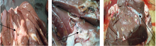
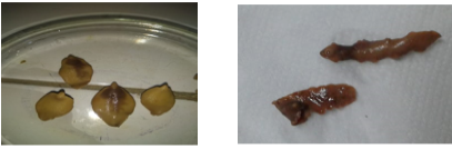
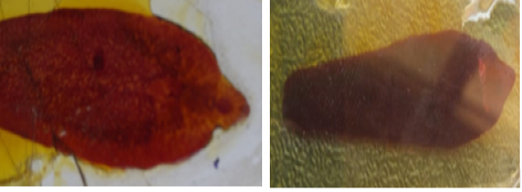
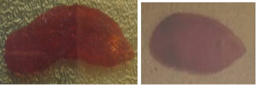
The higher infection was found in buffaloes 28% followed by sheep 26.1%, cattle 23.5%, goats 19.2% and camels 0%. The difference of the incidence among slaughtered animals might be attributed to the grazing style of animals but this difference was not significant with P value >0.05 (Table-1). F. hepatica was the predominant species 27(52.9%) (12 in sheep, 4 in goats, 9 in cattle and 2 in buffaloes), Fasciola gigantica was 18(35.3%) (8 in sheep, 1 in goats, 4 in cattle and 5 in buffaloes), mixed infection was 3(5.9%) in cattle and larval stage was 3 (5.9%) in sheep morphologically (Table-2), our result was lower than that obtained by Usman, (2019) in Nigeria (40.5%) in which higher rate was observed in cattle than sheep and goats, and Ieren et al. (2016) in Nigeria (48%) in which cattle more infected than sheep and goats (males more than females and active rain season than early dry season) and Zemene and Atnaf, (2015) in Ethiopia (21.8%) in which cattle more infected than sheep and goats and in adult animals than young ones, higher infection observed in poor body condition followed by medium body condition. The wide variation in prevalence between various nations could be primarily attributed to variations in climatic conditions, such as rainfall and temperature Fox et al. (2011) as well as other significant factors like grazing in marshy pastures, having access to streams or ponds, having a large herd, and the type of soil Howell et al. (2015). Additionally, human activity (large urbanization, the growth of irrigated regions, and the transformation of natural wetlands into agricultural lands) is linked to the spread of water and food-borne illness Sabourin et al. (2018).
The present study revealed that the occurrence of Fascioliasis in sheep was 23/88 (26.13%), this result was lower than that recorded by Usman, (2019) in Nigeria which was 39.1%, and female sheep recorded higher infection (44.6%) than males (30.7%) in contrast to our result that higher in males (28.57%) than females (20%). Our result was higher than that recorded by Zemene and Atnaf, (2015) in Ethiopia which was 20.14%, and adult sheep more infected than young ones. Also, was higher than that recorded by Jean-Richar et al. (2014) 23% and Ouchene-Khelif et al. (2018) in Algeria 6.5%, the seasonal infection of Fasciola hepatica was highest in winter for sheep.
The infection rate of Fascioliasis was 5/26(19.2%) in examined goats and was higher between older (2-3, >3years) than young goats, in winter than other seasons, among males than females and in freely reared goats (outdoor) than housed animals (indoor). Our result was higher than that recorded by Hassan et al. (2019) in Giza Governorate 0.89%, IrfanUllah et al. (2016) in Pakistan 6.6%, Çelik and ÇELİK, (2018) in Turkey 14.14%, Abah et al. (2019) in Nigeria 6.2%, Abdelazeem et al. (2020) in Egypt 3.5%, Our result also was higher than that recorded by Ouchene-Khelif et al. (2018) in Algeria 2.5%, the seasonal prevalence of Fasciola hepatica was highest in summer and winter for goat. While our result was lower than that recorded by Usman, (2019) 35% but in those study the infection was higher in females than males. According to age, sex, season, and rearing system, our results agreed with Abdelazeem et al. (2020) in Assiut and Sohag Governorates, Upper Egypt who said that the highest infection found between older than young goats, in winter than other seasons, among males than females and in freely reared goats than housed animals, but disagreed with Abah et al. (2019) who revealed that female goats more infected than male goats.
The infection rate of Fascioliasis in cattle was 16/68 (23.5%) and was higher in female 5/18 (27.7%) than male 11/50 (22%), more at age > 4year 5/17 (29.4%) than in age 2–4year 9/36 (25%) and 1–2year 2/15 (13.33%), in rainy season than dry ones, in winter 7/25 (28%), 4/18 (22.2%) in autumn, 2/9 (22.2%) in summer and 3/16 (18.75%) in spring. In free rang system (outdoor) 13/53 (24.5%) than housed system (indoor) 3/15 (20%), infected animals had pasty feces, pale mucus membrane, rough coat, weakness and 5 infected females not pregnant. Infection more in Shebeen 3/12 (25%) than Benha 9/37 (24.7%) and Toukh 4/19 (21.05%). Infection rate was higher in cattle that slaughtered in shops in village 14/29 (48.3%) than that slaughtered in abattoirs 2/39 (5.1%). Our result was higher than that recorded by Ejeh et al. (2015) in Nigeria 14.6%, Henok and Mekonen, (2011) in Ethiopia 14.6% in which adult recorded higher infection, while was lower than that recorded by Jean-Richar et al. (2014) in the Lake Chad area of Chad 68% in cattle that grazed at lake water which was the source of infection and all infected with F. gigantica, Usman, (2019) in Nigeria 45.7% in which males more infected than females and aged cattle above five years recorded higher infection, Zemene and Atnaf, (2015) in Ethiopia 30.6% in which adult cattle recorded higher infection, Yemisrach and Mekonnen, (2012) in Ethiopia 28.6% but young cattle recorded higher infection than adult ones and Ouchene-Khelif et al. (2018) in Algeria 26.7%, the seasonal infection of Fasciola hepatica was highest in summer and winter for cattle.
The infection rate of Fascioliasis in buffaloes was 7/25 (28%) and was higher in male 6/15 (40%) than female 1/10 (10%), more at age 2–4-year 6/18 (33.3%) than > 4 year 1/7 (14.3%), more in spring 2/5 (40%) than winter 3/10 (30%) and Autumn 2/8 (25%). All infected buffaloes were in free rang system (outdoor) 7/11 (63.6%), infected buffaloes had pasty feces, and some had weakness. Infection more in Benha 5/13 (38.5%) than Toukh 2/6 (33.3%). All infected buffaloes slaughtered in shops in villages. Our result was higher than that recorded by Adane et al. (2019) 20.24% in which the higher infection was recorded in female than male and in old animals than young, Mohammed et al. (2016) 24.4% in which the highest infection found in young than adult animals. Our result in large ruminant (cattle and buffaloes) was 23/93 (24.7%) these was higher than that recorded by Fazly et al. (2015) 6/80 (7.50%) in cattle and buffalo samples and F. gigantica was only identified in the samples.
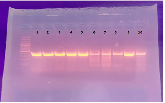
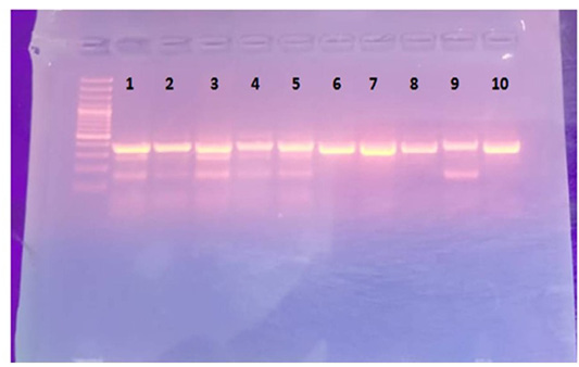
The current study revealed the Fasciola species that infect sheep, goats, cattle, and buffaloes from Qualyobia, Egypt was Fasciola hybrid form (Figures 2 and 3). This agreed with Mosaab et al. (2021) who found Fasciola hybrid form in sheep and buffaloes in Aswan, Egypt, but disagreed with the results of Amer et al. (2011) who found that F. hepatica was predominant in sheep compared to other hosts, also disagreed with Salihu et al. (2022) who siad that the genotype of Fasciola infecting cattle and other ruminant species in Nigeria was F. gigantica. Moreover, our result disagreed with Abdelazeem et al. (2020) who recorded that the Fasciola species that affected cattle and goats was Fasciola hepatica and gigantica based on the sequencing of (ITS-2) of nuclear ribosomal DNA (rDNA). While our result in camels was 0% and this agreed with Ouchene-Khelifi et al. (2018) in Algeria who not found Fascioliasis in camels, and this may be attributed to those camels not grazed as in small and large ruminant.
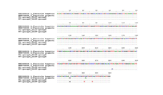
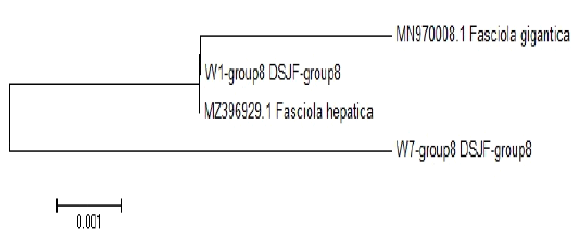
Molecular characterization of the Fasciola species were detected based on partial sequences of ITS2 rDNA and the study revealed that the Fasciola spp. sequences from diverse hosts are almost matching to those of earlier available sequences. It has been shown that Fasciola hybrid form is existing in Qualyobia, Egypt, the hybrid form was recorded for the first time at south valley university, Egypt. These outcomes meet the full agreement with those found by Amer et al. (2011) and Mosaab et al. (2021) who recorded the presence of the three Fasciola species in Egypt including the hybrid form, while the present result differed from that recorded by Arafa et al. (2018) Egypt, Cairo who reported pure F. gigantica (isolate from cow and buffalo). However, El-Tahawy et al. (2018) from Nile Delta, Egypt recorded that the Fasciola in Egypt concerning the application of PCR for differentiation of two species of Fasciola by using certain primers of F. hepatica, they recorded that eight samples were found to have positive bands associated to F. hepatica and 2 negative bands representing F. gigantica. Alignment revealed that our result in W7 isolate (GenBank accession OP245094) was typical identical to found in Egypt (MT423006), (MW600275), (AJ853848), Kenya (MN970008), Nigeria (MN608173), Turkey (KY613944), Iran (KF866247), Germany (KF425321), South Africa (MW793533), Belgium (MW046876), Switzerland (MK321643) and Japan (AB010976) while W1 isolate (GenBank accession OP245092) was typical identical to found in Egypt (MT423007), Kenya (MZ396929), Libya (MT025519), Iran (JF432071), India (MZ413927), Viet Nam (MT429176), Saudi Arabia (MN559388), Spain (MG569979) and China (MH385388).
Alignment analysis confirmed our results and revealed similarity between one isolate (W1) with Fasciola hepatica isolate on GenBank when used primer of F. hepatica and W7 revealed similarity with some nucleotide differences (SNP, INDEL) with F. gigantica on GenBank when used primer of F. gigantica and may be due to the presence of hybrids in our isolates, and this outcome may be due to that our isolate is relatively recently introduced to Qualyobia Province as this the first time reported the Fasciola hybrid.