
Chikungunya virus is Aedes mosquitoe transmitted virus that has typical signs and symptoms of high fever, headache, hemorrhage and body rashes. Although in the past decade millions of people in several continents like Asia, Africa and some islands of Indian Ocean have faced the major outbreaks of Chikungunya virus because of its travel associated febrile nature. An inexplicable paralyzing disease has trapped thousands of people in Karachi region of Pakistan and the symptomatology among suspected cases was compatible with Chikungunya fever. Thus Chikungunya outbreak was reported in Pakistan but actually the recent reports about the occurrence of Chikungunya outbreak in the southern region of Pakistan has been highly controversial. In order to further confirm the circulation of the virus and put an end to the speculative claims for the corresponding outbreak in Pakistan, a study was conducted at the Center of Excellence in Molecular Biology in Lahore. The study included 500 suspected serum samples. All clinical specimens were tested for IgM and IgG specific antibodies against Chikungunya virus using a commercial ELISA kit. Twenty seven (5.4%) and thirty five (7%) samples were IgM and IgG positive, respectively. All antibody positive samples and a subset of negative samples (195) were further subjected to PCR for confirmatory purposes. Two different sets of primers were used for the identification of the viral genome. Thirty samples were found PCR positive and a subset of ten samples was direct sequenced. Sequence analysis showed 94% similarity to Stenotrophomonas maltophilia which is considered to be the second most prevalent bacterial species in mosquitoe’s midgut. This observation may lead towards the confusion that the outbreak is either of Dengue or Chikunguna virus. In conclusion, confirmatory molecular characterization of the viral genome remains controversial and further studies are needed in this respect.
Short Communication · Volume 52, Issue 4, 3 May 2020 · Open Access
Prevalence and Upcoming Threats of Chikungunya Virus in Pakistani Population
· · · ·
Affiliations & Notes ⌄
- 1Division of Molecular Virology, National Centre of Excellence in Molecular Biology, 87-West Canal Bank Road Thokar Niaz Baig, Lahore-53700, University of the Punjab, Lahore, Pakistan Samia Afzal and Sadia Zahid authors contributed equally. , * Corresponding author: samiaraza@live.com
Article Info ⌄
- Journal:
- Pakistan Journal of Zoology
- Published:
- 3 May 2020

From the article
- Results indicated that 27(5.4%) out of 500 and 35(7%) out of 500 samples were IgM and IgG positive, respectively.
- In conclusion, our results showed that serological evidence of acute Chikungunya cases supports the occurrence of clusters of Chikungunya fever in the region, but confirmatory molecular characterization of the viral genome remains controversial.
- Our findings emphasize on the requirement of affordable and susceptible diagnostic tests for early and accurate diagnosis of Chikungunya virus and in this regard, further vigilant studies are required.
Selected from the article's own text.
Abstract
Materials and methods
The study included 500 suspected serum samples collected by the Institute of Public Health (IPH) and Genome centre for Molecular based research and Diagnostic (GCMD). All clinical specimens were tested for IgM and IgG specific antibodies against Chikungunya virus using a commercial ELISA kit (Euroimmun, Luebeck, Germany). All antibody positive samples were further subjected to RT-PCR for confirmatory purposes.
Viral RNA was extracted from serum using Viral Nucleic Acid extraction Kit (Favorgen Biotech Corporation, Australia). cDNA was synthesized from RNA of Chikungunya virus in 30 cycles using 10 μl of RNA with a reaction mixture of 10 μl containing 4 μl 5X first strand buffer (FSB), 0.5 μl 0.1 M Dithiothreitol (DTT), 2 μl 10 mM dNTPs, 1 μl 20 pM antisense primers, 1.3 μl dH2O, 0.2 μl RNA inhibitor (RMI) and 1 μl of Moloney Murine Leukiemia Virus (MMLV) Reverse Transcriptase (RTse) (Invitrogen Biotechnology, USA). The 20 μl reaction mixture was incubated at 37oC for 50 min. followed by 2 min. heat inactivation of M-MLV at 95oC. The samples were then incubated for 2 min. at 22oC.
For PCR amplification, two different sets of primers were used for the identification of the viral genome by using already reported primer sequences for Chikungunya detection. First primer set is of E2 region with product size of (1st round 427bp, 2nd round 172bp) and second primer set is of NSP1 region with product size 239bp as shown in Table I.
Complementary DNA (cDNA) was used as template for amplification, 2μl of cDNA with 8 μl of master mix. PCR mix was made by mixing 1 μl 10X PCR buffer (with ammonium sulphate), 2.4μl MgCl2, 1 μl 500 μM dNTPs, 1 μl pM forward and reverse primer each, 2.4 μl d H2O and 0.2 μl of 5 U of Taq DNA polymerase (Invitrogen Biotechnology, USA). The thermal profile for first round using outer sense and antisense was initial denaturation at 94ºC for 2 min followed by 30 cycles of denaturation at 94ºC for 45 sec, annealing at 64ºC for 35 sec, extension at 72ºC for 2 min and final extension at 72ºC for 10 min. Same thermal profile was used for second round using type specific primers. Only the annealing was done at 56ºC for 45 sec. in 30 cycles. The PCR product was visualized by2% agarose gel, stained with ethidium bromide, visualized under UV light.
A subset of 10 samples was directed to sequencing of chikungunya virus isolates. Briefly, the PCR product was run on 1.2% agarose gel. The specific bands were excised and purified using the QIA quick Gel Extraction Kit (Qiagen, Germany). This gel purified amplicons were used for sequencing PCR reactions. Sequencing PCR products analysed on automated genetic analyzer according to the manufacturer’s instructions (Big Dye Deoxy Terminators; Applied Biosystems, Weiterstadt, Germany). The identity of the sequences was confirmed by Basic Local Alignment Search Tool (BLAST). Sequences that were unique (obtained for the first time) were submitted to GenBank Data Base. The BioEdit v7.0.5 software was used for the translation, the alignment of amino acid and protein sequences.
Results and discussion
Total 500 suspected enrolled samples were used for antibodies detection. Results indicated that 27(5.4%) out of 500 and 35(7%) out of 500 samples were IgM and IgG positive, respectively. These 62 IgM and IgG positive samples were further confirmed by RT-PCR analysis. Out of these 62 samples, 30 were found RT-PCR positive. Results were confirmed by two different sets of primers with product size 172bp for first primer set and 239bp for second primer set (Fig . 1). 10 randomly selected samples
Table I. Primer sets used for the detection of Chikengunya virus.
| Primer sets | Primer name | 5’-3’ sequence | Product size |
| 1 | CHIK-F1 | TAATGCTGAACTCGGGGACC | 427bp |
| CHIK-R1 | ACCTGCCACACCCACCATCGAC | ||
| CHIK-F2 | GATCAGGTTAACCGTGCCGACT | 172bp | |
| CHIK-R2 | CACTGACACAACTACCACAGTCA | ||
| 2 | ChikD-F1 | GCAGACGCAGAGAGGGCCAG | 239bp |
| ChikD-R1 | CGTGCTGCAAGGTAGTTCTC | ||
| ChikD-F1 | GCTATTTGTAAGAACGTCAG | ||
| ChikD-R2 | TACCGTGCTGCGGTCGGGAA |
were used for genomic sequence analysis of Chikungunya virus and the sequence analysis amazed that it showed 94% similarity to Stenotrophomonas maltophilia (Fig . 2) instead of Chikungunya virus.
Previous reported studies showed that Chikungunya infections have normally been connected with the outbreaks and it has also been observed that sometimes unreported infections being the main cause of an outbreak, as in Malaysia in 2006 (Abu-Bakar et al., 2007). The major reason behind the unreported cases of chikungunya is its similarity of clinical symptoms with other infections, like dengue . The rapid diagnostic tests available in most laboratories may sometimes complicate the process of correct diagnosis of Chikungunya, as in the case of IgM antibodies which remain importunate for long time in the patient’s blood . On the other hand, RT-PCR is an advanced diagnostic test, but it is only available in some state of the art laboratories and sequencing facilities are only present in research institutes.
From our current study it is obvious that Aedes mosquitoe’s midgut microbiota has an important role in host - pathogen interaction especially the Stenotrophomonas maltophilia. This bacteria Stenotrophomonas maltophilia is considered to be the second most prevalent bacterial species in mosquitoe’s midgut and enhances the vector competency and disease transmission like dengue and Chikungunya fevers. One of our interesting finding is the observation of Stenotrophomonas maltophilia bacteria, may leads towards the confusion that the outbreak is either of Dengue or Chikunguna virus .
Conclusion
In conclusion, our results showed that serological evidence of acute Chikungunya cases supports the occurrence of clusters of Chikungunya fever in the region, but confirmatory molecular characterization of the viral genome remains controversial. Our findings emphasize on the requirement of affordable and susceptible diagnostic tests for early and accurate diagnosis of Chikungunya virus and in this regard, further vigilant studies are required.
Acknowledgments
The authors wish to thank all the staff of IPH and GCMD who helped in sample collection and transportation.
Statemet of conflict of interest
The authors have declared that they do not have any conflict of interest.
Figures
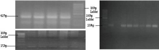
PCR amplification of E2 region through nested PCR (1st Round 427bp, 2nd Round 172bp) and NSP1 region of 239bp in Chikungunya virus.
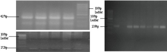
PCR amplification of E2 region through nested PCR (1st Round 427bp, 2nd Round 172bp) and NSP1 region of 239bp in Chikungunya virus.
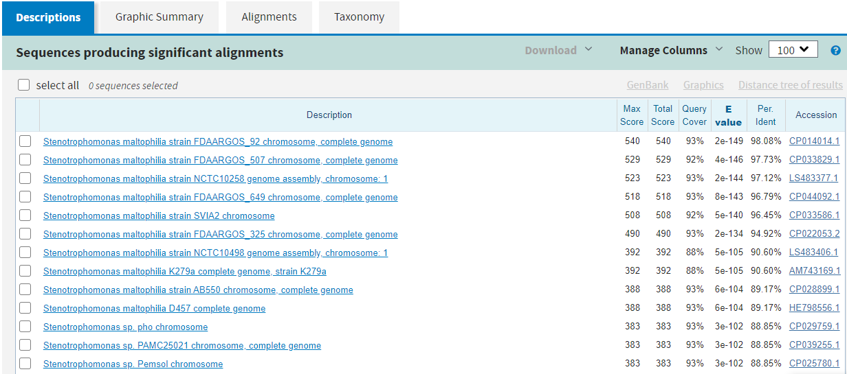
Sequence homology results showing homology with Stenotrophomonas maltophilia.
References
- AbuBakar, S., Sam, I.-C., Wong, P.-F., MatRahim, N., Hooi, P.-S. and Roslan, N., 2007. Emerg. Infect. Dis., 13: 147. https://doi.org/10.3201/eid1301.060617[CrossRef]
- Diallo, M., Thonnon, J., Traore-Lamizana, M. and Fontenille, D., 1999. Am. J. trop. Med. Hyg., 60: 281-286. https://doi.org/10.4269/ajtmh.1999.60.281[CrossRef]
- Gallian, P., Leparc-Goffart, I., Richard, P., Maire, F., Flusin, O., Djoudi, R., Chiaroni, J., Charrel, R., Tiberghien, P. and De Lamballerie, X., 2017. PLoS Neglec. trop. Dis., 11: e0005254. https://doi.org/10.1371/journal.pntd.0005254[CrossRef]
- Knipe, D.M., Howley, P.M., Griffin, D.E., Knipe and Howley, 2001. Fundamental virology. Lippincott Williams and Wilkins Philadelphia.
- Kosasih, H., de Mast, Q., Widjaja, S., Sudjana, P., Antonjaya, U., Ma’roef, C., Riswari, S.F., Porter, K.R., Burgess, T.H. and Alisjahbana, B., 2013. PLoS Neglec. Trop. Dis., 7: e2483. https://doi.org/10.1371/journal.pntd.0002483[CrossRef]
- Kosasih, H., Widjaja, S., Surya, E., Hadiwijaya, S.H., Butarbutar, D.P., Jaya, U.A., Alisjahbana, B. and Williams, M., 2012. Southeast Asian J. trop. Med. Public Hlth., 43: 55.
- Lee, V.J., Chow, A., Zheng, X., Carrasco, L.R., Cook, A.R., Lye, D.C., Ng, L.-C. and Leo, Y.-S., 2012. PLoS Neglec. trop. Dis., 6: e1786. https://doi.org/10.1371/journal.pntd.0001786[CrossRef]
- Mallhi, T., Khan, Y., Khan, A., Tanveer, N. and Qadir, M., 2017. Microbes Infect., 19: 13-14. https://doi.org/10.1016/j.nmni.2017.05.008[CrossRef]
- Shahid, M., Amin, I., Afzal, S., Fatima, Z. and Idrees, M., 2019. Pakistan J. Zool., 51: 1971-1974. http://dx.doi.org/10.17582/journal.pjz/2019.51.5.sc4[Full text][CrossRef]
- Pfeffer, M., Linssen, B., Parker, M. and Kinney, R., 2002. J. Vet. Med. Ser. B., 49: 49-54. https://doi.org/10.1046/j.1439-0450.2002.00535.x[CrossRef]
- Powers, A.M., Brault, A.C., Tesh, R.B. and Weaver, S.C., 2000. J. Gen. Virol., 81: 471-479. https://doi.org/10.1099/0022-1317-81-2-471[CrossRef]
- Powers, A.M. and Logue, C.H., 2007. J. Gen. Virol., 88: 2363-2377. https://doi.org/10.1099/vir.0.82858-0[CrossRef]
- Pulmanausahakul, R., Roytrakul, S., Auewarakul, P. and Smith, D.R., 2011. Int. J. Infect. Dis., 15: e671-e676. https://doi.org/10.1016/j.ijid.2011.06.002[CrossRef]
- San-Ho, P., Ng, M.M.L. and Chu, J.J.H., 2010. Virol. J., 7: 13. http://www.virologyj.com/content/7/1/13
- Staples, J.E. and Fischer, M., 2014. N. Engl. J. Med., 371: 887-889. https://doi.org/10.1056/NEJMp1407698[CrossRef]
- Yadav, K.K., Bora, A., Datta, S., Chandel, K., Gogoi, H.K., Prasad, G. and Veer, V., 2015. Parasite Vector, 8: 641. https://doi.org/10.1186/s13071-015-1252-0[CrossRef]
Services
Article usage statistics
Loading…