
Global health is continuously being threatened by a barrage of ravaging viral pathogens circulating in Africa a continent ravaged with poverty, hunger and poor health care system. These viral diseases have been reported to cause significant loss in labour, time and national economies of affected countries, deaths and morbidity among infected populace. Therefore, identifying emerging and reemerging viruses, the current trend and clinical challenges these viruses pose to human health in the continent is strategic in the development of countermeasures to curb the rather alarming situation. This mini-review utilized diverse search platforms using structured search strategies to undertake a systematic assessment of relevant literatures to present concise information on the subject. Interestingly, with few exceptions most regions of Africa share common viral pathogens of clinical relevance, namely Monkey pox virus, Ebola virus, Zika virus, Chikungunya virus, Dengue virus, Lassa virus, Measles virus, Yellow fever virus. Although the challenges found were enormous however, deliberate and conscientious government policies and health sector funding are critical indices for the control of emerging and reemerging viruses in Africa.
Mini-Review · 17 November 2025 · Open Access
Current Trends and Challenges of Some Emerging and Re-Emerging Viruses in Africa: A Systematic Minireview
· · · ·
Affiliations & Notes ⌄
- 1Department of Molecular Biology and Genomics, Global Health and Infectious Diseases Control Institute, Nasarawa State University Keffi, Nigeria
- 2Department of Pharmacology and Therapeutics, College of Medicine, Federal University of Health Sciences Otukpo, Benue State, Nigeria
- 3Applied Biology/Microbiology Department, School of Applied Science, Federal Polytechnic Nasarawa, Nasarawa State, Nigeria
- 4Institute of Human Virology, Nigeria. *Correspondence | Jeremiah John Oloche, Department of Molecular Biology and Genomics, Global Health and Infectious Diseases Control Institute, Nasarawa State University Keffi, Nigeria
- 5Email: joloche1012@gmail.com
Article Info ⌄
- Journal:
- Hosts and Viruses
- Published:
- 17 November 2025

From the article
- In conclusion, it is evident from the literature reviewed that the burden of emerging and re-emerging viral infections are more in low income countries especially Africa while the technical knowhow needed in combating these viruses are more in developing countries.
- It is evident also that Africa is characterized by the greatest infectious disease burden as well as by the weakest public health infrastructure in the world; further, efforts to establish public health infrastructures that are effective may take a period of years, even decades.
- Emerging infectious diseases should be identified as priority diseases.
- We are persuaded that the most important step towards the elimination of existing burden of infectious diseases in Africa is a massive increase in the number of qualified personnel, including both physicians and scientists.
Selected from the article's own text.
Abstract
Introduction
Infectious diseases have contributed up to 50% of world’s disease burden and among the leading causes of human morbidity and mortality with consequent high healthcare expenditure especially in low income countries including Africa ; Devendra et al., 2019). The emergence of high-threat disease pathogens such as viruses and bacteria which were previously under control and newly discovered elements has increased in recent years on a global scale . Emerging infectious disease (EID) is one that is caused by a pathogen that has not previously been identified in a particular population or geographical area. On a similar note, reemerging infectious diseases are caused by dissemination of pathogens that was prevalent in a community but under control or the discovery of an infectious etiology in previously diseases or detected in a new geographical area or the acknowledgment of diseases that have long been but were unrecognized entities.
Viral infections make up a sizable portion of newly and reemerging infectious diseases and they are becoming increasingly prevalent Sachidananda and Shrikara (2022) however, combined information on these viruses in one article are inexistent or at best rare. Available reviews disseminate information on only a single viral pathogen in an article. Hence the need of a mini-review that will provide readers and the scientific community with succinct, but adequate information that would foster comprehensive policy decisions and necessary preparation for the management of emerging and re-emerging viruses instead of the current isolated treatment.
Materials and Methods
This paper is a systematic mini-review of published manuscripts on emerging and re-emerging viral diseases in Africa identified through extensive literature search. Additionally, this mini-review was preceded by an initial literature to see if any high quality publication on the subject was in circulation. Subsequently, accessible electronic literatures were searched with restrictions to relevant subject headings, terms and keywords, by the name of viral diseases in Africa. Databases searched include: PubMed, Google Scholar, Web of Science, Science Direct and Scopus. The reference lists of all relevant articles were hand-searched to identify further any additional studies that may not have been captured by extensive searches. The article titles and abstracts were screened, followed by a comprehensive assessment of the whole text.
Results and Discussion
Emerging and re-emerging viruses in Africa in 21st century reported to account for most clinically significant infections include Monkey pox virus, Ebola virus, Dangue virus, Zika virus, Lassa virus, Influenza virus, yellow fever virus, Rift Valley fever virus, West Nile fever virus, SARS-Cov2, Chikungunya virus, Measles virus (Mahendra et al., 2022). However, this mini-review focuses on eight of the viruses.
Trends of emerging and re-emerging viral diseases
Monkey pox virus
Monkey pox (Mpox) is a zoonotic disease caused by Orthopoxvirus, a virus very close to that causing smallpox in humans. This virus is endemic to central and western African countries. It first emerged and was identified in 1958 as a pathogen of Macaca cynomolgus and then in 1970 was described as a human pathogen in Bokenda, Equateur Province of the Democratic Republic of Congo. The virus is capable of infecting humans that had not been vaccinated against smallpox, which confers cross-protection against Mpox , other primates and rodents. Most of the reported cases were related to animal-to-human transmission and were associated with the handling and eating of infected animals, but several cases of human-to-human transmission occurred. Since 2000, human monkey pox cases have been reported in the Central African Republic, Republic of the Congo, DRC, South Sudan, Nigeria, Liberia and Sierra Leone. In 2017, Nigeria experienced the largest documented outbreak around 40 years after the last confirmed cases of monkey pox ; .
The period of incubation after infection with Mpox virus ranges from 7 to 14 days during which the infected person is asymptomatic . Subsequently, a 1–4 days prodromal phase in which the infected subject becomes contagious, presenting with typical symptoms (Figure 1) such as fever, fatigue, myalgia, headaches and lymphadenopathy ; ; ; ; . This followed by the rash on the face, decline in fever and the onset of cutaneous macular lesions that evolve to popular, vesicular and, pustular lesions. Potential life-threatening complications such as encephalitis and diarrhea causing bronchopneumonia or pharyngeal abscesses could also occur. Severe corneal damage resulting to definitive vision loss ; has also been observed.
The trend and re-emergence of Mpox cases and fatalities in Africa member states within a period of 2022-2024 as reported by the WHO is alarming, and represents 62.3% of new cases globally (WHO, 2024; . At September 8, 2024, a total of 8,179 laboratory-confirmed cases, including 55 deaths, had been reported. In 2024 alone, available data at September 8, 5,776 confirmed cases and 32 deaths have been reported across 15 countries. The Democratic Republic of the Congo (5,160 cases), Burundi (385 cases), and Nigeria (55 cases) account for the majority of cases in 2024 (WHO, 2024).
The recent upsurge of cases of Mpox in Africa and other non-endemic areas has been hypothesized to be due to discontinuation of mass vaccination against smallpox during the 1980s. The smallpox vaccine has been proven to confer about 85% cross-immunity against Mpox virus hence, cessation resulting in increased human susceptibility to the virus and enhanced virus transmissibility through development of host immune evasion mechanisms ; Gomes et al., 2022). In addition, the acquisition of non-synonymous mutations associated with coding regions of the viral genome that harbor predicted host recognition elements necessary for adaptation of the virus ; .
The Mpox virus has been reported by the World Health Organization ; WHO, 2024) to exist as clade I and clade II with Clade I having sub clades Ia and Ib and clade II has sub clades IIa and IIb. Although sub-clades Ia and Ib still pose health risks in 2024 however, sub clade IIb is known to have caused a global outbreaks in 2022 and 2023 (WHO, 2024; .
Ebola virus
Ebola virus (Filoviridae) is one the viruses that has caused severe human epidemics identified since 1976. Ebola virus diseases also known as Ebola haermorrhagic fever is caused by the pathogen, Ebola virus . The disease is transmissible from animal reservoirs to humans likely via direct contact with ill, dead or killed wild animals mainly collected as bush meat . The virus then spread in the human population through human-to-human transmission ; .
Ebola disease re-emerged in the forest zones along the Gabon and Democratic Republic of the Congo (DRC) border in 2001. The world’s largest ever outbreak of Ebola fever probably started in December 2013 in rural Guinea after the infection of the suspected index case, a 2-year-old child ; ; . The subsequent epidemic crossed into Sierra Leone and Liberia with quick escalation of number of cases with a total of 881 healthcare workers infected, and 513 deaths. Ebola virus disease is marked with high case-morbidity and fatality rate . However, in central Africa, the trend of the outbreaks were localized remote rural epidemics involving relatively few cases, thus allowing targeted intervention in the affected villages ; WHO, 2025). On the other hand, the West African epidemic was an urban epidemic that occurred in a highly mobile population, leading to rapid spread of the virus. In DRC, new outbreaks still occur regularly, such as the two declared in 2018 ; WHO, 2025).
Beside the 2014–2016 Ebola virus epidemic, other Ebola virus outbreaks occurred in Africa including the 2018–2020 outbreak in the DRC which recorded 3481 cases and 2299 deaths, the 2021 outbreak in Guinea ; Keita et al., 2021; ; WHO, 2022; . Furthermore, the re-emergence of Sudan virus outbreak, one of the species of Ebola virus was reported in the Mubende District of western Uganda in 2022. Patients presenting with symptoms (Figure 2) such as; fever, gastrointestinal signs, haemorrhagic manifestations and multiple organ dysfunction syndrome (suspected viral hemorrhagic fever) were identified and isolated, blood samples were tested and the Sudan Ebola virus was confirmed ; . The outbreak recorded a total of 142 confirmed cases (plus 22 probable) and 55 confirmed deaths marked Uganda’s sixth Ebola outbreak, five of which have been caused by the Sudan Ebola virus ; CDC, 2022).
Zika virus
Zika virus is a virus belonging to the family Flavivirus and transmitted by Aedes mosquitoes ; . The virus was first detected in April 1947 in a febrile sentinel rhesus macaque monkey in the Zika forest on the shores of Lake Victoria of Uganda ; . The first human case was diagnosed in 1962–1963 in Uganda. Overall, data currently support a silent transmission of Zika virus among humans, mosquitoes and animals throughout tropical Africa for more than 70 years without reports of epidemic ; . The first (2007) and second (2013) epidemics occurred in Micronesia and Polynesia, respectively, all in Yap Island in the Pacific ; . The first outbreak of Zika virus in Africa was detected in 2015 in Cape Verde. Zika virus exhibited two unique features among the arboviruses: sexual transmission and congenital central nervous system malformations. Sexual transmission was first suspected in an American citizen returning from Senegal and then was confirmed after its emergence in the Americas .
Zika virus infection may present with only mild symptoms or no symptoms at all ; , but can cause serious congenital abnormalities and other adverse outcomes if infection occur during pregnancy ; . The main symptoms are eruptive skin rash (exanthema), arthralgia, non-purulent conjunctivitis and headache ; ; . The exanthem may be morbilliform with or without tiny papules, may start on the upper portion of the body and descend downward with progression of illness . Other symptoms such as low grade fever retro-orbital pain, abdominal pain, nausea, vomiting, diarrhoea and constipation, may also occur . Diagrammatic presentation of transmission and symptoms of Zika virus disease is shown in Figure 3.
The link between Zika virus and severe malformations of the central nervous system (particularly microcephaly) has been demonstrated by many studies in Brazil and has been reported retrospectively in French Polynesia. Genetic differences between African and Asian/American strains of Zika virus and appearance of the ‘pathogenic clone’ may explain the emergence of Zika virus infections in South America and Asia .
Chikungunya virus
Chikungunya virus, an Alphavirus (family Togaviridae), first reported in Tanzania in 1952, is transmitted by Aedes mosquitoes . The virus is associated with fever, rash and arthralgias. Severe joint pains can persist for a long time. From 1960 to 1990, epidemics were recorded in the Democratic Republic of Congo, Central African Republic, Malawi, Uganda, Burundi, Angola, Guinea, South Africa and Nigeria ; . In 2004, a large epidemic involving almost half a million cases was reported in Kenya. The epidemic reached the southwestern Indian Ocean region, India and Southeast Asia thus making it a global threat . From 2004 to 2007, several outbreaks were reported in Guinea, Tanzania, Sudan, Gabon and Cameroon. Two outbreaks were also reported on La Reunion Island in 2009 and 2010. In 2011, an epidemic hit the DRC. The circulation of chikungunya was also reported in Senegal in the area of Kedougou in 2015 .
After an incubation period of 2 to 10 days, chikungunya virus infection causes joint pain, which is often very debilitating and mainly affects the wrists, fingers, ankles, feet and knees, and more rarely the hips and shoulders . The joint pain is often accompanied by headache together with fever, severe muscle pain, a skin rash on the torso and limbs, conjunctivitis, and the inflammation of one or more cervical lymph nodes. Bleeding gums and nosebleeds have also been reported . In some cases, severe neurological manifestations can occur, including meningoencephalitis and peripheral neuropathy. These mainly affect elderly or immunocompromised people, as well as newborn infants who were infected in utero at the same time as their mother. The symptoms of chikungunya are shown in Figure 4.
Dengue virus
Dengue is the most important mosquito-borne viral disease in humans and is caused by infection with Dengue virus [DENV] . Dengue virus is of the family Flaviviridae that is transmitted to humans through a bite of infected Aedes mosquitoes . Due to mutations of the virus, the severity of the infection varies from time to time Kularatne and Dalugama (2022). In the past four decades, dengue has caused a significant impact on human health and national economies. Approximately 390 million people are infected with DENV annually with 96 million developing clinical symptoms that lead to hospitalizations of half a million and 25,000 deaths, annually (WHO, 2024).
One of the main factors which cause the spread of the disease globally is inefficient vector control management resulting from speedy urbanization with improper infrastructure planning, personnel travel leading to the spreading of the disease into new environments when they travel from one place to another . It was later hypothesized that transmission of dengue into Africa was believed to occur from the New World during slave trade . Going forward, the disease assumed more severe forms thereby attracting the peak of global attention that it deserves before the COVID-19 pandemic . Dengue is caused by an RNA virus that has four different serotypes, DENV1, 2, 3 and DENV4.
The common hallmark symptom of the disease is high fever (104°F/40°C) associated with severe headache, often described as break-bone fever, Other symptoms frequently reported as characteristic of dengue disease are retro-orbital pain, aches and pains throughout the body, skin rash which may develop few days after the onset of fever, dehydration, nosebleeds and bleeding gums may occur. Severe dengue symptoms often occur after the fever has gone away. The symptoms include; severe abdominal pain, persistent vomiting, rapid breathing, bleeding gums or nose, fatigue, restlessness, blood in vomit or stool, being very thirsty, pale and cold skin and feeling weak ; . Figure 5 is pictorial presentation of prevailing symptoms during the febrile phase of Dengue fever virus infection.
Lassa virus
Lassa virus (LV) is endemic in West African countries especially Nigeria, Sierra Leone, Guinea and Liberia where the large populations of rodent genus Mastomys serves as a reservoir and vector ; . Lassa virus is the cause of Lassa fever known as acute viral haemorrhagic fever (VHF) with high case fatality rates. Although it is suggested by genetic tracing that zoonotic transfers of LV to humans have been in existence for centuries ; however, the index patient with Lassa fever was first described in 1969 in Lassa, Nigeria. Currently there are no approved vaccines or treatment for Lassa fever but, ribavirin a guanosine nucleoside analogue is used for the treatment of the VHF ; .
In 1972, there was an outbreak of Lassa fever in the Eastern Province of Sierra Leone with peri-domestic rodent Mastomys natalensis as the major reservoir of LV . Infected rodents or sporadic human cases have been reported in other West African countries, including Mali, Togo and Benin ; Patassi et al., 2016). The infected rodents transmit the virus to offspring vertically, while infected rodents to humans and other rodents, transmission is horizontal , 2014).
According to the Centre for Disease Control and Prevention (CDC, 2024), an estimated 100,000– 300,000 infections occur each year in West Africa with approximately 5,000 deaths annually, a figure adjudge to be low probably due to the lack of standardized surveillance system for LV, and its potential misdiagnoses for other infectious diseases that are also endemic in this region, such as malaria ; CDC, 2019). Furthermore, Lassa fever is a seasonal disease and accounts for about 10-16% of annual hospital admissions in some areas of Sierra Leone and Liberia . Lassa fever has the second highest global burdens among all known viral hemorrhagic fevers, second only to Dengue fever which has an estimated 390 million infections per year, 96 million of which present with clinical symptoms (Bhatt et al., 2013; although most LV infected individuals develop immune response strong enough to control the infection.
Notably, the trend of Lassa fever outbreaks in Nigeria has changed from irregular to regular annual occurrence with increase in yearly confirmed cases and deaths, attributed to increased clinical awareness and improved diagnostic capacity with reduction in case fatality ; NCDC, 2021; . Recently the Nigeria Centre for Disease Control (NCDC) reported that in 2024 that there were 573 confirmed cases and 108 deaths with a case fatality rate (CFR) of 18.8%, which is higher than the CFR for the same period in 2023 (16.4%). In the report 64% of all the confirmed cases occurred in Ondo, Edo and Bauchi, while 36% were from 21 states, indicating a nationwide outbreak. The Lassa fever outbreak in Nigeria continues to worsen, with a total of 4,726 cases reported since the first week of 2024. Unfortunately, 142 deaths have been recorded, and 31 healthcare workers have been affected across 27 states, including the FCT. The hardest-hit states include Bauchi, Taraba, Edo, Ondo, Plateau, Benue, Cross River, Rivers, Anambra, and Ebonyi, spanning 123 Local Government Areas. While Lassa fever is endemic in Nigeria, the case fatality rate (CFR) of 18.5% has raised significant concerns, particularly regarding late diagnosis and reporting, which have contributed to the higher fatality rate. When compared to the 2023 outbreak, the number of suspected cases in 2024 has risen, with 4,726 cases reported so far, up from 3,361 during the same period in 2023 (NCDC, 2024).
The incubation period of Lassa fever is between the range of 1 and 21 days (NCDC, 2004). About 20% of infected persons present with severe illness that may include; bleeding (from the gums, nose, or different regions), respiratory distress, swelling of the face and neck, convulsions, tremors, encephalitis and shock and multiorgan dysfunction that might eventually lead to death ; ; . The remaining 80% are either asymptomatic or present only mild symptoms (Figure 6) such as fever, headache, weakness, malaise, sore throat, nausea and vomiting, diarrhoea, cough ; ; ; ; ; . However, but case fatalities as high as 50–70% have been reported during some outbreaks in Nigeria . Bilateral or unilateral sensory-neural deafness has been reported in about 33.2% of survivors ; ; ; . Lassa fever causes spontaneous abortion and high fatality rates during the third trimester in pregnant women ; ; .
Measles virus
Measles is a communicable disease caused by a member of the genus Morbillivirus ; CDC, 2020). Before the introduction of measles vaccine in 1963 and widespread vaccination, major epidemics of measles occurred almost every two to three years and caused approximately 2.6 million deaths per year. Despite the availability of safe and clinically effective live attenuated vaccines currently in use, measles remain one of the major diseases that cause high morbidity and mortality largely among unvaccinated children . Recently, over a thousand children were reportedly infected in a measles outbreak in North-East Nigeria .
The disease accounted for approximately 140,000 global deaths in children below 5 years in 2018, with most of the reported cases occurring in Africa and the Eastern Mediterranean regions (WHO, 2019). It is estimated that 128 000 deaths due to measles occurred in 2021 mostly among children under the age of five years. The reemergence of MV infection in developed and developing countries despite regional elimination and availability of clinically effective measles vaccines is cause for a great concern (WHO, 2019; ; .
Measles disease is prevalent in unvaccinated individuals with a single index case capable of causing 12–18 secondary infections in a healthy population ; . The measles virus (MV) infects the central nervous system leading to serious neurological disorders and can cause complications resulting in death. Although the majority of MV infections occur in children, however, there are no age restrictions. The symptoms (Figure 7) of measles appear 7 to 14 days post infection beginning with high fever (may spike to more than 104°), cough, runny nose (coryza), red, watery eyes (conjunctivitis). Subsequently, within 2 – 3 days of onset of symptoms, tiny white spots (Koplik spots) may appear inside the mouth, followed by measles rash appearing as flat red spots that appear on the face at the hairline which then spreads to the neck, trunk, arms, legs, and feet in 3 to 5 days ; ; CDC, 2020, 2024).
Yellow fever virus
Yellow fever is a preventable and an epidemic-prone viral disease that is transmitted from the infected vectors such as mosquitoes, ticks and other arthropods to humans through bites. The causative agent of YF, a mosquito-borne yellow fever virus (YFV), is an enveloped positive sense RNA virus of the genus Flavivirus ; . YF is endemic in subtropical and tropical parts of Africa and Central and South America because of the high presence of the vector, Aedes species, while humans serve as the primary host ; . Research has shown that YF originated in Africa and spread to the Americas through the slave trade in the 15th–16th centuries ; .
The re-emergence of yellow fever has been reported across several states in Nigeria since September 2017. Most recently, a total of 1,312 suspected cases were reported in 367 Local Government Areas (LGAs) across 36 States and the Federal Capital Territory within the period of 1 January to 31 August 2021 . A total of 31 out of 45 blood samples sent to the laboratory for investigation were positive with 12 of the positive cases having a history of yellow fever vaccination. Case fatality ratio of 11% was recorded among unvaccinated infected individuals. Furthermore, 287 laboratory-confirmed cases reported in Nigeria with a case fatality of 2.7% amongst of 7894 reported cases between September 2017 and September 2019. Outbreaks were confirmed in 55 LGAs with most of the outbreaks across four major epicenters in Kwara/Kogi, Edo, Ebonyi and Bauchi states .
In Cameroun, of the 20,261 yellow fever suspected patient’s samples that were collected and tested in 2015, 360 with a major YF IgM positive for yellow fever IgM antibodies. The trend continued throughout 2015 with a majority of cases occurring during the latter part of the year which was followed by a decrease in cases in 2016 . Available data in some African countries show the incidence of yellow fever in 100,000 population to range from < 1 case in Nigeria, < 3 cases in Uganda, 13 cases in Democratic Republic of the Congo, 27 cases in Kenya, 40 cases in Ethiopia, 46 cases in Gambia, 1267 cases in Senegal, and 10,350 cases in Ghana. Case fatality rate attributed to yellow fever outbreaks in Ghana and Nigeria were 10% and 86%, respectively ; .
A follow-up on yellow fever cases in Nigeria shows gaps in population immunity against yellow fever virus. The World Health Organization-WHO (2021), estimated that the national immunization coverage for yellow fever was 54% in 2020 and below the threshold of 80% necessary to protect against outbreaks. Available data indicate that in Anambra, Benue, Delta, Enugu, Imo, Niger, Ondo, Osun and Oyo states that reported positive cases, the routine immunization coverage declined between 2018 and 2020 and was below 80% in 2020. Garske et al. (2014) in a modeling study based on African data sources estimated the burden of yellow fever during 2013 to be 84,000–170,000 severe cases and 29,000–60,000 deaths. In 2023 there were sporadic reports of 12 cases of Yellow fever in Uganda with no fatality (CDC, 2024).
The incubation period of YF is 3–6 days after a bite Yellow fever infected mosquito (CDC, 2024). This is followed by flu-like symptoms, joint pain, body ache, epigastric pain, and nausea and vomiting. After 1–2 days relapse, about 20–60% of infected individuals progress to severe stage of the disease presenting with symptoms (Figure 8) including exhaustion, hemorrhagic fever, thrombocytopenia, severe liver disease with bleeding and jaundice which causes yellow skin coloration, fatal renal failure, vascular disease, and death . ; CDC, 2024).
Counter-measures against emerging and re-emerging viruses in Africa
The negative health implications associated with incidences of major emerging and re-emerging viruses in Africa cannot be over emphasized. The lack of approved drugs for the treatment of diseases caused by these viral pathogens underscores the all important relevance of the knowledge and strict adherence to WHO outlined control measures as a clear path to mitigate effects on the African populace . Table 1 shows major control measures for the identified major emerging and re-emerging viruses in Africa.
Factors contributing to the emergence and re-emergence viruses in Africa
Several factors influence the emergence and re-emergence of viruses have been identified: microbial adaptation and change, human susceptibility to infection, climate and weather, changing ecosystems, human demographics and behavior, economic development and land use, international travel and commerce, technology and industry. Other factors are breakdown of public health systems, poverty and social inequality, war and famine, lack of political will and intent to change the trend. Unfortunately, most of these factors are inherent in most countries of Africa. In addition, improved disease assessment through improved public health surveillance could also contribute to the apparent onset and reappearance of some diseases .
Table 1: Control measures for major emerging and re-emerging viral diseases in Africa.
| S. | Disease | Control measures | Treatment | Reference |
| 1. | Monkey pox | Strict hygiene measures is the primary prevention strategy. Vaccines such as MVA and LC16m8 have shown efficacy | Tecovirimat, Immunoglobulins, such as vaccinia immunoglobulin, can be used as preventive treatments for immune-compromised patients | Jadhav et al., 2025. Cuerel et al., 2022. Adler et al., 2022. Patel et al., 2023 |
| 2, | Ebola | Vaccination. [Zabdeno (Ad26.ZEBOV) and Mvabea (MVA-BN-Filo) for individuals ≥1 year]. Reduce risk of wild-life to human transmission | Two FDA-approved treatments are currently available to treat Ebola disease Inmazeb and Ebanga. Supportive care is necessary for survival | Liu et al., 2022. FDA, 2020. Jain et al., 2020 |
| 3. | Zika | No approved vaccine, Vector control | No virus-specific therapeutic interventions, Treatment is symptomatic | Quanquin et al., 2017. Muthuraj et al., 2023 |
| 4. | Chikungunya | Vaccination (VIMKUNYA and IXCHIQ, restricted to age ≥60 years) | No specific treatment. Treatment is symptomatic. Acetaminophen is the recommended drug for relief of joint pain. NSAIDS are contraindicated | CDC, 2024. Hakim and Aman, 2022); Cavalcanti et al., 2022. |
| 5. | Dengue | Vaccination with either Dengvaxia or QDENGA | No specific treatment. Treatment is symptomatic. Acetaminophen is the recommended drug for relief of fever and pain. NSAIDS are contraindicated | WHO, 2024. Natali et al., 2021 |
| 6. | Lassa | No approved vaccine, rat control, great cleanliness is required | No approved drug for treatment. Ribavirin with supportive care are often given to patients with Lassa fever | Basha et al., 2023. Ibukun, 2020. Raabe et al., 2017 |
| 7. | Measles | Vaccination (MMR and MMRV are often used for vaccination of children) | No specific treatment. Treatment is symptomatic | Endalamaw et al., 2024. CDC, 2024. Rabaan et al., 2022 |
| 8. | Yellow fever | Vaccination (YF Vax) and strict vector control | No specific treatment. Treatment is symptomatic | Amanna et al., 2024. Malik et al., 2023 |
- Similarity in early clinical symptoms: One of the challenges in diagnosing emerging and re-emerging viral diseases such as Lassa fever in West Africa is the similarities in initial clinical presentations with other febrile illnesses that are endemic in the region. This leads to delay in the diagnosis, isolation and treatment. Most often than not, Lassa fever is diagnosed in patients after failure of anti-malarial and antibiotic therapies .
- Inadequate health facilities: In many African countries, the ratio to population of health care facilities is very low. These results in overwhelming of few facilities by the populace per time. These hospitals also do not have up to date equipment for early and proper diagnosis and treatment of patients. These lead to either late or misdiagnoses of cases. Secondly there is poor referral system of specimen for urgent and proper diagnosis from one facility to another. In addition, absence of validated and rapid diagnostic tool necessary for early detection and treatment. Definitive methods of diagnosis of emerging and re-emerging viral diseases require virus isolation which is almost impractical method in endemic areas due to the absence of high bio-safety level-4 laboratories (BSL-4). The definitive diagnosis of these viral diseases requires the use of polymerase chain reaction (PCR), enzyme-linked immunosorbent assay (ELISA), immunoblot, and magnetic bead-based assays however, they are mainly used for research purpose rather than for diagnosis ; .
- Improper funding of health care: The health care systems in majority of African countries are poorly funded. In 2001, it was discovered that the average spending on health per capita was $36 with some countries spending as low as $2. This is contrary to European countries which spend as high as $4800 . This makes tackling these diseases very difficult in Africa.
- Inadequate well trained health care personnel: Due to the general poverty and economic hardship in Africa, qualified medical personnel prefer to work abroad than in Africa. This leads to brain drain of medical experts hence imposing serious challenge in achieving success.
- Lack of political will by Government: African Government lack the political will when it comes to health care. Priority is given to other sectors of governance rather than health. The health is then left to survive by the help from donor agencies and individuals of good will.
Conclusions and Recommendations
In conclusion, it is evident from the literature reviewed that the burden of emerging and re-emerging viral infections are more in low income countries especially Africa while the technical knowhow needed in combating these viruses are more in developing countries.
It is evident also that Africa is characterized by the greatest infectious disease burden as well as by the weakest public health infrastructure in the world; further, efforts to establish public health infrastructures that are effective may take a period of years, even decades. Emerging infectious diseases should be identified as priority diseases. The challenge will be to combine surveillance and epidemic preparedness and response activities for these priority diseases. Evidently this task is quite difficult because the infrastructure and level of support for surveillance, research and training on emerging infectious diseases in Africa are limited. Laboratory-based surveillance and targeted research surveys to identify common sources of infection in different community types would allow a unified approach to tackle this enormous challenge. We are persuaded that the most important step towards the elimination of existing burden of infectious diseases in Africa is a massive increase in the number of qualified personnel, including both physicians and scientists.
Novelty Statement
This mini-review utilized a one-stop approach and provide readers with succinct, but adequate information that would foster comprehension of basic knowledge on emerging and re-emerging viruses in Africa on the go. Our approach is in contrast with the regular systematic reviews that present detail isolated review on individual emerging or re-emerging virus.
Author’s Contribution
All authors participated equally in literature search, drafting and review of the manuscript.
Generative AI and AI-assisted technology statement
Authors declare that generative AI and AI-assisted technology were sparsely used for this review.
Conflicts of interest
The authors have declared no conflict of interest.
Figures
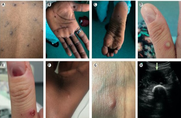
Skin and soft tissue manifestations of Monkey pox virus infection. Features included: A, D= vesicular or pustular lesions; B, C= macular lesions involving the palms and soles; D, E= a subungual lesion; F, G= more subtle papules and smaller vesicles; H= a deep abscess (Cuerel et al., 2022).
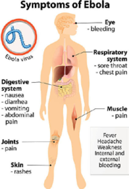
Pictorial presentation of symptomatology of Ebola virus disease Adopted from https://www.istockphoto.com/fr/photos/ebola-symptoms
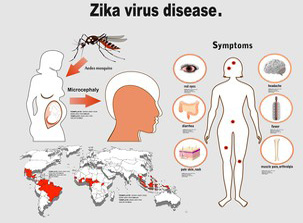
Diagrammatic presentation of transmission and symptoms of Zika virus disease (Adopted from https://www.shutterstock.com/image-vector/zika-virus-symptoms-infographics-figures-text-370996097).
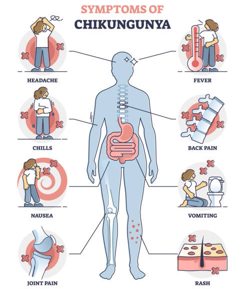
Schematic presentation of symptoms of Chikungunya virus disease (https://images.app.goo.gl/uCjtMMBXLpPzKTD2A).
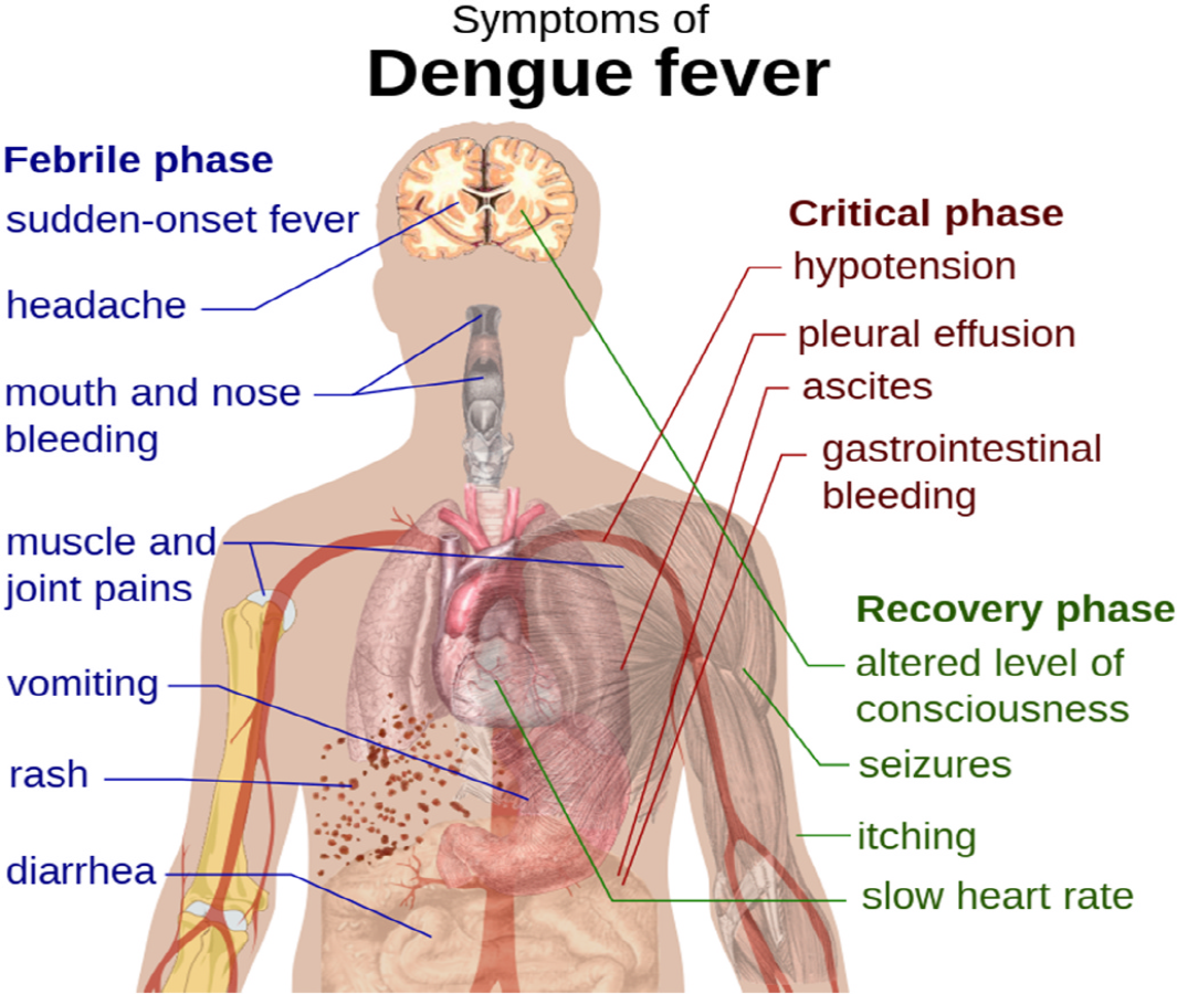
Schematic depiction of the symptoms of dengue fever (Haggstrom, 2014).
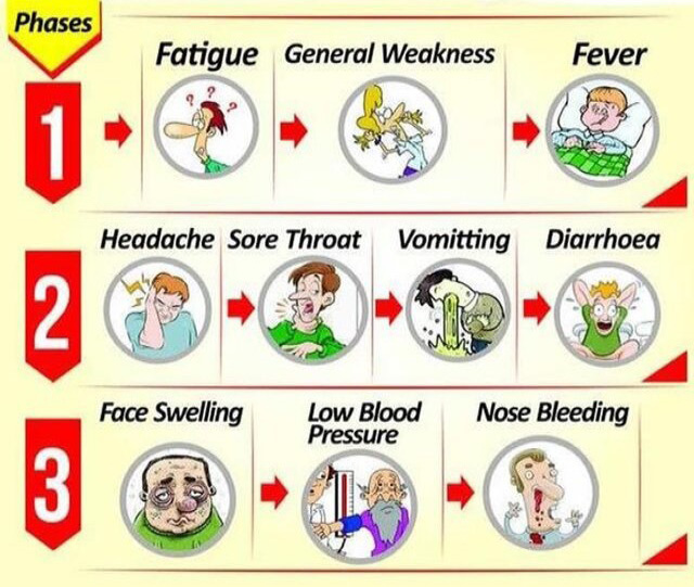
Symptoms of Lassa fever. Red arrow indicates prevailing symptom with disease progression (Basha et al., 2023).
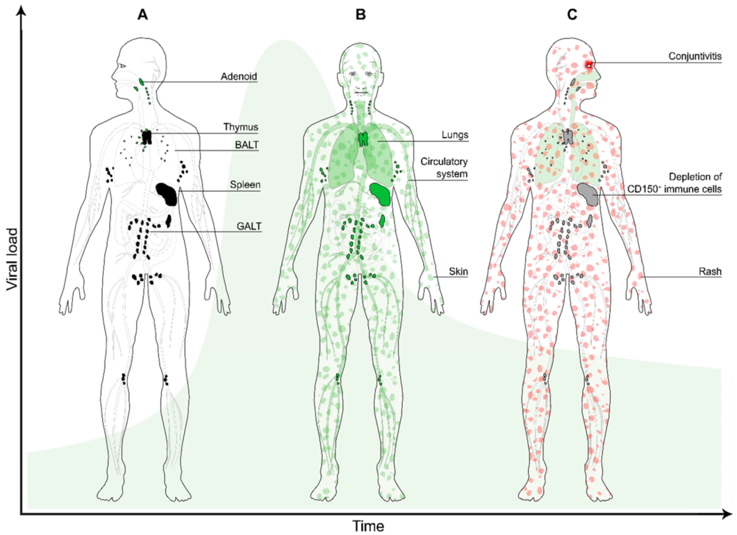
Clinical symptoms of Measles virus diseases. A, B and C represents progression in clinical symptoms with increasing viral load post exposure (Laksono et al., 2016)
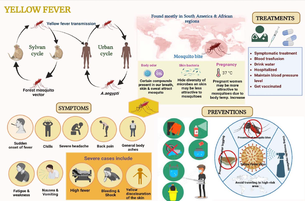
Schematic diagram of Transmission, symptoms prevention and treatment of yellow fever virus (Malik et al., 2023).
References
- Adler, H., Gould, S., Hine, P., Snell, L.B., Wong, W., Houlihan, C.F., Osborne, J.C., Rampling, T., Beadsworth, M.B., Duncan, C.J. Dunning, J., Fletcher, T.E., Hunter, E.R., Jacobs, M., Khoo, S.H., Newsholme, W., Porter, D., Porter, R.J., Ratcliffe, L., Schmid, M.L., Semple, M.G., Tunbridge, A.J., Wingfield, T. and Price, N.M., 2022. Clinical features and management of human monkeypox: A retrospective observational study in the UK. Lancet Infect Dis., 22:11531162.
- Agbonlahor, D.E., Akpede, G.O., Happi, C.T. and Tomori, O., 2021. 52 Years of Lassa Fever Outbreaks in Nigeria, 1969–2020: An epidemiologic analysis of the temporal and spatial trends. Am. J. Trop. Med. Hyg., 105(4): 974. https://doi.org/10.4269/ajtmh.20-1160[CrossRef]
- Akpede, G.O., Asogun, D.A., Okogbenin, S.A., Dawodu, S.O., Momoh, M.O., Dongo, A.E., Ike, C., Tobin, E., Akpede, N., Ogbaini-Emovon, E., Adewale, A.E., Ochei, O., Onyeke, F., Okonofua, M.O., Atafo, R.O., Odia, I., Adomeh, D.I., Odigie, G., Ogbeifun, C., Muebonam, E., Ihekweazu, C., Ramharter, M., Colubri, A., Sarbeti, P.C., Happi, C.T., Gunther, S. and Agbonlahor, D.E., 2019. Caseload and case fatality of lassa fever in Nigeria, 2001-2018: A specialist center’s experience and its implications. Front Publ. Health, 7: 170. https://doi.org/10.3389/fpubh.2019.00170[CrossRef]
- Amanna, I.J., Thomas, A., Engelmann, F., Hammarlund, E., Raue, P., Bailey, A.L., Poore, E.A., Quintel, B.K., Lewis, A.D., Axthelm, M.K., Johnson, A.L., Colgin, L.M., Diamond, M.S., Messaoudi, I. and Slifka, M.K., 2024. Development of a hydrogen peroxide-inactivated vaccine that protects against viscerotropic yellow fever in a non-human primate model. Cell Rep. Med., 5(7): 101655. https://doi.org/10.1016/j.xcrm.2024.101655[CrossRef]
- Andersen, K.G., Shapiro, B.J., Matranga, C.B., Sealfon, R., Lin, A.E., Moses, L.M., Folarin, O.A., Goba, A., Odia, I., Ehiane, P.E., Momoh, M., England, E.M., Winnicki, S., Branco, L.M., Gire, S.K., Phelan, E., Tariyal, R., Tewhey, R., Omoniwa, O., Fullah, M., Fonnie, R., Fonnie, M., Kanneh, L., Jalloh, S., Gbakie, M., Saffa, S., Karbo, K., Gladden, A.D., Qu, J., Stremlau, M., Nekoui, M., Finucane, H.K., Tabrizi, S., Vitti, J.J., Birren, B., Fitzgerald, M., McCowan, C., Ireland, A., Berlin, A.M., Bochicchio, J., Tazon-Vega, B., Lennon, N.J., Ryan, E.M., Bjornson, Z., Milner, D.A., Jr, Lukens, A.K., Broodie, N., Rowland, M., Heinrich, M., Akdag, M., Schieffelin, J.S., Levy, D., Akpan, H., Bausch, D.G., Rubins, K., McCormick, J.B., Lander, E.S., Gunther, S., Hensley, L., Okogbenin, S.; Viral Hemorrhagic Fever Consortium; Schaffner, S.F., Okokhere, P.O., Khan, S.H., Grant, D.S., Akpede, G.O., Asogun, D.A., Gnirke, A., Levin, J.Z., Happi, C.T., Garry, R.F. and Sabeti, P.C., 2015. Clinical sequencing uncovers origins and evolution of Lassa Virus. Cell., 162(4):738-50. doi: 10.1016/j.cell.2015.07.020. PMID: 26276630; PMCID: PMC4537774.[CrossRef]
- Asogun, D.A., Gunther, S., Akpede, G.O., Ihekweazu C. and Zumla, A., 2019. Lassa fever: Epidemiology, clinical features, diagnosis, management and prevention. Infect. Dis. Clin. N. Am., 33: 933–951. https://doi.org/10.1016/j.idc.2019.08.002[CrossRef]
- Basha, Sk.M., Venugopalaiah, P. and Chandra, Y.P., 2023. A review on Lassa fever. Int. J. Clin. Pharmacokinet. Med. Sci., 3(3): 96-103. https://doi.org/10.26452/ijcpms.v3i3.542[CrossRef]
- Bassey, B.E., Braka, F., Onyibe, R., Kolude, O.O., Oluwadare, M., Oluwabukola, A., Omotunde, O., Iyanda, O.A., Tella, A.A. and Olanike, O.S., 2022. Changing epidemiology of yellow fever virus in Oyo State, Nigeria. BMC Publ. Health, 22: 467. https://doi.org/10.1186/s12889-022-12871-0[CrossRef]
- Baud, D., Gubler, D.J., Schaub, B., Lanteri, M.C. and Musso, D., 2017. An update on Zika virus infection. Lancet., 390(10107):2099-2109. doi: 10.1016/S0140-6736(17)31450-2. PMID: 28647173[CrossRef]
- Belhadi, D., Baied, M.E., Mulier, G., Malvy, D., Mentre, F. and Laouenan, C., 2022. The number of cases, mortality and treatments of viral hemorrhagic fevers: A systematic review. PLOS Negl. Trop. Dis., 16(10): e0010889. https://doi.org/10.1371/journal.pntd.0010889[CrossRef]
- Bhatt, S., Gething, P.W., Brady, O.J., Messina, J.P., Farlow, A.W., Moyes, C.L., Drake, J.M., Brownstein, J.S., Hoen, A.G., Sankoh, O., Myers, M.F., George, D.B., Jaenisch, T., Wint, G.R., Simmons, C.P., Scott, T.W., Farrar, J.J. and Hay, S.I., 2013. The global distribution and burden of dengue. Nature., 496(7446):504-7. doi: 10.1038/nature12060. PMID: 23563266; PMCID: PMC3651993.[CrossRef]
- CDC. 2019. Lassa fever. https://africacdc.org/disease/lassa-fever/ Assessed May 9, 2025.
- Cavalcanti, L., Pereira, M.R. and Franca, O., 2022. A review on chikungunya virus epidemiology, pathogenesis and current vaccine development. Viruses, 14(5): 969. https://doi.org/10.3390/v14050969[CrossRef]
- Center for Disease Control, 2004. Lassa fever fact sheet. Atlanta, GA: US Department of Health and Human Services, CDC. Available at: http://www.cdc.gov/ncidod/dvrd/spb/mnpages/dispages/lassaf.htm
- Centers for Disease Control and Prevention (U.S.). Chikungunya Virus (https://www.cdc.gov/chikungunya/index.html). Last reviewed 3/2024. Accessed 5/6/2025.
- Centre for Disease Control, Measles, 2020. US Department of Health and Human Services, Washington, DC, USA.
- Cuerel, A., Favre, G., Vouga, M. and Pomar, L., 2022, Monkeypox and Pregnancy: Latest Updates. Viruses, 14: 2520. https://doi.org/10.3390/v14112520[CrossRef]
- Cummins, D., McCormick, J.B., Bennett, D., Samba, J.A., Farrar, B., Machin, S.J. and Fisher-Hoch, S.P., 1990. Acute sensorineural deafness in Lassa fever. JAMA., 264(16):2093-6. PMID: 2214077.
- Cunha Moizeis, R.N., Fernandes, M., Guedes, M., Bezerra Pereira, H.W., Ferreira Lanza, D.C., Galvao, A. and Fernandes, J.V. 2018. Chikungunya fever: A threat to global public health. Pathog. Glob. Health, 112(4): 182. https://doi.org/10.1080/20477724.2018.1478777[CrossRef]
- Damon, I.K., 2011. Status of human monkeypox: Clinical disease, epidemiology and research. Vaccine, 29(Suppl. 4): D54–D59. https://doi.org/10.1016/j.vaccine.2011.04.014[CrossRef]
- Derrington, S.M., Cellura, A.P., McDermott, L.E., Gubitosi, T., Sonstegard, A.M., Chen, S. and Garg, A., 2016. Mucocutaneous findings and course in an adult with Zika virus infection. JAMA Dermatol., 152(6):691-3. doi: 10.1001/jamadermatol.2016.1433. PMID: 27168495.[CrossRef]
- Devendra, G., Chai, M., Sumida, E., Chen, R., Gozun, M., Chow, D. and Miller, F., 2025. Differences in COVID-19 Fatality Rates among Ethnic Groups, Hawaii, USA, 20202022. Emerging Infectious Diseases., 31(10):1942-1949. https://doi.org/10.3201/eid3110.241070[CrossRef]
- Dick, G.W., Kitchen, S.F. and Haddow, A.J., 1952. Zika virus. I. Isolations and serological specificity. Trans. R. Soc. Trop. Med. Hyg., 46: 509–520. https://doi.org/10.1016/0035-9203(52)90042-4[CrossRef]
- Duffy, M.R., Chen, T.H., Hancock, W.T., Powers, A.M., Kool, J.L., Lanciotti, R.S., Pretrick, M., Marfel, M., Holzbauer, S., Dubray, C., Guillaumot, L., Griggs, A., Bel, M., Lambert, A.J., Laven, J., Kosoy, O., Panella, A., Biggerstaff, B.J., Fischer, M. and Hayes, E.B., 2009. Zika virus outbreak on Yap Island, Federated States of Micronesia. N Engl J Med., 360(24):2536-43. doi: 10.1056/NEJMoa0805715. PMID: 19516034.[CrossRef]
- Elliott, K.C., Mattapallil, J.J. 2024. Zika Virus-A reemerging neurotropic arbovirus associated with adverse pregnancy outcomes and neuropathogenesis. Pathogens, 13(2): 177. https://doi.org/10.3390/pathogens13020177[CrossRef]
- Emanuel, J., Marzi, A. and Feldmann, H. 2018. Filoviruses: Ecology, molecular biology, and evolution. Adv. Virus Res. 100: 189–221. https://doi.org/10.1016/bs.aivir.2017.12.002[CrossRef]
- Endalamaw, D., Nibret, E., Munshea, A., Mekonnen, F., Tadesse, S., Zeru, T., Tefera, M. and Yismaw, G., 2024. Measles vaccine effectiveness in African children: A systematic review and meta-analysis. BMC Infect. Dis., 24: 1330. https://doi.org/10.1186/s12879-024-10239-w[CrossRef]
- Fenollar, F. and Mediannikov, O., 2018. Emerging infectious diseases in Africa in the 21st century. New Microbes New Infect., 26: S10-S18. https://doi.org/10.1016/j.nmni.2018.09.004[CrossRef]
- Ferreira, F.C.d.S.L., Bastos Camacho, L.A. and Villela, D.A.M., 2022. Occurrence of yellow fever outbreaks in a partially vaccinated population: An analysis of the effective reproduction number. PLoS Negl. Trop. Dis., 16: e0010741. https://doi.org/10.1371/journal.pntd.0010741[CrossRef]
- Fichet-Calvet, E., Becker-Ziaja, B., Koivogui, L. and Gunther, S., 2014. Lassa serology in natural populations of rodents and horizontal transmission. Vector Borne Zoonotic Dis., 14: 665–674. https://doi.org/10.1089/vbz.2013.1484[CrossRef]
- Fichet-Calvet, E., Lecompte, E., Koivogui, L., Daffis, S. and Ter Meulen, J., 2008. Reproductive characteristics of Mastomys natalensis and Lassa virus prevalence in Guinea, West Africa. Vector Borne Zoonotic Dis., 8: 41–48. https://doi.org/10.1089/vbz.2007.0118[CrossRef]
- Fine, P.E.M., Jezek, Z., Grab, B. and Dixon, H., 1988. The transmission potential of monkeypox virus in human populations. Int. J. Epidemiol., 17: 643–650. https://doi.org/10.1093/ije/17.3.643[CrossRef]
- Firth, C., Kitchen, A., Shapiro, B., Suchard, M.A., Holmes, E.C. and Rambaut, A., 2010. Using time-structured data to estimate evolutionary rates of double-stranded DNA viruses. Mol. Biol. Evol., 27: 2038–2051. https://doi.org/10.1093/molbev/msq088[CrossRef]
- Garry, R.F., 2023. Lassa fever - the road ahead. Nat Rev Microbiol., 21(2):87-96. doi: 10.1038/s41579-022-00789-8. Epub 2022 Sep 12. PMID: 36097163; PMCID: PMC9466315.
- Garske, T., Van Kerkhove, M.D., Yactayo, S., Ronveaux, O., Lewis, R.F., Staples, J.E., Perea, W., Ferguson, N.M., 2014. Yellow Fever Expert Committee. Yellow Fever in Africa: estimating the burden of disease and impact of mass vaccination from outbreak and serological data. PLoS Med., 11(5):e1001638. doi: 10.1371/journal.pmed.1001638. PMID: 24800812; PMCID: PMC4011853.[CrossRef]
- Garske, T., Van Kerkhove, M.D., Yactayo, S., Ronveaux, O., Lewis, R.F., Staples, J.E., Perea, W. and Ferguson, N.M., 2014. Yellow fever expert committee (2014). Yellow Fever in Africa: Estimating the burden of disease and impact of mass vaccination from outbreak and serological data. PLoS Med., 11: e1001638. https://doi.org/10.1371/journal.pmed.1001638[CrossRef]
- Gay, N.J., 2004. The theory of measles elimination: Implications for the design of elimination strategies. J. Infect. Dis., 189: S27–S35. https://doi.org/10.1086/381592[CrossRef]
- Grifn, D.E. and Bellini, W.J. 1996. Measles virus, in Virology, B.N. Fields, D.M. Knipe, and P.M. Howley, Eds., Lippincott-Raven, Philadelphia, PA, USA. pp. 1267–1312.
- Gubler, D.J., Vasilakis, N. and Musso, D., 2017. History and emergence of Zika virus. J. Infect. Dis., 216(Suppl. 10): S860–867. https://doi.org/10.1093/infdis/jix451[CrossRef]
- Gunther, S. and Lenz, O., 2004. Lassa virus. Crit. Rev. Clin. Lab. Sci., 357.
- Guzman, M.G. and Martinez, E., 2024. Central and peripheral nervous system manifestations associated with dengue illness. Viruses, 16(9): 1367. https://doi.org/10.3390/v16091367[CrossRef]
- Haggstrom, M., 2014. Medical gallery of mikael haggstrom. Wiki J. Med., 1(2), https://doi.org/10.15347/wjm/2014.008[CrossRef]
- Hakim, M.S. and Aman, A.T., 2022. Understanding the biology and immune pathogenesis of Chikungunya virus infection for diagnostic and vaccine development. Viruses, 15(1): 48. https://doi.org/10.3390/v15010048[CrossRef]
- Halani, S., Tombindo, P.E., Miranda, R.N., Erdman, L.K., Whitehead, C., Bielecki, J.M., Ramsay, L., Ximenes, R., Boyle, J., Krueger, C., Willmott, S., Morris, S.K., Murphy, K.E. and Sander, B., 2021. Clinical manifestations and health outcomes associated with Zika virus infections in adults: A systematic review. PLoS Neglect. Trop. Dis., 15(7): e0009516. https://doi.org/10.1371/journal.pntd.0009516[CrossRef]
- Harapan, H., Michie, A., Sasmono, R.T. and Imrie, A., 2020. Dengue: A minireview. Viruses, 12(8): 829. https://doi.org/10.3390/v12080829[CrossRef]
- Harapan, H., Ophinni, Y., Megawati, D., Frediansyah, A., Mamada, S.S., Salampe, M., Bin Emran, T., Winardi, W., Fathima, R., Sirinam, S., Sittikul, P., Stoian, A.M., Nainu, F. and Sallam, M., 2022. Monkeypox: A Comprehensive Review. Viruses., 14(10):2155. doi: 10.3390/v14102155. PMID: 36298710; PMCID: PMC9612348.[CrossRef]
- Hutin, Y.J., Williams, R.J., Malfait, P., Pebody, R., Loparev, V.N., Ropp, S.L., Rodriguez, M., Knight, J.C., Tshioko, F.K., Khan, A.S., Szczeniowski, M.V. and Esposito, J.J., 2001. Outbreak of human monkeypox, Democratic Republic of Congo, 1996 to 1997. Emerg Infect Dis., 7(3):434-8. doi: 10.3201/eid0703.010311. PMID: 11384521; PMCID: PMC2631782.[CrossRef]
- Ibukun, F.I., 2020. Inter-lineage variation of lassa virus glycoprotein epitopes: A challenge to lassa virus vaccine development. Viruses, 12(4): 386. https://doi.org/10.3390/v12040386[CrossRef]
- Interim Centers for Disease Control and Prevention (CDC) Guidance for Use of Smallpox Vaccine, Cidofovir, and Vaccinia Immune Globulin (VIG) for Prevention and Treatment in the Setting of an Outbreak Monkeypox Infections. Available online: https://stacks.cdc.gov/view/cdc/25058 (accessed on 8 May, 2025).
- Isidro, J., Borges, V., Pinto, M., Ferreira, R., Sobral, D., Nunes, A., Santos, J.D., Borrego, M.J., Nuncio, M.S., Pelerito, A., Cordeiro, R., and Gomes, J.P.O. Dos Santos., 2022. Multi-country outbreak of Monkeypox virus: genetic divergence and first signs of microevolution. p. 18. Available from: https:// virological.org/t/multi-country-outbreak
- Jacob, S.T., Crozier, I., Hewlett, A., Kraft, C.S., Vega, L., Soka, M.J., Wahl, V., Griffiths, A., Bollinger, L. and Kuhn, J.H., 2020. Ebola virus disease. Nature reviews. Disease Primers, 6(1): 13. https://doi.org/10.1038/s41572-020-0147-3[CrossRef]
- Jadhav, V., Paul, A., Trivedi, V., Bhatnagar, R., Bhalsinge, R. and Jadhav, S.V. 2025. Global epidemiology, viral evolution, and public health responses: A systematic review on Mpox (1958–2024). J. Glob. Health, 15: 04061. https://doi.org/10.7189/jogh.15.04061[CrossRef]
- Jain, S., Khaiboullina, S.F. and Baranwal, M., 2020. Immunological perspective for ebola virus infection and various treatment measures taken to fight the disease. Pathogens, 9(10): 850. https://doi.org/10.3390/pathogens9100850[CrossRef]
- Jain, S., Martynova, E., Rizvanov, A., Khaiboullina, S. and Baranwal, M., 2021. Structural and functional aspects of ebola virus proteins. Pathogens., 10:1330. doi: 10.3390/pathogens10101330[CrossRef]
- Jezek, Z., Szczeniowski, M., Paluku, K.M. and Mutombo, M., 1987. Human monkeypox: Clinical features of 282 patients. J. Infect. Dis., 156: 293–298. https://doi.org/10.1093/infdis/156.2.293[CrossRef]
- Joseph Pergolizzi, J., LeQuang, J.A., Fleischer, C., Joseph Pergolizzi, I., Pergolizzi, C. and Raffa, R.B., 2020. The Zika virus: Lurking behind the COVID-19 pandemic? J. Clin. Pharma. Therapeut., 46(2): 267. https://doi.org/10.1111/jcpt.13310[CrossRef]
- Joseph, S. and Campbell, K.P., 2021. Lassa fever virus Binds Matriglycan: A polymer of alternating xylose and glucuronate on α-Dystroglycan. Viruses, 13: 1679. https://doi.org/10.3390/v13091679[CrossRef]
- Kakai, C.G., Okunromade, O.F., Dan-Nwafor, C.C., Chabi, A.I.B., Martial, G.T.P., Dalhat, M.M., Ward, S., Tante, O., Nguku, P.M., Hamadi, A., Ilori, E., Lokossou, V., Brito, C., Ojo, O.E., Kone, I., Agbeko, T.T., Ihekweazu, C. and Merrill, R.D., 2020. Improving Cross-Border Preparedness and Response: Lessons Learned from 3 Lassa Fever Outbreaks Across Benin, Nigeria, and Togo, 2017-2019. Health Secur., 18(S1):S105-S112. doi: 10.1089/hs.2019.0080. PMID: 32004125; PMCID: PMC10823436.[CrossRef]
- Kawuki, J., Musa, T.H. and Yu, X., 2021. Impact of recurrent outbreaks of Ebola virus disease in Africa: a meta-analysis of case fatality rates. Publ. Health, 195(2021): 89-97. https://doi.org/10.1016/j.puhe.2021.03.027[CrossRef]
- Kayem, N.D., Benson, C., Aye, C.Y.L., Barker, S., Tome, M., Kennedy, S., Ariana, P., Horby, P., 2020. Lassa fever in pregnancy: a systematic review and meta-analysis. Trans R Soc Trop Med Hyg., 114(5):385-396. doi: 10.1093/trstmh/traa011. PMID: 32125412; PMCID: PMC7197258.[CrossRef]
- Keita, A.K., Koundouno, F.R., Faye, M., Düx, A., Hinzmann, J. Diallo, H., Ayouba, A., Le Marcis, F., Soropogui, B., Ifono, K., 2021. Resurgence of Ebola virus in 2021 in Guinea suggests a new paradigm for outbreaks, Nature, 597(7877): 539–543. https://doi.org/10.1038/s41586-021-03901-9[CrossRef]
- Kok, B.H., Lim, H.T., Lim, C.P., Lai, N.S., Leow, C.Y. and Leow, C.H., 2023. Dengue virus infection. A review of pathogenesis, vaccines, diagnosis and therapy. Virus Res., 15(324): 199018. https://doi.org/10.1016/j.virusres.2022.199018[CrossRef]
- Kola, O., 2021. Nigeria: 1,158 displaced children infected by measles. https://www.aa.com.tr/en/africa/nigeria-1- 158-displaced-children-infected-by-measles/2203817.
- Kornbluh, R. and Davis R., 2020. Global trends in measles publications. Pan Afr. Med. J., 35(Suppl 1): 14. https://doi.org/10.11604/pamj.supp.2020.35.1.18508[CrossRef]
- Kraemer, M.U.G., Pigott, D.M., Hill, S.C., Vanderslott, S., Reiner, R.C. Jr, Stasse, S., Brownstein, J.S., Gutierrez, B., Dennig, F., Hay, S.I., Wint, G.R.W., Pybus, O.G., Castro, M.C., Vinck, P., Pham, P.N., Nilles, E.J. and Cauchemez, S., 2020. Dynamics of conflict during the Ebola outbreak in the Democratic Republic of the Congo 2018-2019. BMC Medicine. 18(1):113. doi: 10.1186/s12916-020-01574-1. PMID: 32336281; PMCID: PMC7184697.
- Kularatne, S.A. and Dalugama, C., 2022. Dengue infection: Global importance, immunopathology and management. Clin. Med. (Lond), 22(1): 9-13. https://doi.org/10.7861/clinmed.2021-0791[CrossRef]
- Kularatne, S.A. and Dalugama, C., 2022. Dengue infection: Global importance, immunopathology and management. Clinical Medicine (Lond)., 22(1):9-13. doi: 10.7861/clinmed.2021-0791. PMID: 35078789; PMCID: PMC8813012
- Laksono, B.M., De Vries, R.D., McQuaid, S., Duprex, W.P. and De Swart, R.L., 2016. Measles virus host invasion and pathogenesis. Viruses, 8(8): 210. https://doi.org/10.3390/v8080210[CrossRef]
- Lassa Fever, CDC, 2019. Retrieved 2024 Mar 5, from https://www.cdc.gov/vhf/lassa/index.html
- Legrand, J., Grais, R.F., Boelle, P.Y., Valleron, A.J. and Flahault, A., 2006. The dynamics of Ebola epidemics. Epidemiol. Infect., 135: 610–621. https://doi.org/10.1017/S0950268806007217[CrossRef]
- Lim, J.K., Seydou, Y., Carabali, M., Barro, A., Dahourou, D.L., Lee, K.S., Nikiema, T., Namkung, S., Lee, S., Shin, M.Y., Bonnet, E., Kagone, T., Kaba, L., Edwards, T., Some, A., Yang, J.S., Alexander, N., Yoon, K. and Ridde, V., 2019. Clinical and epidemiologic characteristics associated with dengue during and outside the 2016 outbreak identified in health facility-based surveillance in Ouagadougou, Burkina Faso. PLoS Negl. Trop. Dis., 13(12): e0007882. https://doi.org/10.1371/journal.pntd.0007882[CrossRef]
- Liu, H., Hu, T., Wong, S.H. and Lin, T., 2022. Therapeutic strategies against Ebola virus infection. Viruses, 14(3): 579. https://doi.org/10.3390/v14030579[CrossRef]
- Lukashevich, I.S., Paessler, S. and De La Torre, J.C. 2019. Lassa virus diversity and feasibility for universal prophylactic vaccine. F1000Res. 8: 134. https://doi.org/10.12688/f1000research.16989.1[CrossRef]
- Malik, S., Pandey, I., Kishore, S., Sundarrajan, T., Nargund, S.L., Ghosh, A., Emran, T.B., Chaicumpa, W. and Dhama, K., 2023. Yellow fever virus, a mosquito-borne flavivirus posing high public health concerns and imminent threats to travellers– an update. Int. J. Surg., 109: 134 –137. https://doi.org/10.1097/JS9.0000000000000086[CrossRef]
- Mazzola, L.T. and Kelly-Cirino, C., 2019. Diagnostics for Lassa fever virus: A genetically diverse pathogen found in low-resource settings. Biomed. J. Glob. Health, 4 (Suppl2): e001116. https://doi.org/10.1136/bmjgh-2018-001116[CrossRef]
- Mbala-Kingebeni, P., Aziza, A., Di Paola, N., Wiley, M.R., Makiala-Mandanda, S., Caviness, K., Pratt, C.B., Ladner, J.T., Kugelman, J.R., Prieto, K., Chitty, J.A., Larson, P.A., Beitzel, B., Ayouba, A., Vidal, N., Karhemere, S., Diop, M., Diagne, M.M., Faye, M., Faye, O., Aruna, A., Nsio, J., Mulangu, F., Mukadi, D., Mukadi, P., Kombe, J., Mulumba, A., Villabona-Arenas, C.J., Pukuta, E., Gonzalez, J., Bartlett, M.L., Sozhamannan, S., Gross, S.M., Schroth, G.P., Tim, R., Zhao, J.J., Kuhn, J.H., Diallo, B., Yao, M., Fall, I.S., Ndjoloko, B., Mossoko, M., Lacroix, A., Delaporte, E., Sanchez-Lockhart, M., Sall, A.A., Muyembe-Tamfum, J.J., Peeters, M., Palacios, G. and Ahuka-Mundeke., S., 2019. Medical countermeasures during the 2018 Ebola virus disease outbreak in the North Kivu and Ituri Provinces of the Democratic Republic of the Congo: a rapid genomic assessment. Lancet Infect Dis., 19(6):648-657. doi: 10.1016/S1473-3099(19)30118-5. Epub 2019 Apr 15. PMID: 31000464.[CrossRef]
- McCollum, A.M. and Damon, I.K., 2014. Human monkeypox. Clin. Infect. Dis. 58: 260–267. https://doi.org/10.1093/cid/cit703[CrossRef]
- McCormick, J.B., King, I.J., Webb, P.A., Johnson, K.M., O’Sullivan, R., Smith, E.S., Trippel, S. and Tong, T.C., 1987. A case-control study of the clinical diagnosis and course of Lassa fever. J Infect Dis., 155(3):445-55. doi: 10.1093/infdis/155.3.445. PMID: 3805772.
- Merson, L., Bourner, J., Jalloh, S., Erber, A., Salam, A.P., Flahault, A. and Olliaro, P.L. 2021. Clinical characterization of Lassa fever: A systematic review of clinical reports and research to inform clinical trial design. PLoS Negl. Trop. Dis., 15(9): e0009788. https://doi.org/10.1371/journal.pntd.0009788[CrossRef]
- Monath, T.P., Newhouse, V.F., Kemp, G.E., Setzer, H.W. and Cacciapuoti, A., 1974. Lassa virus isolation from Mastomys natalensis rodents during an epidemic in Sierra Leone. Science, 185: 263–265. https://doi.org/10.1126/science.185.4147.263[CrossRef]
- Monath, T.P. and Vasconcelos, P.F.C. 2015. Yellow fever. J. Clin. Virol. 64: 160–173. https://doi.org/10.1016/j.jcv.2014.08.030[CrossRef]
- Murphy, H.L. and Ly, H. 2021. Pathogenicity and virulence mechanisms of Lassa virus and its animal modeling, diagnostic, prophylactic, and therapeutic developments. Virulence, 12(1): 2989–3014. https://doi.org/10.1080/21505594.2021.2000290[CrossRef]
- Muthuraj, P.G., Krishnamoorthy, C., Anderson-Berry, A., Hanson, C. and Natarajan, S.K., 2023. Novel Therapeutic Nutrients Molecules That Protect against Zika Virus Infection with a Special Note on Palmitoleate. Nutrients, 15(1): 124. https://doi.org/10.3390/nu15010124[CrossRef]
- Muthuraj, P.G., Krishnamoorthy, C., Anderson-Berry, A., Hanson, C. and Natarajan, S.K. 2023. Novel therapeutic nutrients molecules that protect against zika virus infection with a special note on palmitoleate. Nutrients, 15(1): 124. https://doi.org/10.3390/nu15010124[CrossRef]
- Mwanyika, G.O., Mboera, L.E.G., Rugarabamu, S., Ngingo, B., Sindato, C., Lutwama, J.J., Paweska, J.T. and Misinzo, G., 2021. Dengue virus infection and associated risk factors in Africa: A Systematic review and meta-analysis. Viruses, 13: 536. https://doi.org/10.3390/v13040536[CrossRef]
- Natali, E.N., Babrak, L.M. and Miho, E., 2021. Prospective artificial intelligence to dissect the Dengue immune response and discover therapeutics. Front. Immunol., 12: 574411. https://doi.org/10.3389/fimmu.2021.574411[CrossRef]
- National Center for Emerging and Zoonotic Infectious Diseases (NCEZID) https://www.cdc.gov/ebola/outbreaks/index.html 2022
- NCDC, 2017. An update of monkeypox outbreak in Nigeria. (Nigeria centre for disease control, 2017), (available at https://ncdc.gov.ng/diseases/sitreps/?cat=8).
- Nigeria Centre for Disease Control (NCDC) file:///C:/Users/user/Downloads/Lassa%20fever%20Sitrep%20Week%2011%202024.int.pdf 2024.
- Nigeria Centre for Disease Control, 2016/2017 Lassa fever Outbreak in Nigeria: Epi Week 51: 24 December, 2017. 2017.
- Nigeria Centre for Disease Control, 2018. Lassa fever outbreak situation report: Epi Week 52: 31st December 2018. 2018.
- Nigeria Centre for Disease Control, 2019. Disease situation reports. An update of Lassa fever outbreak in Nigeria for Week 52 –December 2019. Abuja, Nigeria: 2019.
- Nigeria Centre for Disease Control, 2020. Lassa fever situation report: Epi Week 53: 28 December 2020–03 January 2021.
- Nomhwange, T., Jean Baptiste, A.E., Ezebilo, O., Oteri, J., Olajide, L., Emelife, K., Hassan, S., Nomhwange, E.R., Adejoh, K., Ireye, F., Nora, E.E., Ningi, A., Bathondeli, B. and Tomori, O., 2021. The resurgence of yellow fever outbreaks in Nigeria: A 2-year review 2017–2019. BMC Infect. Dis., 21: 1054. https://doi.org/10.1186/s12879-021-06727-y[CrossRef]
- Nwaiwu, A.U., Musekiwa, A., Tamuzi, J.L., Sambala, E.Z. and Nyasulu, P.S., 2021. The incidence and mortality of yellow fever in Africa: A systematic review and meta-analysis. BMC Infect. Dis., 21: 1089. https://doi.org/10.1186/s12879-021-06728-x[CrossRef]
- O’Toole, A. and Rambaut, A., 2022. Update to observations about putative APOBEC3 deaminase editing in the light of new genomes from USA.
- Ogbu, O., Ajuluchukwu, E. and Uneke, C.J., 2007. Lassa fever in West African sub region: An overview. J. Vector Borne Dis., 44: 1–11.
- Okogbenin, S., Okoeguale, J., Akpede, G., Colubri, A., Barnes, K.G., Mehta, S., Eifediyi, R., Okogbo, F., Eigbefoh, J., Momoh, M., Rafiu, M., Adomeh, D., Odia, I., Aire, C., Atafo, R., Okonofua, M., Pahlman, M., Becker-Ziaja, B., Asogun, D., Okokhere, P., Happi, C., Gunther, S., Sabeti, P.C. and Ogbaini-Emovon, E., 2019. Retrospective cohort study of Lassa Fever in pregnancy, Southern Nigeria. Emerg Infect Dis., 25(8):1494500. doi: 10.3201/eid2508.181299. PMID: 31310586; PMCID: PMC6649346
- Okokhere, P., Colubri, A., Azubike, C., Iruolagbe, C., Osazuwa, O., Tabrizi, S., Chin, E., Asad, S., Ediale, E., Rafiu, M., Adomeh, D., Odia, I., Atafo, R., Aire, C., Okogbenin, S., Pahlman, M., Becker-Ziaja, B., Asogun, D., Fradet, T., Fry, B., Schaffner, S.F., Happi, C., Akpede, G., Gunther, S. and Sabeti, P.C., 2018. Clinical and laboratory predictors of Lassa fever outcome in a dedicated treatment facility in Nigeria: a retrospective, observational cohort study. Lancet Infect Dis., 18(6):684-695. doi: 10.1016/S1473-3099(18)30121-X. Epub 2018 Mar 6. Erratum in: Lancet Infect Dis. 18(5):492. doi: 10.1016/S1473-3099(18)30208-1. PMID: 29523497; PMCID: PMC5984133[CrossRef]
- Olawade, D.B., Wada, O.Z., Fidelis, S.C., Oluwole, O.S., Alisi, C.S., Orimabuyaku, N.F. and David-Olawade, A.C. 2024. Strengthening Africa’s response to Mpox (monkeypox): Insights from historical outbreaks and the present global spread. Sci. One Health, 3: 100085. https://doi.org/10.1016/j.soh.2024.100085[CrossRef]
- Oloche, J.J., Oluremi, B.B., Aruwa, C.E. and Sabiu, S., 2023. Molecular modeling identification of key secondary metabolites from Xylopia aethiopica as promising therapeutics targeting essential Measles viral proteins. Evid. Based Complement. Altern. Med., Article ID 1575358, 13 pages. https://doi.org/10.1155/2023/1575358[CrossRef]
- Patassi, A.A., Landoh, D.E., Mebiny-Essoh, T.A., Halatoko, W.A., Assane, H., Saka, B., Naba, M.A., Yaya, I., Edou, K.A., Tamekl, T.A., Banla, A.K., Davi, K.M., Manga, M., Kassankogno, Y. and Salmon-Ceron, D., 2017. Emergence of Lassa Fever Disease in Northern Togo: Report of Two Cases in Oti District in 2016, Case Report. Infect Dis., 8242313, 5 pages, 2017.
- Pal, M., 2022. Human Monkeypox: An Emerging and Re-emerging Infectious Viral Disease. https://doi.org/10.31080/ASMI.2022.05.1045[CrossRef]
- Patel, M., Adnan, M., Aldarhami, A., Bazaid, A.S., Saeedi, N.H., Alkayyal, A.A., Saleh, F.M., Awadh, I.B., Saeed, A. and Alshaghdali, K. 2023. Current insights into diagnosis, prevention strategies, treatment, therapeutic targets, and challenges of monkeypox (Mpox) infections in human populations. Life, 13(1): 249. https://doi.org/10.3390/life13010249[CrossRef]
- Petersen, E., Petrosillo, N. and Koopmans, M., 2018. Emerging infections-an increasingly important topic: Review by the emerging infections task force. Clin. Microbiol. Infect., 24(4): 369-375. https://doi.org/10.1016/j.cmi.2017.10.035[CrossRef]
- Price, M.E., Fisher-Hoch, S.P., Craven, R.B. and McCormick, J.B., 1988. A prospective study of maternal and fetal outcome in acute Lassa fever infection during pregnancy. Br. Med. J., 297: 584–587. https://doi.org/10.1136/bmj.297.6648.584[CrossRef]
- Quanquin, N., Wang, L. and Cheng, G., 2017. Potential for treatment and a zika virus vaccine. Curr. Opin. Pediat., 29(1): 114. https://doi.org/10.1097/MOP.0000000000000441[CrossRef]
- Raabe, V.N., Kann, G., Ribner, B.S., Morales, A., Varkey, J.B., Mehta, A.K., Lyon, G.M. and Vanairsdale, S. 2017. Favipiravir and ribavirin treatment of epidemiologically linked cases of Lassa fever. Clin. Infect. Dis., 35(1): 855–859.
- Rabaan, A. A., Mutair, A. A., Alhumaid, S., Garout, M., Alsubki, R. A., Alshahrani, F. S., Alfouzan, W. A., Alestad, J. H., Alsaleh, A. E., Al-Mozaini, M. A., Koritala, T., Alotaibi, S., Temsah, H., Akbar, A., Ahmad, R., Khalid, Z., Muhammad, J. and Ahmed, N. 2022. Updates on measles incidence and eradication: Emphasis on the immunological aspects of measles infection. Medicina, 58(5): 680. https://doi.org/10.3390/medicina58050680[CrossRef]
- Russell, M.N., Cetron, M.S. and Eidex, R.B., 2006. The US-certified yellow fever vaccination center registry: A tool for travelers, state health departments, and vaccine providers. J. Travel Med., 13: 48–49. https://doi.org/10.1111/j.1708-8305.2006.00011.x[CrossRef]
- Sachidananda, Mallya, P. and Shrikara, M., 2022. Emerging and reemerging viral infections in globe with special emphasis in India - A review. Biomedicine., 42(6):1138-1149
- Shaffer, J.G., Schieffelin, J.S., Momoh, M., Goba, A., Kanneh, L., Alhasan, F., Gbakie, M., Engel, E.J., Bond, N.G., Hartnett, J.N., Nelson, D.K.S., Bush, D.J., Boisen, M.L., Heinrich, M.L., Rowland, M.M., Branco, L.M., Samuels, R.J., Garry, R.F. and Grant, D.S. 2021. The viral hemorrhagic fever consortium. Space-time trends in lassa fever in sierra leone by ELISA Serostatus, 2012-2019. Microorganisms, 9(3): 586. https://doi.org/10.3390/microorganisms9030586[CrossRef]
- Shenge, J.A. and Opayele, A.V., 2020. The Impact and control of emerging and re-emerging viral diseases in the environment: An African perspective. In: Abia, A., Lanza, G. (eds) Current Microbiological Research in Africa, Springer, Cham. https://doi.org/10.1007/978-3-030-35296-7_7[CrossRef]
- Shieh, W.J., Demby, A., Jones, T., Goldsmith, C.S., Rollin, P.E., Ksiazek, T.G., Clarence, J., Peters, C.J. and Zaki, S.R., 2022. Pathology and Pathogenesis of Lassa Fever: Novel Immunohistochemical Findings in Fatal Cases and Clinico-pathologic Correlation, Clinical Infectious Diseases. (10(15):18211830. https://doi.org/10.1093/cid/ciab719[CrossRef]
- Simo Nemg, F.B., Abanda, N.N., Yonga, M.G., Ouapi, D., Samme, I.E., Djoumetio, M.D., Endegue-Zanga, M.C., Demanou, M. and Njouom, R., 2022. Sustained circulation of yellow fever virus in Cameroon: An analysis of laboratory surveillance data, 2010–2020. BMC Infect. Dis., 22: 418. https://doi.org/10.1186/s12879-022-07407-1[CrossRef]
- Simon, F., Parola, P., Grandadam, M., Fourcade, S., Oliver, M., Brouqui, P., Hance, P., Kraemer, P., Mohamed, A.A., de Lamballerie, X., Charrel, R. and Tolou, H., 2007. Chikungunya infection: an emerging rheumatism among travelers returned from Indian Ocean islands, Report of 47 cases. Medicine (Baltimore)., 86(3):123-137. doi: 10.1097/MD/0b013e31806010a5. PMID: 17505252.
- von Magnus, P., Andersen, E.A. and Petersen, K.B. 1958. A pox-like disease in cynomolgus monkeys. Acta Path Microbiol. Scand., 46: 159. https://doi.org/10.1111/j.1699-0463.1959.tb00328.x.[CrossRef]
- WHO, 2024. https://www.who.int/news-room/fact-sheets/detail/dengue-and-severe-dengue. Assessed May 9, 2025.
- World Health Organization, 2019. Measles, World Health Organization, Geneva, Switzerland.
- World Health Organization, 2025. Ebola Virus Disease. Available online: https://www.who.int/health-topics/ebola/#tab=tab_1 (accessed on 9 May 2025).
- World Health Organization, 2025. Monkeypox-monthly epidemiological update. Epidemiological report. 26 August 2024. Available: https://www.who.int/news-room/fact-sheets/detail/monkeypox. Accessed: 3 February 2025.
- WHO. 2022. Democratic Republic of Congo declares new Ebola outbreak in Mbandaka. https://www.afro.who.int/countries/democratic-republic-of-congo/news/democratic-republic-congo-declares-new-ebola-outbreak-mbandaka Assessed May 9, 2025.
- WHO. 2021. Yellow Fever- Nigeria. https://www.who.int/emergencies/disease-outbreak-news/item/2021-DON336 Assessed May 9, 2025.
- Zeller, H., Van, B.W. and Sudre, B., 2016. Chikungunya: Its history in Africa and Asia and its spread to new regions in 2013–2014. J. Infect. Dis., 214(Suppl. 5): S436–440. https://doi.org/10.1093/infdis/jiw391[CrossRef]
Services
Article usage statistics
Loading…