During the study period, 32 horses met the inclusion criteria were included in the present study. The age of the whole population was ranged from 3-11 years (mean age of 7.5±1.3 years). The whole population was belonged to draft breeds. The gender distribution was 19 mare (59.5%), 10 stallion (31%) and 3 geldings (9.5%). The diagnosis was based on the results of clinical examination, laboratory analysis, rectal examination, ultrasonographic examinations and in two cases was supported by postmortem findings. Ten horses (31.25%) were control horses (Group I); diseased horses included 16 horses (50%) with severe colic due to enteritis and/or colitis (Group II) and 6 horses (18.75%) with colic due to impaction of the intestine (Group III) (Table 1).
The horses in the control group (Group I) were apparently healthy horses with no evidence of gastrointestinal disorders, normal clinical examination results and normal laboratory results. Horses with signs of intermittent moderate abdominal pain, loss of appetite, soft feces, straining during defecation, increased peristaltic activity, increased frequency of defecation and in some case enterogastric reflux were assigned to enteritis and/or colitis group (Group II). Horses with signs of reduced fecal output, abdominal distension, stretching, rolling was compatible with impaction of the intestine (Group III) and on rectal examination an impacted intestinal segment was palpated.
The clinical examination was performed for all horses. The results of clinical examination revealed no significant changes in the mean values of temperature (p=0.976) and capillary refill time (p=0.988), and significant changes in heart rate (p=0.036) and respiration rate (p=0.014). Temperature (oC) was 37.6 ± 0.4, 37.9 ± 0.9 and 38.1 ± 1.1 in control horses, horses with enteritis and/or colitis and horses with impaction of the intestine, respectively. Heart rate (beats per min) was 34 ± 5.2, 48 ± 10.2 and 61 ± 7.4 in control horses, horses with enteritis and/or colitis and ho-
Table 1: Selected clinical parameters observed during clinical examination of healthy horses (Group I), horses with colic associated with enteritis and/or colitis (Group II) and horses with colic associated with impaction of the intestine (Group III). Values are expressed as mean ± SEM.
Table 2: Selected hematological parameters measured in clinically healthy horses (Group I), horses with colic associated with enteritis and/or colitis (Group II) and horses with colic associated with impaction of the intestine (Group III). Values are expressed as mean ± SEM.Means having the same superscript are not significantly different from each other.
rses with impaction of the intestine, respectively. Respiration rate (breaths per min) was 12 ± 3.6, 28 ± 4.5 and 34 ± 4 in control horses, horses with enteritis and/or colitis and horses with impaction of the intestine, respectively (Table 1).
The results of CBC showed significant changes in hematocrit %, leucocytic count, neutrophils % and lymphocyte % in horses with colic (group II and III) compared to clinically healthy horses. Meanwhile, other parameters including erythrocyte count, hemoglobin, MCV, MCH, MCHC, monocyte %, eosinophils % and basophils % were not significantly changed in all groups (Table 2).
Table 3: Plasma D-dimer concentration in healthy horses (Group I), horses with colic associated with enteritis and/or colitis (Group II) and horses with colic associated with impaction of the intestine (Group III) before and after therapy, values are expressed as mean ± SEM (Min. – Max.). Means having the same superscript are not significantly different from each other.
There was significant increase in plasma D-dimer concentration in horses with impaction of the intestine (1481.60 ± 114.01 ng/mL) and in horses with enteritis and/or colitis (516.89 ± 46.83 ng/mL) compared to clinically healthy horses (278.02 ± 29.29 ng/mL). In addition, there was significant decrease in plasma D-dimer concentration in horses with enteritis and/or colitis and in horses with impaction of the intestine after therapy compared to before therapy (Table 3).
On ultrasonography, there was significant increase in the intestinal wall thickness in case of enteritis and/or colitis horses with presence of anechoic intestinal contents consistent with excess fluids (Figure 1). In horses with impaction of the intestine there were significant marked increase in the diameter of the intestine, ranged from 4-8.2 cm with decreased motility except in 2 horses that were died, complete atony of intestine were observed in both cases (Figure 2 and 3). This was confirmed in 2 cases that was died after the first clinical examination, postmortem examination was performed, food mass was found engorged in the pelvic flexure of the colon with presence of congested mucosa.
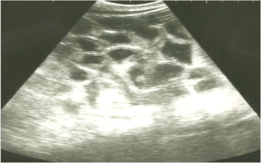
Figure 1: Ultrasonographic image of the left abdomen of horses with colic associated with enteritis and/or colitisusing 3.5 MHz transducer. The intestinal loops appear distended with fluid contents; the intestinal wall is slightly thickenedand corrugated. Increased intestinal motility of the intestinal loops was detected on the screen.
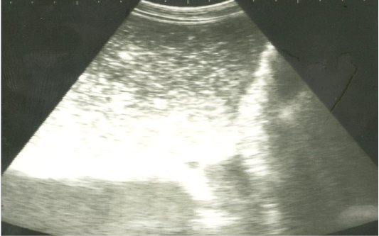
Figure 2: Ultrasonographic image of the left abdomen of horses with colic associated with impaction of the intestineusing 3.5 MHz transducer. There was marked distension of the descending colon with ingested materials that appeared hypo to hyperechoic. The intestinal loop diameter exceeds 8 cm.
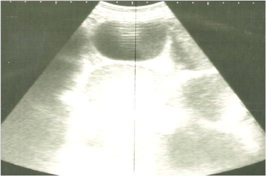
Figure 3: Ultrasonographic image of the left abdomen of horses with colic associated with impaction of the intestine using 3.5 MHz transducer. The intestinal loops were distended with fluid and food particlesand prominent signs of ileus.

