Special Issue: Emerging and Re-Emerging Animal Health Challenges in Low and Middle-Income Countries · Volume 13, Issue 1, 14 October 2025 · Open Access
Prescence and Verification of LasI and LasR Genes in Pseudomonas aeruginosa Causing Urinary Tract Infection in Dogs
·
Affiliations & Notes ⌄
1Department of Microbiology, College of Veterinary Medicine, University of Baghdad, Baghdad, Iraq. *Correspondence | Shatha E. Jasim, Department of Microbiology, College of Veterinary Medicine, University of Baghdad, Baghdad, Iraq
In this study the detection of QS genes LasR and LasI was found in 100% of biofilm forming isolates and 100% missing in isolate that do not contain biofilm (Figure 7).
Sequencing analysis of P. aeruginosa 16S rRNA gene was performed by MEGA6 software and showed the changes in nucleotide sequence compared to other globally known sequences.
In conclusion, this study highlights the biofilm forming abilities, importance of QS genes, and mutation occurrence of P. aeruginosa isolates from canine UTIs.
The identification of LasI/ LasR genes was found in P. aeruginosa isolates resulting in thought that these genes are connected to varying levels of intrinsic virulence and pathogenicity that prompting the need for further studies.
Selected from the article's own text.
Abstract
Pseudomonas aeruginosa is a pathogenic bacterium and has virulence factors under the quorum-sensing (QS) control increasing its pathogenicity. The purpose of this study was to detect some of the QS genes including LasI and LasR in P. aeruginosa isolated from dogs with urinary tract infection (UTI). Additionally, we aimed to determine histopathological changes in mice experimentally infected with bacteria. A group of mice was injected with phosphate buffer saline as a negative control, second group was infected with P. aeruginosa produced biofilm, and third group was infected with P. aeruginosa non produced biofilm. A total of 108 urine samples were collected from UTI dogs, obtained from the veterinary hospital and veterinary clinics. The identification of suspected isolates was confirmed by VITEK-2; and the genetic determination of 16S rRNA, LasI and LasR gene was performed. Detection of biofilm layer by tube method and congo red agar was performed. The results showed that 6/108 (5.5%) of isolates are positive for P. aeruginosa, five isolates (83.33%) produced biofilm layer which carried both LasI and LasR genes, and one isolate 1/6 (16.66%) had no biofilm product and missing LasI and LasR gene. In addition, presence of changes in nucleotide sequence of 16S rRNA in all P. aeruginosa isolates were assessed. LasI gene showed transition mutation in nucleotide sequence when compared to other globally known genes. The histopathological results revealed that the P. aeruginosa isolates produced biofilm layer severely affected mice compared to the isolates from non- biofilm layer. Taken together, molecular surveillance identifies that utilizing the LasR and LasI genes at the same time improves the accuracy of detection of P. aeruginosa producing biofilm.
Pseudomonas aeruginosa is frequently cause localized or system-specific infections, such as urinary tract infection, ears, eyes, and occasionally the respiratory system , mastitis in cattle . It is widely known to cause otitis externa in dogs , wound infections , and isolated from urine of dogs affected with malignant tumor (Sahar et al., 2009). Urinary tract infections (UTIs) are the most prevalent diseases in dogs, which are caused by bacterial pathogens , occurs frequently causing chronic kidney diseases with significant effects on the kidney functions . This bacterium regarded as a significant pathogen because its wide variety of resistance mechanisms ; . It has a vast genome that gives several virulence characteristics, including the ability to form biofilm , adhered to surfaces by extracellular polymer substances (EPS) called matrix, enables bacteria to survive in a harsh condition, and permits the cells within the biofilm to communicate with each other . The microbial biofilm is the most efficient and competing expression of the genome in prokaryotic biofilm cells, and being more protected and metabolically efficient, demonstrating tolerance to many stressors through a signaling system controlled by quorum-sensing (QS) system (Lazar and Chifiriuc, 2010; which is a bacterial cell-cell communication system that uses chemical signals to control gene expression levels depended on the cell density manner, it is crucial for pathogenicity, biofilm formation, and antibiotic resistance . Thes chemical signals known as autoinducers (AIs) that bind to specific receptors . Interestingly the genes that contribute to the composition of this system are about 10% of P. aeruginosa genome and for this status are responsible for most of the cellular physiology processes and virulence phenotypes .
In P. aeruginos, a there are seven genes associated with the occurrence of QS which are rhlA, rhlR, rhlI, lasR, lasI, lasB, phzA1 , and the main highly interconnected QS systems are Las, Rhl, Pqs, and Iqs , which are channels interlinked together that used transcriptional regulators such as LasR, RhlR, PqsR, and IqsR, respectively and connected to specific AIs leading to aggravating the expression of the selected genes that cause bacterial virulence . This bacterium has two AHL-depended on systems known as LasR/LasI and RhlR/RhlI, which regulate the synthesis and signal transduction of the N-(3-oxo-dodecanoyl)-L-homoserine lactone (OdDHL) and N-butanoyl-L-homoserine lactone (BHL), respectively . The QS system has significant role in this bacterium by increasing its pathogenicity .
This study aimed to investigate biofilm production in clinical P. aeruginosa isolates from dogs suffering UTI, and to detect the occurrence of mutations in16S rRNA in all six P. aeruginosa isolates. Additionally, we aim to determine if experimental infection of mice with diverse P. aeruginosa could induce differing histopathological changes.
MATERIALS AND METHODS
Samples collection
Samples were collected from dogs with suspected urinary tract infection (UTI) of different ages, both genders, and different breads, during the period from February 2024 to June 2024, obtained from the veterinary hospital and veterinary clinics located in various areas of Baghdad. This included 108 urine samples from 108 dogs with suspected UTI by spontaneous urination, collected in a sterile container, marked, and kept in cooling box to perform examination and bacterial diagnosing in the microbiology laboratory in the College of Veterinary Medicine in Baghdad.
General urine examination
This method was performed according to Punia et al., 2018 to detect the UTI in dogs. Taking 5 mL of the urine sample into a test tube and centrifuged at 3000 rpm for 7 min. The supernatant was discarded, placed the sediment on a glass slide for examination under a light microscope at 40× magnification.
Isolation and identification
Based on a loopful of urine spread onto blood agar and MacConkey agars (HiMedia, India), incubated aerobically at 37°C for 24 hrs. Then, isolated the colonies on selective media cetrimide agar and pseudomonas agars (HiMedia, India) to get pure growth , after that identified by biochemical tests such as oxidase and catalase, confirmation the isolates using VITEK-2 compact system according to the manufacturer company VITEK-2 technique (BioMerieux, France) was done using a GN colorimetric identification card to conformation the isolates ; .
Biofilm detection methods
Tube method (qualitative assay): A loopful of the isolated bacteria was inoculated into 5 mL of tryptic soy broth supplemented with 1% glucose and incubated at 37°C for 72 h. The tube was washed with normal saline and air-dried. The dried tube was heat-fixed by passing it through a flame three times. Subsequently, the biofilm was stained with 0.1% crystal violet for 1 h, excess stain was removed, and the tube was rinsed with normal saline and dried in an inverted position. Biofilm formation was confirmed by the presence of a visible layer on the inner walls and at the bottom of the tube .
Congo red method: On Congo red agar, P. aeruginosa isolate was cultured for 48 h at 37°C, black colonies indicated the biofilm formation, while the red colonies indicated non-biofilm formation .
Polymerase chain reaction (PCR): DNA was extracted according to the commercial purification system using (Genomic DNA Mini Kit, Korea) to detect 16S rRNA, LasR and LasI genes. The primers designed are mentioned in Table 1, and the optimum conditions of PCR cycles were: firstly one cycle of initial denaturation at 95ᵒC for 5 min, then 35 cycles of denaturation at 95ᵒC for 45 sec, annealing (16S rRNA at 58ᵒC/ LasR and LasI at 57ᵒC)
Table 1: The sequence of primers used in this study.
Primer
Sequence
Primer sequence
GC%
Size of product (bp)
Reference
16s RNA
27F
5'- AGAGTTTGATCCTGGCTCAG- 3'
50.0
1250 bp
(Srinivasanet al., 2015)
1392R
5'- GGTTACCTTGTTACGACTT- 3’
42.1
LasI
F
5'- CGTGCTCAAGTGTTCAAGG-3'
52.63
295 bp
R
5'- TACAGTCGGAAAAGCCCAG-3’
52.63
LasR
F
5'- ATGGCCTTGGTTGACGGT -3'
55.56
700 bp
R
5'- GCAAGATCAGAGAGTAATAAGACC -3’
44.00
for 45 sec, and extension at 72ᵒC for 1 min, at last, final extension at 72ᵒC for 5 min. The PCR products were stained with red stain (Intron, Korea), then electrophoresis on 1.5% agarose and observed under UV trans illuminator. The amplified products were sent for sequencing to Macrogen Inc. (Geumcheon-gu, Seoul, South Korea) using the Sanger’s method. The sequences were compared with the NCBI GenBank database using the basic local alignment search tool (BLAST) to confirm the isolates as P. aeruginosa.
Animal experimental design
A total of 24 mice were divided into three groups each one contains eight mice: first group (G1) of mice was injected with (PBS) as a negative control, second group (G2) mice were infected with biofilm producing P. aeruginosa, third group (G3) mice were infected with non-biofilm producing P. aeruginosa. The dose was given intraperitonially (I/P) , at 0.5 McFarland tube (1.5× 10⁸ CFU/ml) 0.25 ml, then sacrificed on 5th days .
Histological examination
Specimens were taken from internal organs (kidney, liver and spleen) of mice, fixed in 10% buffered neutral formalin. According to a technique of several steps was done for histological examination.
Statistical analysis
Data were analyzed using SAS (Statistical Analysis System - version 9.1). One-way analysis of variance (ANOVA) program was used to detect the percentage of difference groups in study parameters.
RESULTS
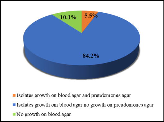
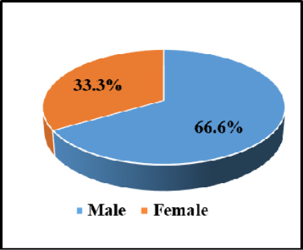
In the present study according to the microscopic analysis, the urine samples from which P. aeruginosa was isolated contained pus and bacteria, that is indicative of a urinary tract infection. The bacterial isolation and identification revealed out of 108 urine samples with UTIs the P. aeruginosa isolates were found in 6/108 (5.5%) (Figure 1). The incidence rate of infection in male was higher than females with a rate of 66.6% and 33.33%, respectively (Figure 2). The rate of infection occurred at a high infection rate 100% in adult > 1 year with no infection rate in young ≤1 year.
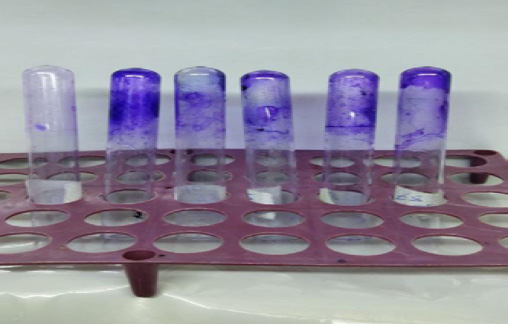
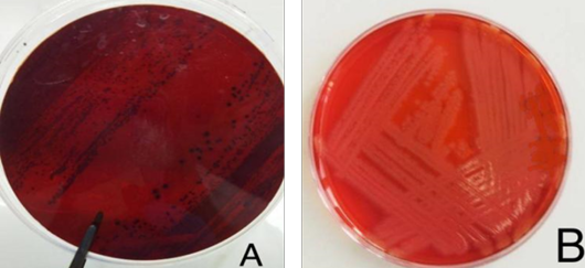
The biofilm formation was performed by using tube method and Congo red agar demonstrated that five out of six isolates (5/6, 83.33%) produced biofilm and only one isolate (1/6, 16.66%) failed to produce. In tube method, the positive results to detect the biofilm formation were indicated by a violet-colored layer adhering to the tube wall, while a colorless tube indicated a negative result (Figure 3). Black colonies on Congo red agar indicated the presence of a biofilm layer, while non-biofilm colonies appeared red color (Figure 4).
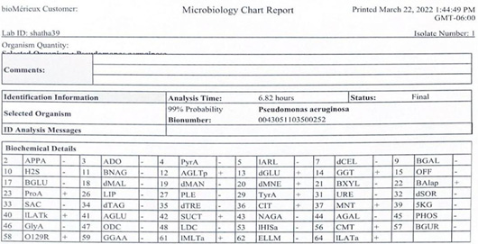
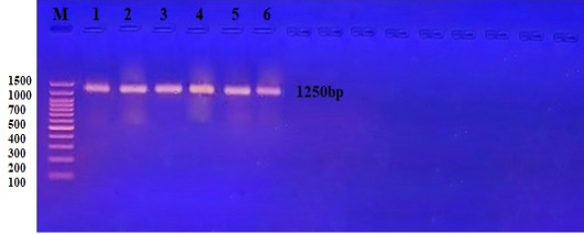
The P. aeruginosa confirmed by VITEK-2 system microbiology showed a match 99% (Figure 5), and the bacteria were genetically diagnosed by PCR and automated DNA sequencing to all isolates of P. aeruginosa amplified directed the 16S rRNA gene as in (Figure 6).
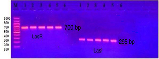
The identity of isolates was subsequently confirmed by sequencing the PCR. BLAST analysis showed 99% similarity to reference sequences of P. aeruginosa, including strain OR793893.1. The sequences have been added to the NCBI GenBank database and are available under the accession codes: PP979721.1, PP979722.1, PP979723.1, PP979724.1, PP979725.1, and PP979726.1. In this study the detection of QS genes LasR and LasI was found in 100% of biofilm forming isolates and 100% missing in isolate that do not contain biofilm (Figure 7).
Sequencing analysis of P. aeruginosa 16S rRNA gene was performed by MEGA6 software and showed the changes in nucleotide sequence compared to other globally known sequences. These changes were transition or transversion and are listed in Table 2.
Sequencing analysis of QS genes LasR and LasI in P. aeruginosa (ID: PP979723.1) was performed in MEGA6 software and has no changes in nucleotide sequence of LasR gene and had 100.
Introduction
Identity when compared to other globally known (ID: CP137940.1). On the other hands, LasI gene showed transition mutation in nucleotide sequence when compared to other globally known (ID: CP137912.1) as listed in Table 3.
Table 2: Genetic variation at 16S rRNA genes of Pseudomonas aeruginosa.
Identities
Sequence ID with submission
Sequence ID with compare
Nucleotide
Location
Type of substitution
No.
99%
ID: PP979721.1
ID: OR793893.1
G\A
133
Transition
1
99%
ID: PP979722.1
ID: OR793893.1
T\A
208
Transvertion
2
G\A
536
Transition
T\A
537
Transvertion
99%
ID: PP979723.1
ID: OR793893.1
T\C
222
Transition
3
A\C
277
Transvertion
99%
ID: PP979724.1
ID: OR793893.1
A\T
252
Transvertion
4
C\T
253
Transition
C\G
563
Transvertion
99%
ID: PP979725.1
ID: OR793893.1
G\T
539
Transvertion
5
99%
ID: PP979726.1
ID: OR793893.1
C\A
563
Transvertion
6
Source: P. aeruginosa; 16S rRNA gene.
Table 3: Genetic variation at LasR and LasI genes of Pseudomonas aeruginosa.
Identities
Predicted effect
Amino acid change
Nucleotide change
Nucleotide
Location
Type of substitution
Gene
No. of gene
100%
----
------
------
------
-----
------
LasR
1
99%
Missense
Q(Glutamine)\ R(Arginine)
CTG\CCG
T\C
4101403
Transition
LasI
2
Source: Pseudomonas aeruginosa (ID: PP979723.1). Sequence ID with compare: 1: LasR=ID: CP137940.1; and 2: LasI=ID: CP137912.1
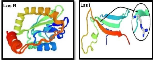
The result of protein conformation of LasR and LasI genes in P. aeruginosa (ID: PP979723.1) showing as three-dimensional (3D) modeling secondary structure, and exposes the location of mutation in only LasI protein as in Figure 8.
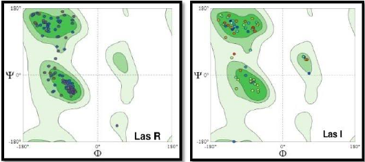
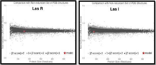
In Ramachandran graph for amino acid in general and local quality estimation with Z score of LasR and LasI genes, the results showed that model evaluation of the amino acid of query protein were in the most favorable energy regions for good quality model (Figure 9). The results also show that the model validation include the local quality estimation with Z score, located within the light or dark gray and blank areas (Figure 10).
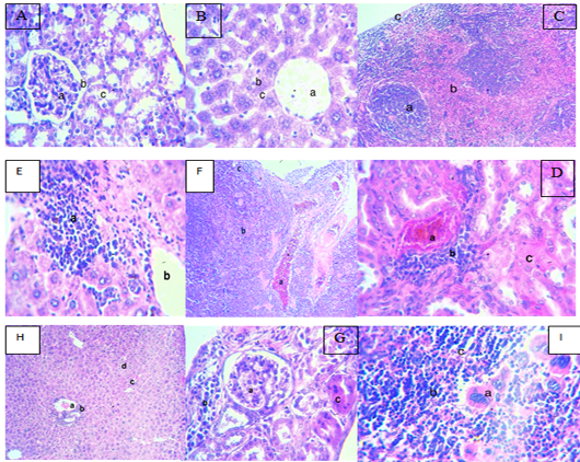
In histological examination of G1 mice injection with PBS as a negative control showed that the kidney with normal appearance of glomerular tuffet surrounded by Boman space, and normal proximal convoluted tubule (Figure 11A). Liver tissue showed normal central vein surrounded by a regular arrangement of normal hepatocytes and sinusoids (Figure 11B). The spleen has a normal (white pulp) consist of lymphoid follicles with central arteriols surrounded by lymphocytes, and the (red pulp) contains lymphocytes with blood vessels, all of them surrounded by a normal capsule (Figure 11C).
In G2 mice infected with P. aeruginosa, produced biofilm there were histological changes in kidney demonstrates severely congested blood vessels, aggregation of mononuclear cells, and cloudy swelling tubules (Figure 11D), the liver showed severe interstitial infiltration with inflammatory cells (lymphocytes and macrophages) with dilated central vein (Figure 11E), in spleen there is an extensive interstitial hemorrhage with the depleted lymphoid follicle, and the presence of necrotic lymphocytes (Figure 11F). In G3 mice infected with P. aeruginosa not produced biofilm there were histological changes in kidney demonstrates enlarged glomeruli surrounded by infiltration of mononuclear cells and tubules cloudy swelling (Figure 11G), the liver showed congested blood vessels, lymphocytic cuffing central vein, sinusoid hemorrhage, and apoptosis (Figure 11H). In spleen there is an increase in megakaryocytic cell, depleted lymphoid follicle with hemorrhage (Figure 11I).
In general, according to the results of this study, only six dogs (6/108, 5.5%) had a UTI caused by P. aeruginosa. This is consistent with another study where they have found 5.3% of dog cases with lower UTI and cystitis were caused by P. aeruginosa isolates. According to another study , P. aeruginosa was present in 4.2% of the dogs that had bladder infections, while isolated 13.4% P. aeruginosa from dogs with UTIs in Italy. The spread of P. aeruginosa in dogs with UTIs varies according to geographic area and underlying medical conditions . In this study, the incidence rate of males was higher than females (66.6%), and (33.3%), respectively, occurred at a high infection rate 100% in (adult > 1 Year). However, the study conducted by , presented that many reports showed UTIs are more prevalent in older female dogs with a mean age of about 7–8 years.
The genotypic results for the 16S rRNA gene confirmed the diagnosis for all isolates, allowing to conclude that all of them were P. aeruginosa, as expected. Also, the detection of QS genes LasR and LasI was found in 100% of biofilm-forming isolates and 100% missing in isolate that do not contain biofilm. The genes LasI and LasR were tested to know the quorum sensing regulatory system, which is essential for coordinating gene expression related to virulence and biofilm formation .
The substitutions in 16S rRNA gene were found in P. aeruginosa in this study showed transition and transversion substitutions at different locations with 99% identity when compared to other globally known ones. Moreover, the substitution in LasI gene found in P. aeruginosa in this study showed one substitution of Q(Glutamine)/R(Arginine) at location 4101403 was a missense mutation and had been 99% identity when compared with (ID: CP137912.1). In general, these genetic changes may result in a high degree of biofilm formation and serve a supporting role in the formation of high levels of pathogenicity in P. aeruginosa. In the study conducted by de Sousa et al. (2023) they suggest that biofilm production is common among P. aeruginosa isolates from dogs and may play a role in the pathogenesis of the disease, and found LasI gene within 90% of P. aeruginosa isolates from dogs and lasR gene was absence, whereas they found 100% of the isolates had lasR gene, and 97.5% had lasI genes.
From previous studies, found limited available data on the prevalence of lasR, lasI virulence genes in P. aeruginosa specifically isolates from dogs, highlighting the importance of this study. Also, some studies investigated the distribution of lasR, lasI genes in P. aeruginosa obtained from humans and the environment , they showed the prevalence of virulence genes and found the genes lasR was present in a significant proportion of carbapenem-resistant P. aeruginosa (CRPA) isolates in 96.7% of the isolates. Another study conducted by (O’Connor et al., 2021) investigated the prevalence of virulence genes in 90 environmental P. aeruginosa and found that lasR was presented in 50% of the isolates. The result of protein conformation of LasR and LasI protein in this study revealed there was no change in 3D secondary structure of lasR and distribution the amino acid of query protein were in the most favorable energy regions for a favorable quality model, and located within the black areas with graph of Z score indicated of an effective model, while LasI 3D structure showed the location of mutation, and distribution the amino acid of query protein were in the most favorable energy regions for good quality model and located within the black areas with graph of Z score, these results were differed from they revealed there is change in 3D structure of LasR protein with distribution the amino acid of query protein were in the unfavorable energy regions, and negative Z-score indicated of a favorable model.
Plokarz et al. (2022); Zhang et al. (2022) have shown that the studying P. aeruginosa biofilms in dogs enables the researchers to gain a better understanding of how these structures contribute to infection and develop more effective treatments revealed that P. aeruginosa possesses four complicated (QS) system used several signals and receptors to regulate virulence and pathogenicity, among which the LasI/ LasR and RhlI/LasR system is regulated by (3-oxo-C12-HSL) and (C4-HSL) respectively, plays a major role in host pathogenesis. The las and rhl quorum-sensing circuitry operate in a hierarchical cascade responsible for regulating the expression of many virulence determinants, secondary metabolites, type 2 secretion system, stationary phase genes and genes involved in biofilm formation . Therefore, the persistence of P. aeruginosa in various environments, including host tissues regulates the synthesis of proteases, toxins, and other exoproducts that contribute to pathogenicity.
The histopathological finding in this study, the G2 mice infected with P. aeruginosa forming biofilm there were histopathological changes in kidney tissue demonstrates severely congested blood vessels, aggregation of mononuclear cells, and cloudy swelling tubules, the liver section showed severe interstitial infiltration with inflammatory cells (lymphocytes and macrophages) with dilated central vein, while spleen tissue shows an extensive interstitial hemorrhage with the depleted lymphoid follicle with the presence of necrotic lymphocytes. In G3 mice infected with P. aeruginosa non-forming biofilm the histopathological changes in kidney tissue demonstrates enlarged glomeruli surrounded by infiltration of mononuclear cells and tubules cloudy swelling, the liver showed congested blood vessels, lymphocytic cuffing central vein, sinusoid hemorrhage, and apoptosis, in addition the spleen section showed an increase in megakaryocytic cell, depleted lymphoid follicle with hemorrhage. These results corresponds to found in their study the severity of lesions induced by four-days old biofilm cells was more as compared to planktonic cells. Although, that planktonic as well as biofilm cells both the cell forms of P. aeruginosa were able to induce chronic renal inflammation in the experimental animals, more significantly, when bacteria and neutrophils interact during the planktonic stage of an infection, the pathogen is typically killed, inflammatory mediators are regulated, neutrophils undergo apoptosis, and acute inflammation is resolved, conversely, the bacterial infection biofilm stage promotes neutrophil necrosis and results in chronic inflammation (Lenda et al., 2019). The reasons for these histopathological changes may be due to the virulence factors of P. aeruginosa that counteract host defenses causing direct damage to the organ tissues or making the bacteria more competitive, this agrees with . Also, this results corresponds to (Alrabia et al., 2024) they showed the presence of cystic dilatation of some renal tubules with cloudy swelling of others, atrophy of some glomeruli with degeneration of some renal tubules, constriction of renal blood vessels, and degenerative changes of some renal tubules were all observed in the kidney tissues of the mice given strain PAO1 injection, also in the liver tissue showed diffuse vacuolation of certain hepatocytes, significant hepatic blood vessel congestion, and focal regions with inflammatory cell infiltration in their liver tissues, and in the spleen there was a localized area of extravasated blood (hemorrhage), splenic blood vessel congestion, and a decrease in white pulp lymphocytes. As well as (Abd Al-Rubai, 2013) appeared kidney aggregation of neutrophils and lymphocytes around congested blood vessels and also between kidney tubules with severe cytoplasmic vacuolation of endothelium cells and others explained congestion of blood vessels with filtration neutrophils between kidney tubules, and showed spleen depletion of white pulp with neutrophil infiltration in the congested red pulp, however, the liver showed granuloma with congestion of the central vein and sinusoids with neutrophils in the lumen as well as necrosis of hepatocytes with an explanation of sinusoids with neutrophils in the lumen added to necrosis hepatocytes (Abd Al-Rubai, 2013). The researchers presented that the spleen of the mice in the P. aeruginosa infected group showed signs of severe inflammation, the red and white pulps were enlarged, and the characteristic germinal center structures were lost. There were also hemorrhages, clogged blood vessels, inflammatory cell infiltration, and many erythrocytes in the red pulp. The study of showed that increased gut LPS produced by P. aeruginosa promotes gut permeability destruction resulting in the direct transfer of LPS to hepatocytes in part via portal veins that promote systemic inflammation (inflammation, apoptosis, and increasing oxidative stressors in hepatocytes), adding to that LecA binds to glycoprotein oligosaccharides on the surface of epithelial and endothelial cells to help P. aeruginosa adhere to host cells . While the researchers revealed that the virulence factors promoted by RpoN are siderophore pyoverdine, and the phenazine compound pyocyanin, these factors are a redox-active molecule that causes oxidative stress that damages target cells. On the other hand, it has been demonstrated that the (T3SS) secretion system effectors ExoS and ExoY, cause regions of cell death, which results in defects in the barrier of the epithelial cells .
CONCLUSION
In conclusion, this study highlights the biofilm forming abilities, importance of QS genes, and mutation occurrence of P. aeruginosa isolates from canine UTIs. The relatively low frequency of P. aeruginosa in dogs UTIs indicates that the epidemiology of this bacterium is not significantly impacted by domestic dogs, probably act as an ecological reservoir for the virulent P. aeruginosa that can be harmful. The identification of LasI/ LasR genes was found in P. aeruginosa isolates resulting in thought that these genes are connected to varying levels of intrinsic virulence and pathogenicity that prompting the need for further studies. The main histopathological finding emphasizing that the biofilm-forming strain caused more severe histological changes than non-biofilm-forming strain.
Acknowledgment
The authors express their sincere gratitude to Baghdad University/College of Veterinary Medicine for the support and facilities provided.
Novelty Statement
The study revealed that the P. aeruginosa isolates produced biofilm layer severely affected mice compared to the isolates from non- biofilm layer. Taken together, molecular surveillance identifies that utilizing the LasR and LasI genes at the same time improves the accuracy of detection of P. aeruginosa producing biofilm.
Author’s Contribution
SEJ: Conceptualization, methodology, validation, writing original draft, writing review and editing.
SMH: Conceptualization, methodology, review and editing, supervisor.
Ethical approval
Ethical approval was obtained through the local committee of animal care in the College of Veterinary Medicine, University of Baghdad (Number 279 on 7/2/2024) before starting this study.
Generative AI and AI-assisted technology statement
The authors declare that no Genrative AI was used in the creation of this manuscript.
Conflict of interest
The authors have declared no conflict of interest.
Figures
Figure 1:.
Incidence rate of UTIs caused by P. aeruginosa isolated from urine dogs
Figure 2:.
Incidence rate of P. aeruginosa from dogs with UTIs according to sex.
Figure 3:.
Tube method for detecting the biofilm formation by P. aeruginosa. A positive result is a violet-colored layer adhering to the tube wall, while a negative result is a colorless tube.
Figure 4:.
Pseudomonas aeruginosa colonies on the Congo red agar. (A) black colonies indicate the biofilm formation. (B) red color colonies indicate non-biofilm formation.
Figure 5:.
VITEK 2 microbiology chart report for P. aeruginosa identification.
Figure 6:.
PCR products with a band size of approximately 1250 bp of 16S rRNA. The products were subjected to electrophoresis on 1.5% agarose at 5 volt/cm2 for 1:30 h. M: DNA ladder (100 bp).
Figure 7:.
PCR product of the band size in LasR and LasI. The product was electrophoresis on 1.5% agarose at 5 volt/cm2. 1x TBE buffer for 1:30 hours. M: DNA ladder (100).
Figure 8:.
The ribbon representation of 3D secondary structure of LasR, and LasI proteins of P. aeruginosa producing biofilm (ID: PP979723.1), the enlarged part of LasI appears the location of mutation.
Figure 9:.
Distribution of the amino acid residues in the Ramachandran graph of LasR and LasI proteins of P. aeruginosa producing biofilm (ID: PP979723.1).
Figure 10:.
Representative Z-score graphs of the generated models, compared to X-ray (gray) and magnetic (dark gray) crystallography models deposited in the PDB. The dots in red refer to the generated models for the LasR and LasI protein of P. aeruginosa biofilm producing isolate (ID: PP979723.1).
Figure 11:.
A: Histological section of normal kidney in G1 mice a- Glomerular tuffet, b-Boman space c-Proximal convoluted tubule. (H and E 400X), B: Histological section of normal liver in G1 mice a- Normal central vein b- hepatocytes c-sinusoids. (H and E 400X), C: Histological section of normal spleen in G1 mice a- Lymphoid follicle consists of central arterioles surrounded by lymphocytes (white pulp) b-Red pulp consists of lymphocytes with blood vessels c-spleen capsule. (H and E 400X), D: Histopathological section of kidney in G2 mice infected with P. aeruginosa produced biofilm. a-Severely congested blood vessels b- Mononuclear cells aggregation surrounding blood vessels c- Tubules cloudy swelling (H and E 400X) , E: Histopathological section of liver in G2 mice infected with P. aeruginosa produced biofilm. a- severe interstitial infiltration in hepatocyte with inflammatory cells (lymphocytes and macrophage) b-dilated central vein (H and E 400X) , F: Histopathological section of spleen in G2 mice infected with P. aeruginosa produced biofilm a- Extensive interstitial hemorrhage b-Depleted lymphoid follicle c-Necrotic lymphocytes. (H and E 200X), G: Histopathological section of kidney in G3 mice infected with P. aeruginosa non produced biofilm. a- Enlargement glomeruli b-Mononuclear cells Infiltration surrounded glomeruli c-Tubule cloudy swelling. (H and E 400X) , H: Histopathological section of liver in G3 mice infected with P. aeruginosa non produced biofilm. a- congested blood vessel b- lymphocytic cuffing central vein c- sinusoids hemorrhage d- apoptosis 200 X (H and E), and I: Histopathological section of spleen in G3 mice infected with P. aeruginosa non produced biofilm. a-Increase in the megakaryocytic cell b- Depleted lymphoid c- Hemorrhage. (H and E 400X).
References
Abd-Al-Rubai MG (2013). The relationship between the site of infection and virulence of Pseudomonas aeruginosa experimental infection in mice. Iraqi J. Vet. Med., 37(2): 284-293.
Abed DA, Hamzah KJ, Abdul-Kareem S, Mehrzad J (2024). Bacteriological and molecular study of some urinary tract infections bacteria in human and cows in Babylon Province. Pak. Vet. J., 44(4).
Al-Barhawee NIK, Al-Rubyee SS (2024). Inhibition of biofilm formation in Agrobacterium tumefaciens by cell-free supernatants of Pseudomonas aeruginosa analyzed by GC-MS. Baghdad Sci. J., 21(7): 2222-2222. https://doi.org/10.21123/bsj.2023.8692[CrossRef]
Ali N, Ahmed ST, Almohaidi AMS (2024). Association of pvc genes expression with Biofilm formation in clinical isolates of Pseudomonas aeruginosa. Baghdad Sci. J., 21(2): 261-261. https://doi.org/10.21123/bsj.2023.7823[CrossRef]
Ali TH (2019). Effect of bacteriocin exract from Bifidobacterium spp. and Lactobacillus casei on the inhibition of the growth and pathogenesis of multiple resistant Pseudomonas aeruginosa isolated from bovine mastitis. Ms.c thesis- College of Veterinary Medicine- Baghdad University, Baghdad, Iraq.
Al-Rabia MW, Asfour HZ, Alhakamy NA, Bazuhair MA, Ibrahim TS, Abbas HA, Seleem NM (2024). Cilostazol is a promising anti-pseudomonal virulence drug by disruption of quorum sensing. AMB Express, 14(1): 87. https://doi.org/10.1186/s13568-024-01740-1[CrossRef]
Al-Rasheed AA, Garba B, Handool KO, Al-Jashamy KA, Odhah MNA, Dirie NI, Daud HM (2023). An in-vivo experimental evaluation of the efficacy of fish-derived antimicrobial peptides against multidrug-resistant Pseudomonas aeruginosa. Pan Afr. Med. J., 46(1). https://doi.org/10.11604/pamj.2023.46.112.38578[CrossRef]
Al-Taee HS, Al-Samarraae IA, Al-Ahmed HI (2019). Antibiotic susceptibility and molecular detection of Pseudomonas aeruginosa isolated from bovine mastitis. Iraqi J. Vet. Med., 43(2): 77-85. https://doi.org/10.30539/iraqijvm.v43i2.536[CrossRef]
Ataya HA, Soliman SM, Marouf S, Alamry K (2023). Incidence, bacterial causes and antibiotic resistance patterns of urinary tract infection in pet animals. J. Appl. Vet. Sci., 8(2): 35-43.
Azemin A, Alias N, Kari A (2022). Identification of Pseudomonas aeruginosa strains isolated from Dorper sheep milk with subclinical-mastitis infection by uniplex PCR using 16S rRNA, lasI/R, gyrB and ecfX genes. Malays. J. Fundament. Appl. Sci., 18(1): 30-42. https://doi.org/10.11113/mjfas.v18n1.2302[CrossRef]
Bhardwaj S, Bhatia S, Singh S, Franco Jr F (2021). Growing emergence of drug-resistant Pseudomonas aeruginosa and attenuation of its virulence using quorum sensing inhibitors: A critical review. Iran. J. Basic Med. Sci., 24(6): 699.
Cary NC (2010). SAS. SAS/STAT users guide for personal computer. Release 9.13. USA: SAS Institute. https://doi.org/10.1073/pnas.1007077107[CrossRef]
Ciocan MOA, Çiftçi A, Carp CM, Mareș M, Guguianu E, Rîmbu CM (2016). Determination of biofilm production in animals originated Pseudomonas aeruginosa strains. Reposit. Lasi Univ. Life Sci., 59: 4.
Ciszek-Lenda M, Strus M, Walczewska M, Majka G, Machul-Żwirbla A, Mikołajczyk D, Marcinkiewicz J (2019). Pseudomonas aeruginosa biofilm is a potent inducer of phagocyte hyperinflammation. Inflamm. Res., 68: 397-413. https://doi.org/10.1007/s00011-019-01227-x[CrossRef]
de Sousa T, Garcês A, Silva A, Lopes R, Alegria N, Hébraud M, Poeta P (2023). The impact of the virulence of Pseudomonas aeruginosa isolated from dogs. Vet. Sci., 10(5): 343. https://doi.org/10.3390/vetsci10050343[CrossRef]
Devnath P, Uddin MK, Aha F (2017). Extraction, purification and charact Pseudomonas aeruginosa extraction. Int. Res. J. Biol. Sci., 6(5): 1-9.
Fernandes S, Borges A, Gomes IB, Sousa SF, Simões M (2023). Curcumin and 10-undecenoic acid as natural quorum sensing inhibitors of LuxS/AI-2 of Bacillus subtilis and LasI/LasR of Pseudomonas aeruginosa. Food Res. Int., 165: 112519. https://doi.org/10.1016/j.foodres.2023.112519[CrossRef]
Furtuna DK, Debora K, Wasito EB (2018). Comparison of microbiological examination by test tube and congo red agar methods to detect biofilm production on clinical isolates. Folia Med. Indones., 54(1): 22-28. https://doi.org/10.20473/fmi.v54i1.8047[CrossRef]
Gellatly SL, Hancock RE (2013). Pseudomonas aeruginosa: New insights into pathogenesis and host defenses. Pathog. Dis., 67(3): 159-173. https://doi.org/10.1111/2049-632X.12033[CrossRef]
Golovkine G, Faudry E, Bouillot S, Elsen S, Attrée I, Huber P (2016). Pseudomonas aeruginosa transmigrates at epithelial cell-cell junctions, exploiting sites of cell division and senescent cell extrusion. PLoS Pathog., 12(1): e1005377. https://doi.org/10.1371/journal.ppat.1005377[CrossRef]
Grishin AV, Krivozubov MS, Karyagina AS, Gintsburg AL (2015). Pseudomonas aeruginosa lectins as targets for novel antibacterials. Acta Nat., (англоязычная версия), 7(25): 29-41. https://doi.org/10.32607/20758251-2015-7-2-29-41[CrossRef]
Hakim AS, Dorgham SM, Abuelhag HA, Sadek EG, Dapgh AN, Youssif NH (2024). Isolation and identification of Pseudomonas aeruginosa obtained from dogs and cats in Great Cairo regarding status of phenotypic antimicrobial resistance pattern. Egypt. Pharm. J., 23(3): 525-531. https://doi.org/10.4103/epj.epj_340_23[CrossRef]
Hattab J, Mosca F, Di Francesco CE, Aste G, Marruchella G, Guardiani P (2021). Occurrence, antimicrobial susceptibility, and pathogenic factors of Pseudomonas aeruginosa in canine clinical samples. Vet. World, 14(4): 978. https://doi.org/10.14202/vetworld.2021.978-985[CrossRef]
Hemmati J, Nazari M, Abolhasani FS, Ahmadi A, Asghari B (2024). In vitro investigation of relationship between quorum-sensing system genes, biofilm forming ability, and drug resistance in clinical isolates of Pseudomonas aeruginosa. BMC Microbiol., 24(1): 99. https://doi.org/10.1186/s12866-024-03249-w[CrossRef]
Hickey C, Schaible B, Nguyen S, Hurley D, Srikumar S, Fanning S, Schaffer K (2018). Increased virulence of bloodstream over peripheral isolates of P. aeruginosa identified through post-transcriptional regulation of virulence factors. Front. Cell. Infect. Microbiol., 8: 357. https://doi.org/10.3389/fcimb.2018.00357[CrossRef]
Khames SASK, Ahmed ST (2024). Effect of chemically synthesis compared to biosynthesized zinc oxide nanoparticles using extract of Vitex agnus on the expression of MexAB-OprM efflux pump genes of multi-drug resistance Pseudomonas aeruginosa. Baghdad Sci. J., 21(12 Suppl.): 4050-4066. https://doi.org/10.21123/bsj.2024.10976[CrossRef]
Lafta IJ, Sadeq Z (2024). Pseudomonas aeruginosa is an effective indicator for screening of quorum sensing inhibition by plant extracts. Iraqi J. Vet. Med., 48(1): 54-62. https://doi.org/10.30539/nzsc0y49[CrossRef]
Lazãr V, Chifiriuc MC (2010). Architecture and physiology of microbial biofilms. Roum Arch. Microbiol. Immunol., 69(2): 95-107.
Lima JLDC, Alves LR, Jacomé PRLDA, Bezerra JP, Maciel MAV, Morais MMCD (2018). Biofilm production by clinical isolates of Pseudomonas aeruginosa and structural changes in LasR protein of isolates non biofilm-producing. Braz. J. Infect. Dis., 22(2): 129-136. https://doi.org/10.1016/j.bjid.2018.03.003[CrossRef]
Mittal R, Kaur A, Joshi K, Nada R, Chhibber S, Harjai K, Sharma S (2009). Experimental non-obstructive chronic renal infection model with planktonic and biofilm cells of Pseudomonas aeruginosa. Am. J. Biomed. Sci., 1(2): 103-114. https://doi.org/10.5099/aj090200103.[CrossRef]
Mohammad TH, Shehab ZH, Tawfiq AA (2024). Growth inhibition of Pseudomonas aeruginosa using products of some probiotic microorganisms and secondary metabolites of Commiphora myrrha extracts estimated by GC-MS technique. Baghdad Sci. J., 21(11): 3463-3475. https://doi.org/10.21123/bsj.2024.9704[CrossRef]
Mohammed BQ, Abdullah AH, Rayshan AR (2024). Molecular identification of several Pseudomonas aeruginosa virulence genes. J. Anim. Health Prod., 12(s1): 67-74. https://doi.org/10.17582/journal.jahp/2024/12.s1.67.74[Full text][CrossRef]
Mohammed HA, Zgair AK (2022). Detection of quorum sensing genes of Pseudomonas aeruginosa isolated from different areas in Iraq. Iraqi J Sci., pp. 4665-4673. https://doi.org/10.24996/ijs.2022.63.11.5[CrossRef]
Mohmoud MH, Al-Dujaily AH (2018). Dipstick urine analysis screening among asymptomatic dogs of k9 units. Iraqi J. Vet. Med., 42(1): 61-64. https://doi.org/10.30539/iraqijvm.v42i1.32[CrossRef]
O’Connor K, Zhao CY, Mei M, Diggle SP (2022). Frequency of quorum-sensing mutations in Pseudomonas aeruginosa strains isolated from different environments. Microbiology, 168(12): 001265. https://doi.org/10.1099/mic.0.001265[CrossRef]
Panpetch W, Tumwasorn S, Leelahavanichkul A (2024). Presence of Pseudomonas aeruginosa in feces exacerbate leaky gut in mice with low dose dextran sulfate solution, impacts of specific bacteria. PLoS One, 19(11): e0309106. https://doi.org/10.1371/journal.pone.0309106[CrossRef]
Park Y, Koo SH (2022). Epidemiology, molecular characteristics, and virulence factors of carbapenem-resistant Pseudomonas aeruginosa isolated from patients with urinary tract infections. Infect. Drug Resist., pp. 141-151. https://doi.org/10.2147/IDR.S346313[CrossRef]
Płókarz D, Czopowicz M, Bierowiec K, Rypuła K (2022). Virulence genes as markers for Pseudomonas aeruginosa biofilm formation in dogs and cats. Animals, 12: 422. https://doi.org/10.3390/ani12040422[CrossRef]
Punia M, Gulia D, Kumar A, Charaya G (2018). Evaluation of physico-chemical and microscopical changes of urine in dogs with urinary tract infection. Haryana Vet., 57(2): 191-193.
Raheem SA, Abdalshheed DA (2023). Evaluating the antibacterial and biofilm activity of Pseudomonas aeruginosa isolated from dog’s wound infections. Iran. J. Ichthyol., 10: 96-104.
Rampacci E, Bottinelli M, Stefanetti V, Hyatt DR, Sgariglia E, Coletti M, Passamonti F (2018). Antimicrobial susceptibility survey on bacterial agents of canine and feline urinary tract infections: weight of the empirical treatment. J. Glob. Antimicrob. Resist., 13: 192-196. https://doi.org/10.1016/j.jgar.2018.01.011[CrossRef]
Razzaq AH, Al-Kubaisi W, Aziz L, Hussain A (2017). Bacteriological and molecular study of Pseudomonas aeruginosa strains isolated from different clinical cases in Erbil and Kurkuk. Iraqi J. Vet. Med., 41(2): 124-130. https://doi.org/10.30539/iraqijvm.v41i2.61[CrossRef]
Rex DAB, Saptami K, Chandrasekaran J, Rekha PD (2023). Pleotropic potential of quorum sensing mediated N-acyl homoserine lactones (AHLs) at the LasR and RhlR receptors of Pseudomonas aeruginosa. Struct. Chem., 34(4): 1327-1339. https://doi.org/10.1007/s11224-022-02115-7[CrossRef]
Saher MH, Abdulrazak MA, Aboud AS, Al-khaban JM, Iesa AM (2009). Study on bacterial isolation from dogs affected with malignant tumor. Iraqi J. Vet. Med., 33(1): 120-131. https://doi.org/10.30539/iraqijvm.v33i1.725[CrossRef]
Sánchez-Jiménez A, Llamas MA, Marcos-Torres FJ (2023). Transcriptional regulators controlling virulence in Pseudomonas aeruginosa. Int. J. Mol. Sci., 24(15): 11895. https://doi.org/10.3390/ijms241511895[CrossRef]
Schuster M, Greenberg EP (2006). A network of networks: Quorum-sensing gene regulation in Pseudomonas aeruginosa. Int. J. Med. Microbiol., 296(2-3): 73-81. https://doi.org/10.1016/j.ijmm.2006.01.036[CrossRef]
Shalata MEM (2022). Screening of potential blockers of Pseudomonas aeruginosa third quorum sensing system; Pseudomonas quinolone signal (PQS). https://hdl.handle.net/20.500.12608/43073
Srinivasan R, Karaoz U, Volegova M, MacKichan J, Kato-Maeda M, Miller S (2015). Use of 16S rRNA gene for identification of a broad range of clinically relevant bacterial pathogens. PLoS One, 10(2): e0117617. https://doi.org/10.1371/journal.pone.0117617[CrossRef]
Suvarna KS, Layton C, Bancroft JD (2018). Bancroft’s theory and practice of histological techniques E-Book. Elsevier health sciences.
Thompson MF, Litster AL, Platell JL, Trott DJ (2011). Canine bacterial urinary tract infections: New developments in old pathogens. Vet. J., 190(1): 22-32. https://doi.org/10.1016/j.tvjl.2010.11.013[CrossRef]
Wu L, Luo Y (2021). Bacterial quorum-sensing systems and their role in intestinal bacteria-host crosstalk. Front. Microbiol., 12: 611413. https://doi.org/10.3389/fmicb.2021.611413[CrossRef]
Yaseen NN, Ahmed DA (2023). Detection of mexB multidrug efflux gene in some local isolates of Pseudomonas aeruginosa. Iraqi J. Sci., pp. 111-118. https://doi.org/10.24996/ijs.2023.64.1.11[CrossRef]
Yu Z, Wang Y, Chen Y, Huang M, Wang Y, Shen Z, Li G (2020). Antimicrobial resistance of bacterial pathogens isolated from canine urinary tract infections. Vet. Microbiol., 241: 108540. https://doi.org/10.1016/j.vetmic.2019.108540[CrossRef]
Zhang Y, Cheng P, Wang S, Li X, Peng L, Fang R, Chen H (2022). Pseudomonas aeruginosa biofilm dispersion by the mouse antimicrobial peptide CRAMP. Vet. Res., 53(1): 80. https://doi.org/10.1186/s13567-022-01097-y[CrossRef]