
Feline parvovirus (FPV) infection, which is common in cats around the world, progresses with leukopenia, gastroenteritis and can lead to death in young animals. The virus is a non-enveloped, single-stranded DNA virus; located in the genus of Carnivore protoparvovirus 1. Feline panleukopenia virus (FPLV) and Canine Parvovirus (CPV-2), which causes the disease, are genetically closely related and show a high genomic similarity. Groups are formed according to the genomic differences of parvoviruses, especially in the VP2 gene. There are 3 groups as G1, G2, G3 in FPV and 3 groups as 2a, 2b and 2c in CPV-2. The present study aimed to determine the presence of infection, to perform molecular characterization of the virus at the VP2 gene level, and to investigate genomic differences. FPLV DNA was detected in 7 (36.84%) of the stool samples collected from 19 cats with diarrhea and vomiting symptoms. Genotyping and phylogenetic analysis of the positive samples were performed. It was observed that five of the FPLV positive samples obtained were in the G3, one sample was in the G1, and one sample was in the CPV-2b group. FPV was investigated for the first time in Balıkesir province and the data of the feline parvovirus sequence was defined to Genbank. Current phylogenetic information about the FPV in Turkey was obtained in the present study.
Research Article · Volume 56, Issue 2, 17 January 2024 · Open Access
Heterogeneity of Feline Parvoviruses Genotypes and Determination of Distinct Genetic Lineages in Circulation in Turkey
·
Affiliations & Notes ⌄
- 1Faculty of Veterinary Medicine, Department of Virology, Balikesir University, 10100 Balıkesir, Turkey. 2Faculty of Veterinary Medicine, Department of Virology, Atatürk University, 25240 Erzurum, Turkey * Corresponding author: zeynepkarapinar@gmail.com
Article Info ⌄
- Journal:
- Pakistan Journal of Zoology
- Published:
- 17 January 2024

From the article
- Infection due to parvovirus in cats is caused by FPLV and CPV-2 genotypes.
- When this situation is evaluated together with the strains previously detected in our country, it shows that the CPV-2b genotype is still circulating in cats in Turkey.
- The fact that the strains obtained in the studies (phylogeny) conducted to investigate the molecular characterization of FPLV in the world and in our country in recent years are mostly in the G3 group suggests that the G3 genotype has begun to dominate among the circulating strains.
- Since cats are susceptible to both CPV and FPV viruses, superinfection and coinfection with more than one strain of parvovirus can occur.
Selected from the article's own text.
Abstract
Introduction
Feline panleukopenia is an infection with high morbidity and mortality, which is caused by Feline panleukopenia virus (FPLV), also known as feline parvo virus (FPV), in domestic or wild cats (subclasses Mustelidae, Procyonidea and Viverridea) and is characterized with fever, vomiting, anorexia, apathy, leukopenia and diarrhea .
FPLV is a single-stranded, non-enveloped DNA virus with a length of 4.5-5.5 kb which is included in the species of Carniivore protoparvovirus 1, family of Parvoviridae and subfamily of Parvovirinae ; . Although FPLV and canine parvovirus (CPV-2) are closely related genetically, they show high genomic similarity. In the case of canine parvovirus, although it is a DNA virus, 3 antigenic variants, defined as CPV-2a, CPV-2b and CPV-2c, have emerged with a single point mutation occurring at amino acid 426 in a very unique way with a single nucleotide change. It has been stated that viruses of this genetic type can also cause gastroenteritis in cats that cannot be clinically differentiated from FPLV ; ; . The VP2 major capsid protein of parvoviruses reveals the antigenic properties of the virus. The amino acid residues on the VP2 protein facilitate the adaptation of the virus to the organism (at amino acid positions 297, 300, 305, 323, 568) . Genotypes are distinguished from each other phylogenetically with the amino acid changes occurring in the VP2 capsid proteins between FPLV and CPV-2 .
FPLV can be transmitted through the fecal-oral route as well as with body fluids, feces, or fomites. The virus, which is highly resistant to environmental conditions due to its non-membrane character, can survive for a long time at infected organic materials .
Since FPLV uses the replication enzymes of the host organism, Replication occurs primarily in rapidly dividing cells at the S-phase, in tissues with high mitotic activity. In addition, infection of the lymphoid tissues causes immunosuppression. Fetal death, abortion and mumification, and fetal nerve damage may occur due to infection during pregnancy .
The clinical course of the disease in cats infected with FPLV varies depending on factors such as age, breed, vaccination, and immune system status. The disease may be per acute and may result in sudden death due to septic shock in cats under 2 months of age without showing any clinical signs. Vaccination is the effective way for prevention against the FPLV infection .
The aim of this study is to reveal the heterogeneity of the strains circulating in Balıkesir province as a pilot region and the strains obtained in studies conducted in our country and in the world with a broad perspective on the scale of Turkey. In addition, another aim of this study is to investigate the phylogenetic differences of all strains included in the study by evaluating the genomic differences of the virus at the VP2 gene level. Therefore, it was aimed to determine the cat/dog parvovirus genetic strains circulating in cats in our country.
Materials and methods
Materials
The material of the study consisted of fecal samples taken from cats in a shelter in Balıkesir. Stool samples were collected from 19 unvaccinated, mixed breed cats with diarrhea and vomiting symptoms. Fresh fecal samples taken from animals were stored at -20 ºC until they were studied.
Methods
Extraction of viral RNA from stool samples; was performed by using a commercial viral nucleic acid isolation kit (Jena Bioscience, Viral RNA+DNA Preparation Kit, Germany) in accordance with the kit’s procedure. H-forward (5’-CAGGTGATGAATTTGCTACA-3’) and H-reverse (5’- CATTTGGATAAACTGGTGGT 3’) primers targeting the partial region (629 bp) of the VP2 gene of FPLV were used for PCR . The obtained PCR products were evaluated by running on a 1.5% agarose gel. The amplified PCR products were purified using a commercial purification kit and these products were subjected to sequence analysis with Sanger method by using the primers (H forward and H reverse) used in the PCR process. The raw data obtained after sequencing were aligned with the clustal W algorithm using BioEdit version 7.0.5 . Nucleotide sequences were compared with different reference FPLV isolates using the BLAST software available in the NCBI database. Phylogenetic analysis was performed with MEGA v6.0 program . For this purpose, maximum likelihood was used. Bootstrap value was determined as 1000 repetitions.
Results and Discussion
Feline panleukopenia, caused by the FPLV, is an important highly contagious disease of domestic and wild cats characterized by high fever, dehydration, vomiting, leukopenia and diarrhea . Environments where a large number of cats and dogs coexist, such as shelters or living in the same houses, increase the risk of infection. Balıkesir province, which has an important place in terms of tourism, livestock breeding and pet sector and where large shelters are located, is in the North Aegean region in the west of Turkey. In this study, fresh stool samples of 19 cats collected from Balıkesir province were investigated by PCR for the presence of FPLV virus. FPLV DNA was detected in 7 (36.84%) of 19 samples obtained from cats. The ages of the sampled cats were generally less than one year old. There are a few studies investigating the disease serologically and virologically in our country. In a study conducted in Afyonkarahisar province, where FPLV was demonstrated serologically in the cat population in Turkey, it has been determined that seropositive cats are generally under 2 years old and when FPLV positive cats are examined by gender, there is no significant difference according to gender . When meta-analytical molecular studies in our country are examined; The prevalence of FPLV was determined as 39.0% (78/146) in Ankara (center of Turkey), 10% (5/50) in Erzurum (eastern of Turkey), 43.75% (7/16) in Mersin (southern of Turkey) ; ; . FPLV DNA was detected in 36.84% (7/19) of the samples provided in our study (Fig. 1). It should be considered that sero epidemiological differences may vary depending on many factors such as the number of animals sampled in the study, population status of cats, housing, nutritional conditions, immune competence, vaccination, and climate changes. Infection due to parvovirus in cats is caused by FPLV and CPV-2 genotypes. The FPLV genotype has not undergone major antigenic and biological changes since it was first identified in the 1920s, and it has a more genetically stable structure. As a result of mutations in the structure of the VP2 protein of CPV-2, different genotypes (CPV-2a/2b/2c) that cause infections in cats have emerged. After the first reports of
cats infected with the CPV-2a genotypes in the late 1980s, CPV-2a/2b has been reported in cats in many countries, including Japan, Turkey (Fig. 2), Germany, India, China ; ; ; ; ; . Recent reports have shown that the CPV-2c genotype can also infect cats and cause severe symptoms . One of the strains we obtained in our study (CatParvo/BAL/P6/CPV2b-OM805994) was included in the CPV-2b genotype. When this situation is evaluated together with the strains previously detected in our country, it shows that the CPV-2b genotype is still circulating in cats in Turkey. Although the FPLV and CPV-2 genotypes have high genetic homology, they have six amino acid differences from each other at the VP2 capsid protein level, and these changes allow the genotypes to be distinguished from each other ; . These amino acid changes in the VP2 protein (mutations in 80(K), 93(K), 103(V), 323(D), 564(N) and 568(A)) alter the antigenicity of both CPV-2 and FPLV, and affect host range ; . In the future, large-scale studies are planned that will include both our province and western Anatolia, aiming to reveal these differences for FPLV infection, a very important feline viral disease that concerns the pet industry. FPLV genotypes are classified worldwide, based on VP2 gene analysis, into three groups according to their nucleotide mutations: G1 (1521A), G2 (1521G) and G3 (246G, 699C and 1602G) ; ; . Sequences of partial VP2 gene obtained from the gene bank and one sample (CatParvo/BAL/P1/FPV/G1-OM805988), which was obtained in our study as a result of the phylogenetic analysis created from the indexes obtained from our study was included in the G1 group, and it showed high similarity with Far East (China, Japan and Korea) strains (Fig. 3). The rest of the strains we identified in our study in terms of FPV (CatParvo/BAL/P2/FPV/G3-OM805989; CatParvo/BAL/P3/FPV/G3-OM805990; CatParvo/BAL/P5/FPV/G3-OM805991; CatParvo/BAL/P4/FPV/G3-OM805992; CatParvo/BAL/P7/FPV/G3-OM805993) were included in G3 group. When other studies conducted in our country are examined, it was revealed in the study of Muz et al., that FPLV strains detected in Turkey are in the G2 and G3 groups . In addition, it has been reported that C1572ntT and C1623ntA nucleotide residues are common within G2 subgroup members, and these changes may be important specific positions between FPLV and CPV-2 strains or between FPLV subgroups . In the study conducted by Dinçer and Timurkan in 2018, it was determined that one of seven strains in FPLV positive cats were in the G1 group and six in the G3 group . In another study conducted in our country, it was shown that FPLV positive samples were highly similar to Asian strains in the G1 group . One of the strains (OM805988) detected in our study was included in the G1 group together with the strains previously detected in Erzurum and Mersin provinces. Animal movements between countries and continents cause viral agents to appear in new geographical areas. It has been considered that one of the reasons why the strains in the G1 and G3 groups detected in the study are circulating in our country may be the movements of domestic and wild carrier animals and animal trade.
Five of the FPLV positive samples (OM805989, OM805990, OM805991, OM805992, OM805993), which were obtained in our study, were found to be in the G3 group together with the strains previously detected in Portugal, Italy, Australia and provinces of Ankara and Mersin. In the phylogenetic analyzes created with FPLV strains until 2011, it was stated that the strains isolated from Italy and Portugal were in the G3 group . The fact that the strains obtained in the studies (phylogeny) conducted to investigate the molecular characterization of FPLV in the world and in our country in recent years are mostly in the G3 group suggests that the G3 genotype has begun to dominate among the circulating strains. It has been reported that in vaccinated cats, the virus can multiply in the intestinal mucosa, shed in the feces, and thus vaccine-induced infections can occur ; . The presence of strains in the G2 group has been determined by previous studies in different parts of the world and also in our country ; ; . Vaccine strains (Purevax and Felocell) are in the G2 group . Our strains obtained in this study were in a different group from the vaccine strains, and no strain was detected in the G2 group in our study.
One sample we obtained in our study was determined to be of CPV-2b (OM805994) origin and was found to be similar to canine parvovirus strains previously detected in Turkey. In the study in which FPLV was investigated in vaccinated and unvaccinated domestic cats, it was determined that the strains obtained were CPV-2a, CPV-2c and FPLV types . On the other hand, Koç and Oğuzoğlu typed one of the three strains obtained from their study as FPLV, and the other two strains within the CPV genotypes . The data obtained are similar to the findings obtained from previous studies conducted in our country. Although FPLV and CPV are related at the genetic level, they show different evolutionary patterns. The FPLV genome is more stable, has evolved randomly, and many strains have been reported to use wild cats and other carnivores as reservoirs ; ; . In addition, the emergence of CPV variants in cats and other carnivores suggests that the disease seen in cats has the potential to change . As a result of the evolution of the CPV-2 genome, different genotypes (CPV2a/2b/2c) have emerged. In some studies, it has been revealed that FPLV is transmitted to dogs through wild carnivores such as mink and fox ; . As a result of the phylogenetic analysis, one of the strains we obtained in our study was included in the CPV-2 genotype. Although parvoviruses are DNA viruses, they have a linear single-stranded DNA. Depending on the viral replication characteristics, the mutation rate may increase in the viral genome and in the VP2 gene, which encodes the capsid protein. In particular, mutations in the capsid protein can cause the virus to escape from the immune system. In addition, it is thought that the virus may undergo different mutations over time to provide host adaptation and to gain tissue tropism in domestic and wild cats.
Phylogenetic analysis of the genomes of FPLV determined that it is not located in the genotypic base of ancient strains isolated in the 1970s-1980s but are found as mixed with recently isolated strains . Amino acid changes at the VP2 gene region reveal different phylogenetic results. It has been reported that FPV, CPV-2a, and CPV-2 are co-circulated in the domestic cat population in Henan Province of China . In another study, it was determined that FPLVs detected in Egypt were 100% similar to Portuguese isolates . In a study which was conducted in Pakistan, it was determined that these strains clustered in the same branch with the strains from Portugal, South Africa and America in the phylogenetic tree that was created with the FPLV strains obtained from the field, it was stated that this result was due to the close ancestral relationship between the lineages . As a result of the comparison of the positive samples obtained in our study with the reference strain (MK671161) and the partial VP2 gene sequences obtained from Gene bank, it was observed that the amino acid residues were largely preserved.
Studies show that FPLV and CPV genotypes circulate together in many regions of the world. Since cats are susceptible to both CPV and FPV viruses, superinfection and coinfection with more than one strain of parvovirus can occur. This may lead to recombination and the emergence of new virulent strains . In parallel with the data obtained from our study, presence of viruses with different genotypes in the same geographic region supports this information in studies conducted in our country and in different parts of the world.
Conclusions
FPLV is a common disease with high morbidity and mortality. FPLV is spread worldwide with a high mortality rate of 25% to 90% and poses a serious threat to the life and health of cats ; . Despite the routine use of effective vaccines, studies have shown that the cat population in many countries is not well protected and the circulation of the virus cannot be prevented by vaccination ; ; ; ; . The emergence of FPV and CPV-like viruses with varying antigenic structures may alter the susceptibility of cats to different field viruses. In addition, it should keep in mind that cats and dogs can be carriers and sources of infection for each other. Evaluation of vaccine efficacy with the support of new epidemiological and molecular studies to be planned and development of vaccines containing currently circulating virus strains are extremely important for the fight against the FPLV infection.
Funding
The study received no external funds.
Statement of conflict of interest
The authors have declared no conflict of interest.
Figures
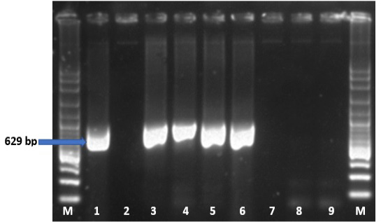
PCR amplification products using FPLV primers. Lane M: 100-bp DNA ladder marker (Fermentas); lane 1, 3, 4, 5, 6: positive amplification PCR products; lane 2: negative control (distilled water); lane 7, 8, 9: negative amplification PCR products.
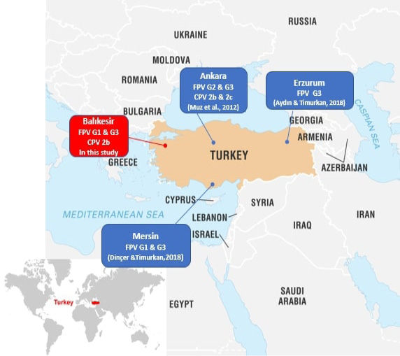
Map showing the provinces where studies on FPV genotyping are carried out in Turkey.
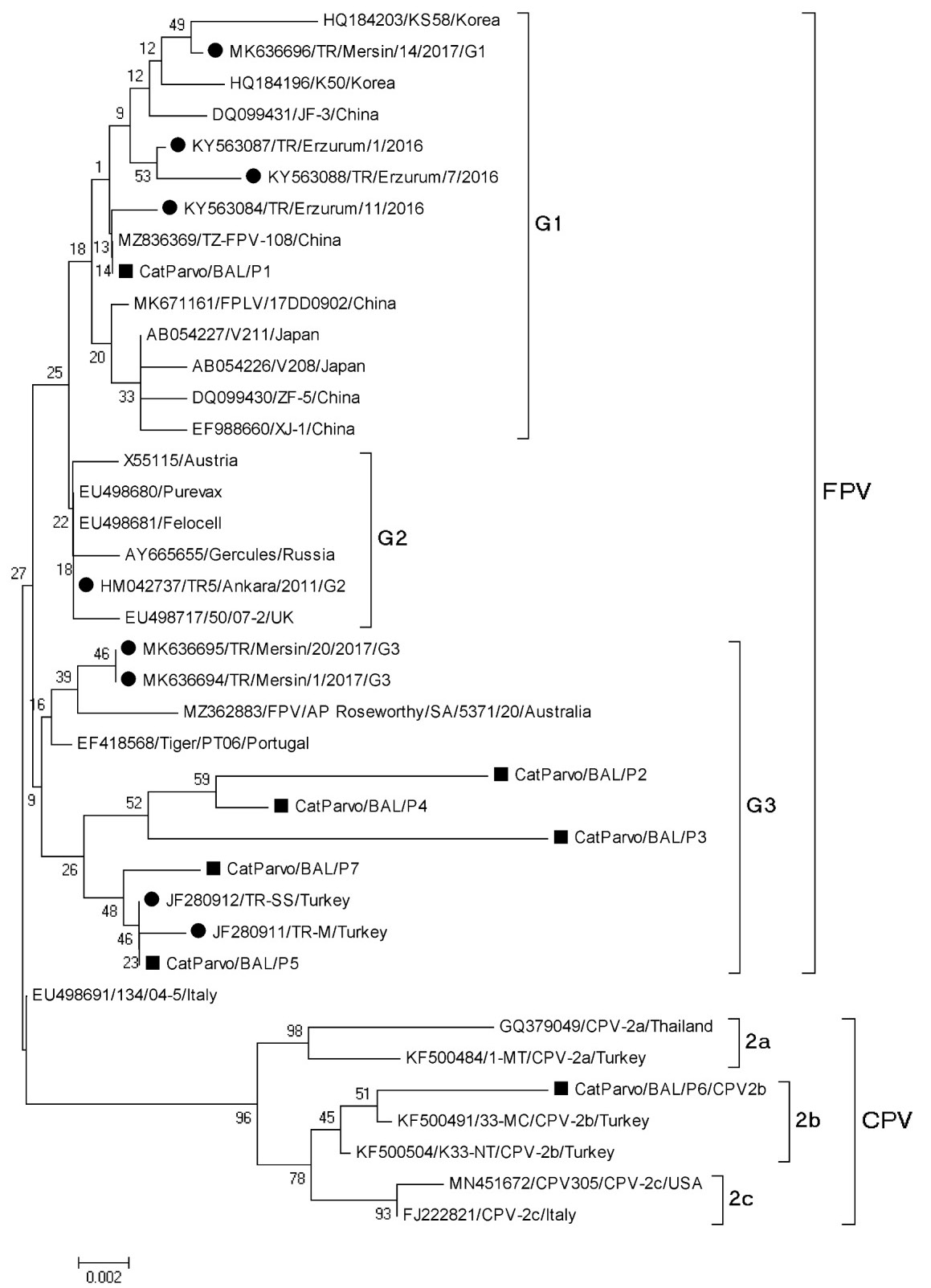
Phylogenetic tree of partial VP2 gene of FPV. New sequences from Balıkesir province are indicated with filled square and other sequences are indicated by the isolate name and country, respectively.
References
- Ahmed, N., Riaz, A., Zubair, Z., Saqib, M., Ijaz, S., Nawaz-ul-Rehman, M.S., Al-Qahtani, A. and Mubin, M., 2018. Molecular analysis of partial VP-2 gene amplified from rectal swab samples of diarrheic dogs in Pakistan confirms the circulation of canine parvovirus genetic variant CPV-2a and detects sequences of feline panleukopenia virus (FPV). Virol. J., 15: 45. https://doi.org/10.1186/s12985-018-0958-y[CrossRef]
- Allison, A.B., Kohler, D.J., Fox, K.A., Brown, J.D., Gerhold, R.W., Shearn-Bochsler, V.I., Dubovi, E.J., Parrish, C.R., and Holmes, E.C., 2013. Frequent cross-species transmission of parvoviruses among diverse carnivore hosts. J. Virol., 87: 2342–2347. https://doi.org/10.1128/JVI.02428-12[CrossRef]
- An, D.J., Jeong, W., Jeoung, H.Y., Yoon, S.H., Kim, H.J., Park, J.Y. and Park, B.K., 2011. Phylogenetic analysis of feline panleukopenia virus (FPLV) strains in Korean cats. Res. Vet. Sci., 90: 163–167. https://doi.org/10.1016/j.rvsc.2010.05.010[CrossRef]
- Awad, R.A., Khalil, W., and Attallah, A.G., 2018. Epidemiology and diagnosis of feline panleukopenia virus in Egypt: Clinical and molecular diagnosis in cats. Vet. World, 11: 578–584. https://doi.org/10.14202/vetworld.2018.578-584[CrossRef]
- Aydın, H. and Timurkan, M.O., 2018. A pilot study on feline astrovirus and feline panleukopenia virus in shelter cats in Erzurum, Turkey. Rev. Méd. Vét., 169: 52-57.
- Barrs, V.R., 2019. Feline panleukopenia: A re-emergent disease. Vet. Clin. North Am. Small Anim. Pract., 49: 651–670.
- Brindhalakshmi, B., Mukhopadhyay, H.K., Antony, P.X., Thanislass, J., Vijayalakshmi, P. and Mangadevi, N., 2016. Isolation and molecular characterization of canine and feline parvovirus strains. An updated review. J. Dairy Vet. Anim. Res., 3: 164-169. https://doi.org/10.15406/jdvar.2016.03.00093[CrossRef]
- Buonavoglia, C., Martella, V., Pratelli, A., Tempesta, M., Cavalli, A., Buonavoglia, D., Bozzo, G., Elia, G., Decaro, N. and Carmichael, L., 2001. Evidence for evolution of canine parvovirus type 2 in Italy. J. Gen. Virol., 82: 3021–3025. https://doi.org/10.1099/0022-1317-82-12-3021[CrossRef]
- Decaro, N., Buonavoglia, D., Desario, C., Amorisco, F., Colaianni, M.L., Parisi, A., Terio, V., Elia, G., Lucente, M.S., Cavalli, A., Martella, V. and Buonavoglia, C., 2010. Characterisation of canine parvovirus strains isolated from cats with feline panleukopenia. Res. Vet. Sci., 89: 275–278. https://doi.org/10.1016/j.rvsc.2010.03.001[CrossRef]
- Decaro, N., Desario, C., Miccolupo, A., Campolo, M., Parisi, A., Martella, V., Amorisco, F., Lucente, M.S., Lavazza, A. and Buonavoglia, C., 2008. Genetic analysis of feline panleukopenia viruses from cats with gastroenteritis. J. Gen. Virol., 89: 2290–2298. https://doi.org/10.1099/vir.0.2008/001503-0[CrossRef]
- Decaro, N., Desario, C., Elia, G., Campolo, M., Lorusso, A., Mari, V., Martella, V. And Buonavoglia, C., 2007. Occurrence of severe gastroenteritis in pups after canine parvovirus vaccine administration: A clinical and laboratory diagnostic dilemma. Vaccine, 25: 1161–1166. https://doi.org/10.1016/j.vaccine.2006.10.020[CrossRef]
- Diakoudi, G., Lanave, G., Capozza, P., Di Profio, F., Melegari, I., Di Martino, B., Pennisi, M.G., Elia, G., Cavalli, A., Tempesta, M., Camero, M., Buonavoglia, C., Bányai, K. and Martella, V., 2019. Identification of a novel parvovirus in domestic cats. Vet. Microbiol., 228: 246–251. https://doi.org/10.1016/j.vetmic.2018.12.006[CrossRef]
- Dinçer, E. and Timurkan, M.Ö., 2018. Mersin ilinde feline panleukopenia virüs (FPLV) enfeksiyonunun tespiti ve filogenetik analizi. Fırat Üniv. Sağlık Bilim. Derg. Vet., 32: 213–217.
- Gür, S., and Avdatek, K., 2016. A serological investigation for feline panleukopenia virus in cats in Afyonkarahisar. Kocatepe Vet. J., 9: 165–170. https://doi.org/10.5578/kvj.26547[CrossRef]
- Hall, T.A., 1999. BioEdit: A user-friendly biological sequence alignment editor and analysis program for Windows 95/ 98/NT. Nucl. Acids Symp. Ser., 41: 95-98.
- Hoelzer, K. and Parrish, C.R., 2010. The emergence of parvoviruses of carnivores. Vet. Res., 41: 39. https://doi.org/10.1051/vetres/2010011[CrossRef]
- Horiuchi, M., Yamaguchi, Y., Gojobori, T., Mochizuki, M., Nagasawa, H., Toyoda, Y., Ishiguro, N. and Shinagawa, M., 1998. Differences in the evolutionary pattern of feline panleukopenia virus and canine parvovirus. Virology, 249: 440–452. https://doi.org/10.1006/viro.1998.9335[CrossRef]
- Kim, Y.J., Yoon, S.W., Jang, J.H., Jeong, D.G., Lee, B.J. and Kim, H.K., 2021. Genetic characterization of feline parvovirus isolate Fe-P2 in Korean cat and serological evidence on its infection in wild leopard cat and Asian badger. Front. Vet. Sci., 8: 650866. https://doi.org/10.3389/fvets.2021.650866[CrossRef]
- Koç, B.T. and Oğuzoğlu, T.Ç., 2016. The investigation of feline parvoviruses (FPVs) into two different phylogenetic lineages in Turkey. J. Appl. Biol. Sci., 10: 4-7.
- Leal, É., Liang, R., Liu, Q., Villanova, F., Shi, L., Liang, L., Li, J., Witkin, S.S. and Cui, S., 2020. Regional adaptations and parallel mutations in Feline panleukopenia virus strains from China revealed by nearly-full length genome analysis. PLoS One, 15: e0227705. https://doi.org/10.1371/journal.pone.0227705[CrossRef]
- Li, X., Wu, H., Wang, L., Spibey, N., Liu, C., Ding, H., Liu, W., Liu, Y. and Tian, K., 2018. Genetic characterization of parvoviruses in domestic cats in Henan province, China. Transb. Emerg. Dis., 65: 1429–1435. https://doi.org/10.1111/tbed.13014[CrossRef]
- Mochizuki, M., Horiuchi, M., Hiragi, H., San Gabriel, M.C., Yasuda, N. and Uno, T., 1996. Isolation of canine parvovirus from a cat manifesting clinical signs of feline panleukopenia. J. clin. Microbiol., 34: 2101–2105. https://doi.org/10.1128/jcm.34.9.2101-2105.1996[CrossRef]
- Muz, D., Oğuzoğlu, T.C., Timurkan, M.O. and Akın, H., 2012. Characterization of the partial VP2 gene region of canine parvoviruses in domestic cats from Turkey. Virus Genes, 44: 301–308. https://doi.org/10.1007/s11262-011-0703-8[CrossRef]
- Niu, J.T., Yi, S.S., Hu, G.X., Guo, Y.B., Zhang, S., Dong, H., Zhao, Y.L. and Wang, K., 2018. Prevalence and molecular characterization of parvovirus in domestic kittens from Northeast China during 2016–2017. Jpn. J. Vet. Res., 66: 145–155.
- Oğuzoğlu, T.C., Muz, D., Timurkan, M.O., Maral, N. and Gurcan, I.S., 2013. Prevalences of feline coronavirus (FCoV), feline leukaemia virus (FeLV), feline immunodeficiency virus (FIV) and feline parvovirus (FPLV) among domestic cats in Ankara, Turkey. Rev. Méd. Vét., 164: 511-516.
- Parrish, C.R., 1999. Host range relationships and the evolution of canine parvovirus. Vet. Microbiol., 69: 29–40. https://doi.org/10.1016/S0378-1135(99)00084-X[CrossRef]
- Parrish, C.R., Aquadro, C.F., Strassheim, M.L., Evermann, J.F., Sgro, J.Y. and Mohammed, H.O., 1991. Rapid antigenic-type replacement and DNA sequence evolution of canine parvovirus. J. Virol., 65: 6544–6552. https://doi.org/10.1128/jvi.65.12.6544-6552.1991[CrossRef]
- Parthiban, M., Aarthi, K.S., Balagangatharathilagar, M. and Kumanan, K., 2014. Evidence of feline panleukopenia infection in cats in India. Virus Dis., 25: 497–499. https://doi.org/10.1007/s13337-014-0231-y[CrossRef]
- Patterson, E.V., Reese, M.J., Tucker, S.J., Dubovi, E.J., Crawford, P.C. and Levy, J.K., 2007. Effect of vaccination on parvovirus antigen testing in kittens. J. Am. Vet. Med. Assoc., 230: 359–363. https://doi.org/10.2460/javma.230.3.359[CrossRef]
- Stuetzer, B. and Hartmann, K., 2014. Feline parvovirus infection and associated diseases. Vet. J., 201: 150–155. https://doi.org/10.1016/j.tvjl.2014.05.027[CrossRef]
- Tamura, K., Stecher, G., Peterson, D., Filipski, A. and Kumar, S., 2013. MEGA6: Molecular evolutionary genetics analysis version 6.0. Mol. Biol. Evol., 30: 2725-2729. https://doi.org/10.1093/molbev/mst197[CrossRef]
- Timurkan, M. and Oğuzoğlu, T., 2015. Molecular characterization of canine parvovirus (CPV) infection in dogs in Turkey. Vet. Ital., 51: 39–44.
- Truyen, U. and Parrish, C.R., 2013. Feline panleukopenia virus: its interesting evolution and current problems in immunoprophylaxis against a serious pathogen. Vet. Microbiol., 165: 29–32. https://doi.org/10.1016/j.vetmic.2013.02.005[CrossRef]
- Truyen, U., 1999. Emergence and recent evolution of canine parvovirus. Vet. Microbiol., 69: 47–50. https://doi.org/10.1016/S0378-1135(99)00086-3[CrossRef]
- Truyen, U., Platzer, G. and Parrish, C.R., 1996. Antigenic type distribution among canine parvoviruses in dogs and cats in Germany. Vet. Rec., 138: 365–366. https://doi.org/10.1136/vr.138.15.365[CrossRef]
- Van Brussel, K., Carrai, M., Lin, C., Kelman, M., Setyo, L., Aberdein, D., Brailey, J., Lawler, M., Maher, S., Plaganyi, I., Lewis, E., Hawkswell, A., Allison, A.B., Meers, J., Martella, V., Beatty, J.A., Holmes, E.C., Decaro, N. and Barrs, V.R., 2019. Distinct lineages of feline parvovirus associated with epizootic outbreaks in Australia, New Zealand and the United Arab Emirates. Viruses, 11: 1155. https://doi.org/10.3390/v11121155[CrossRef]
- Yi, S., Liu, S., Meng, X., Huang, P., Cao, Z., Jin, H., Wang, J., Hu, G., Lan, J., Zhang, D., Gao, Y., Wang, H., Li, N., Feng, N., Hou, R., Yang, S. and Xia, X., 2022. Feline panleukopenia virus with G299E substitution in the VP2 protein first identified from a captive giant panda in China. Front. Cell Infect. Microbiol., 11: 820144. https://doi.org/10.3389/fcimb.2021.820144[CrossRef]
- Zhang, Q., Niu, J., Yi, S., Dong, G., Yu, D., Guo, Y., Huang, H. and Hu, G., 2019. Development and application of a multiplex PCR method for the simultaneous detection and differentiation of feline panleukopenia virus, feline bocavirus, and feline astrovirus. Arch. Virol., 164: 2761–2768. https://doi.org/10.1007/s00705-019-04394-8[CrossRef]
Services
Article usage statistics
Loading…