Special Issue: Emerging and Re-Emerging Animal Health Challenges in Low and Middle-Income Countries · Volume 13, Issue 1, 31 August 2025 · Open Access
Bacterial Etiology of Otitis Externa in Cats
·
Affiliations & Notes ⌄
1Department of internal and preventive medicine, College of veterinary medicine, University of Fallujah, Anbar, Iraq. *Correspondence | Mustafa Salah Hasan, Department of internal and preventive medicine, College of veterinary medicine, University of Fallujah, Anbar, Iraq
PCR analysis demonstrated high detection rates for all targeted bacterial species: S. aureus (15/16, 93.8%), coagulase-negative Staphylococcus (8/8, 100%), P. aeruginosa (10/10, 100%), E. coli (9/9, 100%), and K. pneumoniae (2/2, 100%) (Table 5; Figure 1).
This study provides the first comprehensive analysis of bacterial etiology in feline otitis externa within the Iraqi context, revealing a complex microbial landscape dominated by clinically significant pathogens.
The age-related trends observed, particularly the declining prevalence of S. aureus with increasing age, suggest important epidemiological patterns that may inform targeted therapeutic approaches.
The absence of significant geographic and gender-based variations suggests that host-pathogen interactions may have a greater influence on bacterial colonization patterns than demographic factors within our study region.
Selected from the article's own text.
Abstract
Otitis externa is a common inflammatory condition affecting the external ear canal of domestic cats, often complicated by secondary bacterial infections. Understanding the bacterial etiology is crucial for effective therapeutic management. To identify and characterize bacterial pathogens associated with otitis externa in cats and evaluate their distribution patterns across demographic and geographic variables. A cross-sectional study was conducted on 100 cats presenting with clinical signs of otitis externa from Baghdad and Anbar provinces. Ear swabs were collected for bacterial isolation and identification using conventional microbiological methods, followed by PCR confirmation using species-specific primers. Five bacterial species were identified including Staphylococcus aureus (35.6%), Pseudomonas aeruginosa (22.2%), Escherichia coli (20%), coagulase-negative Staphylococcus (17.8%), and Klebsiella pneumoniae (4.4%). No significant differences were observed between geographic regions (p = 0.856). Age analysis revealed a decreasing trend in S. aureus prevalence with increasing age (p = 0.114). PCR confirmation achieved detection rates of 93.8% to 100% across all species. S. aureus and P. aeruginosa were the predominant bacterial pathogens in feline otitis externa. The high prevalence of these potentially zoonotic organisms emphasizes the importance of proper diagnosis and treatment protocols in veterinary practice.
Otitis externa, or external ear infection, is the most common dermatologic pathology in small animal practice, with domestic cats being more affected worldwide . Otitis externa is an inflammatory condition of the external ear canal, defined as the area from the external acoustic meatus to the tympanic membrane, with clinical signs of erythema, edema, pain, and purulent exudate . Estimates of the prevalence of otitis externa in general practice cat populations range from 2% to 6.6%, while estimates of up to 13% have been reported in specialty referral patients ; . This suggests that otitis externa is often a substantial clinical burden in small animal veterinary medicine (Gotthelf, 201٤; Ali et al., ٢٠٢٤; .
Feline otitis externa is a multifactorial condition with diverse pathogenesis, integrating the influence of predisposing factors, the primary cause, and perpetuating factors that may contribute to the establishment or maintenance of the inflammatory process . Predisposing factors include anatomical abnormalities (i.e. stenotic ear canals, excessive hair within the ear canal), breed differences (i.e. heredity seen in breeds such as Persian, Himalayan), or environmental factors (i.e. high humidity, excessive moisture, or poor ventilation in the ear canal conducive to microbial growth) .
Primary causes of otitis externa in cats include a range of conditions, but parasitic infestations are a particular cause of feline cases. Otodectes cynotis, commonly referred to as ear mites, are implicated in ~50% of all otitis externa cases in cats, especially in young animals or when cats have access to the outdoors . Allergic reactions, specifically food allergies and atopic dermatitis, are another prevalent primary cause; studies have found that as many as 43% of cats with food allergies will concurrently develop otitis externa . Foreign bodies, although less common in cats than in dogs, can occasionally induce otitis externa, especially grass awns and other plant materials .
The usual progression from primary inflammation to complicated otitis externa involves secondary bacterial colonization and infection, which is an essential factor in the progression of the disease . The typical feline ear canal has a delicate microbial environment that mainly composed of gram-positive cocci, with Staphylococcus species being the most commonly cultured organisms from healthy ears , however, when there is disruption to the ear canal environment due to inflammation, elevated temperatures, altered pH, and moisture, the regular microbial community shifts to opportunistic bacterial overgrowth .
S. aureus is one of the most clinically significant bacterial pathogens in feline otitis externa; the prevalence of S. aureus varies between regions, with estimates ranging from 15% to 40% ; . S. aureus is a gram-positive coccus that possesses numerous virulence factors, including adhesins that facilitate attachment to disrupted epithelium, toxins that contribute to tissue damage, and enzymes that enable invasion and persistence (Foster, 2005). In addition, S. aureus also presents with zoonotic potential, as infected cats may serve as a source of disease for humans, particularly in homes with immunocompromised individuals .
P. aeruginosa is another significant bacterial pathogen involved in feline otitis externa, particularly in the chronic and recurrent type . This opportunistic pathogen flourishes within the humid, warm environment of the inflamed ear canal and possesses innate antimicrobial resistance mechanisms as well as biofilm-forming capabilities . It has been demonstrated in studies that infections by P. aeruginosa associated with otitis externa elicit more severe clinical signs, a longer treatment course, and a higher propensity for recurrence than infections caused by other bacterial pathogens .
E. coli has a similarly recognized association in feline otitis externa and has been reported at recent isolation rates of 10% - 25% from infected ears . E. coli species are historically classified as commensal organisms in the gastrointestinal tract; in rare cases, these pathogenic strains of E. coli can establish an infection in the external ear canal, typically when pre-existing immune compromise or concurrent systemic illness is present . E. coli detected in ear infections may also indicate fecal contamination of potential interest to feline caregivers, especially in multi-cat settings, to emphasize the importance of proper hygiene .
Coagulase-negative Staphylococcus species are often regarded as commensal; following the same pattern as previously identified bacteria, they can assume a pathogenic role concerning otitis externa in vulnerable cats, such as those with an underlying allergic condition or in states of immunocompromise . Coagulase-negative Staphylococcus species are increasingly recognized for their ability to form biofilms and exhibit antimicrobial resistance, which contributes to treatment failures and the development of chronic infections . K. pneumoniae is noted less frequently than E. coli or a coagulase-negative Staphylococcus and is classified in veterinary dermatology as an emerging concern due to its association with severe infections and multidrug-resistant patterns . The distribution of bacterial pathogens in otitis externa geographically varies considerably due to factors such as climate, population density, patterns of antimicrobial use, and hygiene practices . In the Middle East, particularly in Iraq, specific data on the bacterial etiology of feline otitis externa are scarce, despite the presence of numerous domestic cats and the veterinary burden of skin and ear diseases (Al-Gburi al., 2021; .
The accurate identification of bacterial pathogens is crucial for optimal therapeutic management of otitis externa. Culture-based approaches have been used in bacteriological examinations and are reliable; however, they require time, and in some cases, may not detect fastidious or pathogenic organisms that are present in low numbers . Molecular diagnostic techniques, such as polymerase chain reaction (PCR) using species-specific primers, have been used since the 1990s and have transformed bacterial identification by being rapid, sensitive, and specific for target organisms . The current gold standard for bacterial identification utilizes the 16S ribosomal RNA gene, enabling researchers to identify species-specific variable regions .
Age-associated patterns of susceptibility to feline otitis externa have been demonstrated in several studies, where younger cats are at a higher risk of developing the disease, possibly due to lower immunological maturity or greater complications resulting from a parasitic load, as well as increased behavioral risks from external or contaminated environments , . Older cats may develop otitis externa in association with neoplasia, immune-mediated diseases, or metabolic diseases such as hyperadrenocorticism .
The gender predisposition for feline otitis externa is controversial, with some studies reporting a higher incidence in male cats, while others find no difference between the sexes ; . More consistency is observed in the predisposition for breeds of, predominantly long-haired cats, with breeds such as Persian, Himalayan, and Maine Coon cats reported to display a higher incidence, potentially due to their anatomical conformation, including a narrow canal and more hair in the external ear .
The economic consequences of otitis externa are greater than the initial treatment costs for the condition . It incorporates additional fees for other diagnostic procedures, extra visits, and/or management of complications (otitis media or facial nerve paralysis). Moreover, the frequency of recurrent treatment typically associated with bacterial otitis externa reinforces the risk of developing antimicrobial resistance and ongoing treatment costs, even though otitis externa is, to a significant extent, a preventable disease .
Preventative methods for feline otitis externa focus on ear hygiene, prompt management of incidents for predisposing conditions, and environmental modifications to minimize risk factors . However, it is contingent on who understands the bacterial pathogens in that area, as well as the risk factors associated with bacterial otitis externa .
The purpose of this study is to help address the knowledge gap regarding bacterial etiology of otitis externa in cats, particularly in an Iraqi context, and authoritatively provide necessary data for evidence-based therapeutic decision-making regarding the cat and contribute to a global understanding of this essential condition in cats, particularly in terms of bacterial identification from the traditional microbiological perspective in comparison to molecular confirmation. This study was designed to examine streams of epidemiological data and incorporate demographic and geographic patterns of bacterial otitis externa into the understanding of the condition.
Materials and Methods
Study design and setting
A cross-sectional observational study was conducted between October 2024 and April 2025 across multiple veterinary clinics and animal hospitals in Baghdad and Anbar provinces, Iraq. The study protocol was approved by the Institutional Review Board of the College of Veterinary Medicine at the University of Fallujah, and informed consent was obtained from all pet owners before sample collection.
Study population and sample size
The study population consisted of domestic cats presenting with clinical signs suggestive of otitis externa. A sample size calculation was performed using the formula for cross-sectional studies with finite populations, considering a 95% confidence interval, a 5% margin of error, and an expected prevalence of 30% based on previous regional studies. The calculated minimum sample size was 81 cats, which was increased to 100 to account for potential sample losses during processing.
Inclusion and exclusion criteria
Inclusion criteria
Domestic cats of any breed, age, and sex
Clinical signs of otitis externa, including ear discharge, odor, erythema, or pruritus
Owner consent for sample collection and participation
No antimicrobial therapy within the preceding 14 days
Exclusion criteria
Cats with ruptured tympanic membranes confirmed by otoscopic examination
Recent ear cleaning within 48 hours of sample collection
Concurrent systemic antimicrobial therapy
Cats with neurological signs suggesting inner ear involvement
Clinical examination and data collection
All cats underwent a comprehensive clinical examination by qualified veterinarians. Otoscopic examination was performed using standard veterinary otoscopes to assess ear canal morphology, degree of inflammation, type and quantity of discharge, and tympanic membrane integrity. Clinical data recorded included signalment (age, sex, breed), geographic location, duration of clinical signs, previous treatments, and concurrent medical conditions.
Age categories were defined as: Kittens (<6 months), juveniles (6-12 months), and adults (>12 months). Breed classification included Sherazi (Persian type), Himalayan, and local mixed breeds. The geographic distribution encompassed urban and suburban areas within Baghdad and Anbar provinces.
Sample collection
Ear swabs were collected using sterile cotton-tipped applicators designed explicitly for bacterial sampling. The sampling technique involved gently inserting the swab into the external ear canal without forcing it past any obstructions, followed by gentle rotation to collect adequate material from the infected site. Care was taken to avoid contamination from the external ear pinna or surrounding skin.
Two swabs were collected from each affected ear: one for immediate bacterial culture and identification, and a second for molecular analysis. Samples were labeled with unique identifiers and transported to the laboratory within 2 hours of collection in Stuart’s transport medium to maintain bacterial viability.
Bacterial isolation and identification
Primary culture
Ear swabs were inoculated onto multiple types of media, including blood agar, MacConkey agar, and mannitol salt agar plates. Plates were incubated at 37°C under both aerobic and microaerophilic conditions for 24 to 48 hours. Bacterial growth was assessed daily, and isolates were sub-cultured for purification and identification.
Bacterial identification
Preliminary identification was performed using standard microbiological techniques, including Gram staining, catalase testing, oxidase testing, and coagulase testing for staphylococci. Biochemical identification was completed using commercially available identification systems and manual biochemical panels, following the manufacturer’s instructions.
S. aureus identification was confirmed by positive catalase and coagulase tests, as well as the fermentation of mannitol. Coagulase-negative Staphylococcus species were identified by positive catalase and negative coagulase tests, with further speciation using additional biochemical tests.
P. aeruginosa identification was based on gram-negative rod morphology, positive oxidase test, characteristic blue-green pigmentation, and distinctive grape-like odor. E. coli was identified by its gram-negative rod morphology, positive indole test, methyl red test, Voges-Proskauer test, and citrate test (IMViC pattern).
K. pneumoniae identification was confirmed by gram-negative rod morphology, positive capsule formation, lactose fermentation, and a negative indole test.
Molecular confirmation by PCR
DNA extraction
Bacterial DNA was extracted from pure cultures using the boiling method. Briefly, bacterial colonies were suspended in sterile distilled water, heated at 100°C for 10 minutes, and centrifuged at 12,000 rpm for 5 minutes. The supernatant containing DNA was used directly for PCR amplification.
PCR amplification
Species-specific PCR was performed for each bacterial isolate using validated primer sets as shown in Table 1.
PCR reactions were performed in 25 μL volumes containing 12.5 μL master mix, 1 μL each primer (10 pmol/μL), 2 μL template DNA, and nuclease-free water. Thermal cycling conditions included initial denaturation at 95°C for 5 minutes, followed by 35 cycles of denaturation (95°C, 30 seconds), annealing (55-60°C depending on primers, 30 seconds), and extension (72°C, 1 minute), with final extension at 72°C for 7 minutes.
Table 1: Primer used in present study.
Primer name
Seq.
Annealing Temp. (°C)
Product Size (bp)
Klpn_16s-F
GACGATCCCTAGCTGGTCTG
60
95
Klpn_16s-R
GTGCAATATTCCCCACTGCT
E.coli_16s-F
GTTAATACCTTTGCTCATTGA
340
E.coli_16s-R
ACCAGGGTATCTAATCCTGTT
acr-F
TTGATTCACCAGCGCGTATTGTC
55
570
acr-R
AGGTATCTGCTTCAATCAGCG
fbp-F
CCTACCTGTTGGTCTTCGACCCG
58
284
fbp-R
GCTGATGTTGTCGTGGGTGAGG
S. aureus 16s-F
-F 5'-GGCCGTGTTGAACGTGGTCAAATCA-3'
S. aureus 16s-R
R5’ -TIACCATTTCAGTACCTTCTGGTAA-3'
55
270
Gel electrophoresis
PCR products were analyzed by agarose gel electrophoresis using 1.5% agarose gels stained with ethidium bromide. Electrophoresis was performed at 100 V for 60 minutes, and the results were visualized under UV transillumination. A 100 bp DNA ladder was used as a molecular weight marker.
Quality control
Quality control measures included the use of positive and negative controls for all PCR reactions, duplicate testing of 10% of samples, and blind re-identification of selected isolates. Laboratory personnel were blinded to clinical information during bacterial identification procedures.
Statistical analysis
Data were analyzed using SPSS version 26.0. Descriptive statistics were calculated for all variables, including frequencies, percentages, means, and standard deviations. Chi-square tests or Fisher’s exact tests were used to compare categorical variables, while continuous variables were analyzed using appropriate parametric or non-parametric tests depending on data distribution. Statistical significance was set at p<0.05.
Associations between bacterial species and demographic variables (age, sex, breed) and geographic location were assessed using cross-tabulation and chi-square analysis. Age-related trends were evaluated using chi-square tests for trend. All statistical tests were two-tailed, and 95% confidence intervals were calculated where appropriate.
Results
Study population characteristics
Table 2 show the study population consisted of 100 feline subjects, with a nearly equal gender distribution (54% male, 46% female). Breed composition was predominantly Sherazi (90%), with smaller representations of Himalayan (8%) and local breeds (2%). The age distribution showed 42% of kittens under 6 months, 38% of juveniles (6-12 months), and 20% of adults (>12 months), establishing a representative sample across the various developmental stages.
Table 2: Demographic characteristics of study population.
Category
Subgroup
Count
Percentage
Sex
Male
54
54%
Female
46
46%
Breed
Sherazi
90
90%
Himalayan
8
8%
Local breed
2
2%
Age Groups
<6 months
42
42%
6-12 months
38
38%
>12 months
20
20%
Bacterial isolation results
A total of 45 bacterial isolates were recovered from the 100 ear samples, representing five distinct bacterial species. S. aureus was the most frequently isolated organism (n=16, 35.6% of isolates), followed by P. aeruginosa (n=10, 22.2%), E. coli (n=9, 20%), coagulase-negative Staphylococcus (n=8, 17.8%), and K. pneumoniae (n=2, 4.4%).
Geographic distribution
Geographic analysis shown in Table 3, revealed no significant regional differences in bacterial isolation rates. The total number of bacterial isolates mirrored the sample distribution (Baghdad: 68.9% vs Anbar: 31.1%, p = 0.856), indicating that geographic location did not significantly affect bacterial prevalence patterns.
Age-related distribution
Age-related trends in bacterial isolation (Table 4) showed S. aureus prevalence decreased with age: 62.5% (<6mo) vs 25.0% (6-12mo) vs 12.5% (>12mo), though not statistically significant (p=0.114). Klebsiella was exclusively isolated in the 6-to 12-month age group (100% of cases, p = 0.099).
Table 3: Bacterial isolation by geographic region.
Region
S. aureus (n=16)
Coag. Staph (n=8)
Pseudomonas (n=10)
E. coli (n=9)
Klebsiella (n=2)
Total Isolates
Baghdad
11 (68.8%)
6 (75.0%)
7 (70.0%)
6 (66.7%)
1 (50.0%)
31 (68.9%)
Anbar
5 (31.2%)
2 (25.0%)
3 (30.0%)
3 (33.3%)
1 (50.0%)
14 (31.1%)
p-value
0.920
0.724†
1.000†
0.892†
1.000†
0.856
†Fisher’s exact test p-values
Table 4: Age group differences in bacterial isolation.
Age group
S. aureus
Coag. Staph
Pseudomonas
E. coli
Klebsiella
<6 months
10 (62.5%)
3 (37.5%)
4 (40.0%)
6 (66.7%)
0 (0%)
6-12 months
4 (25.0%)
3 (37.5%)
4 (40.0%)
2 (22.2%)
2 (100%)
>12 months
2 (12.5%)
2 (25.0%)
2 (20.0%)
1 (11.1%)
0 (0%)
p-value
0.114
0.604†
0.873†
0.188†
0.099†
†Fisher’s exact test p-values
PCR confirmation results
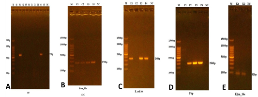
PCR analysis demonstrated high detection rates for all targeted bacterial species: S. aureus (15/16, 93.8%), coagulase-negative Staphylococcus (8/8, 100%), P. aeruginosa (10/10, 100%), E. coli (9/9, 100%), and K. pneumoniae (2/2, 100%) (Table 5; Figure 1).
Table 5: PCR confirmation results.
Bacterial species
Number tested
PCR positive
Detection rate (%)
S. aureus
16
15
93.8
Coagulase-negative Staph
8
8
100
P. aeruginosa
10
10
100
E. coli
9
9
100
K. pneumoniae
2
2
100
Discussion
The current study provides valuable insights into the bacterial nature of otitis externa in cats in the Iraqi context. This research can shed light on the bacterial phenomena associated within the context of a complex microbial environment, which includes potentially pathogenic organisms with significant clinical and zoonotic concerns. The occurrence of S. aureus as the most crucial bacterial pathogen (35.6%) aligns well with the global perspective, as this organism frequently isolated and reported in the majority of feline otitis externa cases ; .
The high frequency of S. aureus in our cat population is noteworthy, especially given that it is known to be both a commensal and pathogenic organism in cats. This Gram-positive coccus possesses numerous virulence factors that facilitate adhesion to damaged epithelium, tissue invasion, and survival in the ear canal environment . The decrease in frequency and severity of S. aureus in older cats, as observed in our study, although not statistically significant (p = 0.114), suggests that there may be an age-dependent frequency of S. aureus in different populations, which warrants further exploration. Other studies in various geographical areas have suggested that some younger cats may be at a greater risk due to their immature immune systems and behaviors ; .
Furthermore, the zoonotic potential of S. aureus is another clinical concern because infected cats serve as a context for human transmission, especially in households with close contact . Transmission of methicillin-resistant S. aureus (MRSA) to owners through pets has been discussed in the literature . Documentation of MRSA strain transmission from cats to their owners underscores the importance of good infection control practices in treatment plans. The relatively high isolation rate found in our study suggests that feline otitis externa may be an important, yet under-recognized, source of staphylococcal exposure for households in Iraq. P. aeruginosa was the second most prevalent pathogen (22.2% of isolates) identified in our study and is well-known for its association with chronic and recurrent cases of otitis externa. The heritable aspects of this gram-negative organism are concerning, including its inherent resistance to antimicrobials and ability to form biofilms in the ear canal ; . Aerobic bacteria, such as P. aeruginosa, are typically associated with more severe cases of otitis externa, requiring longer-term treatments and higher recurrence rates compared to other bacterial pathogens . In our study, it was apparent that P. aeruginosa is not age-dependent, as evidenced by our cases, which showed a normal distribution across age groups (p = 0.873), unlike the age-dependent pattern observed with S. aureus.
The presence of P. aeruginosa in the environment, coupled with its innate ability to survive in various situations, makes this organism particularly troublesome in the veterinary realm . This organism is capable of producing numerous virulence factors that contribute to tissue damage and chronic inflammation typically associated with pseudomonal otitis externa . Management of P. aeruginosa otitis externa generally requires targeted antimicrobial therapy based on susceptibility testing due to the organism’s inherent resistance profile, making empirical treatment often egregious .
Notably, E. coli was isolated from 20% of the samples, which signifies a noteworthy finding, as this organism is increasingly recognized as a pertinent pathogen associated with otitis externa, particularly in cases of underlying immune compromise or concurrent gastrointestinal disease . While historically regarded as a commensal of the intestinal tract, virulent strains of E. coli can cause infection at extra-intestinal sites, including the external ear canal . In ear specimens, E. coli may also suggest the potential for fecal contamination, particularly in the presence of multiple cats or poor hygiene practices) Al-Mayahi and Hussain (2021). Also, pathogenic E. coli was isolated from other animals by same authors ; . This organism was resembling to another bacteria called Salmonella which were isolated from pet animals ; .
The age distribution of E. coli, with a higher prevalence in younger cats as evidenced by 66.7% in <6 months versus 11.1% in >12 months, although not statistically significant (p= 0.188), indicates a potential role of behavioral or immunological factors that may predispose younger animals to infection. This may reflect differential environmental exposures in kittens or developmental differences in local immune responses .
A 17.8% incidence of coagulase-negative Staphylococcus species was observed, underscoring the growing clinical relevance of these organisms in feline otitis externa. Although coagulase-negative Staphylococcus species have traditionally been viewed as innocuous commensals, there is growing evidence regarding their role in chronic infections, especially in cats with immunocompromised conditions and underlying allergic conditions ; . Coagulase-negative Staphylococcus species can form biofilms and subsequently become resistant to antimicrobial agents, likely contributing to treatment failures and chronic infections, making their recognition clinically significant . The low rate of K. pneumoniae (4.4% of isolates) differs from international reports, which found much higher rates; however, differences in environmental exposures and patterns of antimicrobial use may account for this discrepancy . Nonetheless, the exclusive isolation of this organism in the 6–12-month age group, although not statistically significant (p = 0.099), is certainly unusual and warrants further consideration. K. pneumoniae is increasingly associated with severe infections and multidrug resistance; therefore, its isolation in a low-prevalence area is clinically relevant .
The lack of significant geographic differences in bacterial isolation prevalence between Baghdad and Anbar provinces (p = 0.856) is surprising, considering the differences in geographic features (environmental conditions, population density, and access to veterinary care) in these regions. This suggests that the patterns of bacterial colonization in cats with otitis externa may be influenced more by host factors and pathogen features, rather than geographic characteristics, within the context of Iraq . This is in contrast to studies in other regions, which have reported significant geographic variations in bacterial prevalence, possibly due to local environmental conditions or variations in antimicrobial use patterns in the areas studied ; .
The lack of significant variations in isolation rates between males and females suggests that sex may not play a crucial role in bacterial colonization in cats with otitis externa, a finding supported by other studies ; . Similarly, the lack of significant differences based on breed is limited by the predominance of Sherazi cats (90%) in the study population, which may suggest that breed does not significantly influence bacterial susceptibility in this population.
The high PCR confirmation rates reported in this study (93.8-100% for all species) reflect the accuracy of standard microbiology identification and demonstrate that molecular confirmation provides additional necessary support for bacterial species identification and use. The integration of phenotypic and genotypic methods enhances confidence in testing and facilitates the accurate identification of a pathogen ; . The only discordant isolation for S. aureus (93.8% confirmed by PCR) could be related to technical issues in the PCR amplification process or lack of awareness of genetic variation in specific nucleotide sequences.
Using species-specific genetic markers confirmed by PCR offers advantages over conventional identification alone, including increased sensitivity, specificity, and a faster turnaround time compared to traditional methods . The 16S rRNA gene targets we used to identify S. aureus and E. coli have been extensively validated for bacterial taxonomy, based on their universal presence and unique variable regions for each species . The fbp gene target for P. aeruginosa and the acr gene for Staphylococcus species have also demonstrated high specificities for identifying bacteria ; . The clinical ramifications of our findings encompass not only therapeutic considerations but also infection control, zoonotic disease risks, and antimicrobial stewardship. The opportunistic, potentially pathogenic bacteria we isolated indicated that S. aureus and P. aeruginosa were of particular concern, suggesting that appropriate antimicrobial therapy should be based on culture and sensitivity testing instead of relying on empirical treatment . Due to the zoonotic potential of many of the identified organisms, it is essential to observe appropriate hygiene and take protective measures when handling infected cats.
The public health significance of identifying bacterial pathogens in domestic pet cats raises awareness of zoonotic potential risks to the general population, particularly immunocompromised individuals, the elderly, and young children, who are most vulnerable due to their proximity to pets ; . The significance of antimicrobial resistance in these organisms also complicates treatment choices and highlights the importance of rational antimicrobial use in veterinary medicine .
Conclusion
This study provides the first comprehensive analysis of bacterial etiology in feline otitis externa within the Iraqi context, revealing a complex microbial landscape dominated by clinically significant pathogens. S. aureus and P. aeruginosa emerged as the most prevalent bacterial species, accounting for over half of all isolates, with additional contributions from E. coli, coagulase-negative Staphylococcus, and K. pneumoniae.
The age-related trends observed, particularly the declining prevalence of S. aureus with increasing age, suggest important epidemiological patterns that may inform targeted therapeutic approaches. The absence of significant geographic and gender-based variations suggests that host-pathogen interactions may have a greater influence on bacterial colonization patterns than demographic factors within our study region.
The high PCR confirmation rates validate the accuracy of conventional microbiological methods while demonstrating the value of molecular diagnostics in bacterial identification. These findings provide essential baseline data for evidence-based therapeutic decisions and contribute to the global understanding of feline otitis externa etiology.
The zoonotic potential of the identified bacterial species underscores the importance of implementing proper infection control measures and emphasizes the need for One Health approaches in managing companion animal diseases. Future research should focus on antimicrobial susceptibility patterns, treatment outcomes, and longitudinal studies to better understand the dynamics of bacterial infections in feline otitis externa.
Acknowledgments
Authors thank the staff at the University of Fallujah for the support to conduct this study.
Novelty Statement
This study is the first to comprehensively investigate the bacterial causes of otitis externa in cats in Iraq, identifying *Staphylococcus aureus* and *Pseudomonas aeruginosa* as the most prevalent pathogens. By combining conventional microbiology with PCR confirmation, it provides reliable data on bacterial prevalence, zoonotic risks, and age-related trends, offering crucial insights for veterinary treatment and public health in the region.
Author’s Contribution
Mustafa Salah Hasan: Researcher, statistical analysis. Ahmed Ali Ahmed: Practical work, writing, editing and sampling.
Funding
No funding was obtained to conduct this study.
Data availability
Data are available upon request
Generative AI or AI-assisted Technology Statement
The author(s) declare that no Genrative AI was used in the creation of this manuscript.
Conflict of interest
The authors declare no conflict of interest.
Figures
Figure 1:.
Results of the amplification of the acrgene of Staphylococcus spp. (A), 16S gene of S. aureus (B), E. coli 16S gene of E. coli (C), the fbp gene of P. aeruginosa (D) and Klpn_16s gene of K. pneumoniae (D). The sample species were fractionated on a 1.5% agarose gel by electrophoresis and stained with Ethidium Bromide. M: 100bp ladder marker. Lanes S1-C5 resemble 570 bp PCR products, NC: negative control.
References
Abdulgafor AB, Owain MS, Hasan MS, Hussein MA, Aboud QM, Al-Zobaie AJ (2018). Liver, kidney function tests and oxidative damage during and after treatment of Salmonella typhimurium infection in experimental local rabbits. Int. J. Pharma. Qual. Assur., 9(4): 377-380. https://doi.org/10.25258/ijpqa.v9i4.14536[CrossRef]
Ahmed SF, Hassan NK (2020). Comparison of phenotypic and genotypic methods for bacterial identification in veterinary diagnostics. Iraqi J. Biotechnol., 19(3): 67–78.
Al-Dabbagh AM, Hussein RH, Al-Mayahi ZK (2021). Age-related susceptibility patterns in bacterial infections of domestic animals in northern Iraq. Vet. Res. Commun., 45(2): 167–175.
Al-Gburi KM, Al-Mayahi ZK (2021). Age-dependent bacterial susceptibility patterns in feline infections. Iraqi J. Vet. Sci., 35(3): 567–573. https://doi.org/10.33899/ijvs.2020.126748.1368[CrossRef]
Al-Gburi KM, Hamzah ST (2021). Survey of skin diseases in cats attending veterinary clinics in Baghdad. Iraqi J. Vet. Med., 45(1): 23–29.
Ali AS, Kadhim NA, Rasool EMA, Al-Erjan M, Lahhob QR, Mudhafar M (2024). Harnessing CRISPR-Cas9 gene editing for the eradication of inherited retinal diseases in purebred dogs: A path to preservation and health. J. Anim. Health Prod., 12(Special Issue 1): 145–156.
Al-Jundiy MM, Mahmood SS (2022). Biofilm formation and antimicrobial resistance in P. aeruginosa isolated from veterinary sources in Iraq. Iraqi J. Vet. Med., 46(1): 78–85.
Al-Mayahi RK, Ali NH (2019). Pathogenic E. coli in companion animals: Virulence factors and clinical significance. Vet. Res. Forum, 10(4): 287–294.
Al-Mayahi ZK, Hussein AA (2021). Fecal contamination indicators in companion animal infections. Iraqi J. Publ. Health, 5(2): 123–130.
Al-Mohana AM, Hamza DA, Rasheed MS, Abbas BA (2019). Bacterial isolates from otitis externa in dogs and cats in Baghdad, Iraq. Iraqi J. Vet. Sci., 33(2): 267–272.
Al-Rubaie AS, Mohammed HH (2020). Environmental factors influencing bacterial distribution in Iraqi veterinary clinics. Environ. Microbiol. Rep., 12(4): 445–452.
Al-Rubaie AS, Mohammed HH (2020). Regional variations in bacterial prevalence among domestic animals in Iraqi provinces. Vet. Med. Int., 3847526.
Al-Saffar FJ, Al-Marjani MF, Al-Garawi ZS (2021). Zoonotic potential of S. aureus isolated from companion animals in central Iraq. J. Infect. Dev. Count., 15(7): 966–973.
Al-Sailawi HA, Hadi AA, Raheem HA, Mudhafar M, Dhahi SJ, Lahhob QR (2024). Impact of serratiopeptidase vs. N-acetyl cysteine (NAC) on skin grafting healing in albino male rabbits. Adv. Anim. Vet. Sci., 12(10): 1941-1947. https://doi.org/10.17582/journal.aavs/2024/12.10.1941.1947[Full text][CrossRef]
August JR (1988). Otitis externa: A disease of multifactorial etiology. Vet. Clin. North Am. Small Anim. Pract., 18(4): 731–742. https://doi.org/10.1016/S0195-5616(88)50076-1[CrossRef]
Beco L, Guaguère E, Lorente Méndez C, Noli C, Nuttall T, Vroom M (2013). Suggested guidelines for using systemic antimicrobials in bacterial skin infections: Part 2—Antimicrobial choice, treatment regimens and compliance. Vet. Rec., 172(6): 156–160. https://doi.org/10.1136/vr.101070[CrossRef]
Bensignor E, Grandemange E (2006). Comparison of an antifungal agent with a mixture of antifungal, antibiotic, and corticosteroid agents for the treatment of Malassezia dermatitis in dogs. Vet. Rec., 158(6): 193–195. https://doi.org/10.1136/vr.158.6.193[CrossRef]
Bjarnsholt T, Jensen PØ, Fiandaca MJ, Pedersen J, Hansen CR, Andersen CB, Pressle T, Givskov M, Høiby N (2009). P. aeruginosa biofilms in the respiratory tract of cystic fibrosis patients. Pediatric Pulmonol., 44(6): 547–558. https://doi.org/10.1002/ppul.21011[CrossRef]
Bond R, Saijonmaa-Koulumies LE, Lloyd DH (1995). Population sizes and frequency of Malassezia pachydermatis at aural and other sites of dogs. J. Appl. Microbiol., 78(3): 289–293.
Boost MV, So SY, Perreten V (2011). Low rate of methicillin-resistant coagulase-positive staphylococcal colonization of veterinary personnel in Hong Kong. Zoon. Publ. Health, 58(1): 36–40. https://doi.org/10.1111/j.1863-2378.2009.01286.x[CrossRef]
Bourély C, Cazeau G, Jarrige N, Leblond A, Mad J Y, Haenni M, Gay E (2019). Antimicrobial resistance patterns of bacteria isolated from dogs with otitis. Epidemiol. Infect., 147: e121. https://doi.org/10.1017/S0950268818003278[CrossRef]
Buckley LM, McEwan NA, Nuttall T (2000). Trichophyton terrestre as a cause of feline dermatophytosis. Vet. Rec., 147(7): 189–190.
Chomel BB, Sun B (2011). Zoonoses in the bedroom. Emerg. Infect. Dis., 17(2): 167–172. https://doi.org/10.3201/eid1702.101070[CrossRef]
Clarridge JE III (2004). Impact of 16S rRNA gene sequence analysis for identification of bacteria on clinical microbiology and infectious diseases. Clin. Microbiol. Rev., 17(4): 840–862. https://doi.org/10.1128/CMR.17.4.840-862.2004[CrossRef]
Cole LK, Kwochka KW, Kowalski JJ, Hillier A (1998). Microbial flora and antimicrobial susceptibility patterns of isolated pathogens from the horizontal ear canal and middle ear in dogs with otitis media. J. Am. Vet. Med. Assoc., 212(4): 534–538. https://doi.org/10.2460/javma.1998.212.04.534[CrossRef]
Driscoll JA, Brody SL, Kollef MH (2007). The epidemiology, pathogenesis and treatment of P. aeruginosa infections. Drugs, 67(3): 351–368. https://doi.org/10.2165/00003495-200767030-00003[CrossRef]
Fernández G, Barbero P, Blanco JL, Martín E, García ME (2014). Analysis of clinical and microbiological findings in feline otitis externa. Vet. Dermatol., 25(5): 364-e58.
Fey PD, Olson ME (2010). Current concepts in biofilm formation of Staphylococcus epidermidis. Future Microbiol., 5(6): 917–933. https://doi.org/10.2217/fmb.10.56[CrossRef]
Gotthelf LN (2004). Primary factors of otitis externa in dogs and cats. Vet. Clin. North Am. Small Anim. Pract., 34(2): 459–468. https://doi.org/10.1016/j.cvsm.2003.10.006[CrossRef]
Griffin CE (2006). Otitis techniques to improve practice. Clin. Tech. Small Anim. Pract., 21(3): 96–105. https://doi.org/10.1053/j.ctsap.2006.05.002[CrossRef]
Guardabassi L, Schwarz S, Lloyd DH (2004). Pet animals as reservoirs of antimicrobial-resistant bacteria. J. Antimicrob. Chemoth., 54(2): 321–332. https://doi.org/10.1093/jac/dkh332[CrossRef]
Habeeb SM, Al-Mayahi ZK, Hussein NR (2020). Prevalence and antimicrobial susceptibility of S. aureus isolated from companion animals in Baghdad. Vet. World, 13(8): 1606–1612.
Hariharan H, Coles M, Poole D, Lund L, Page R (2006). Update on antimicrobial susceptibilities of bacterial isolates from canine and feline otitis externa. Can. Vet. J., 47(3): 253–255.
Hasan MS, Abdulgafor AB, Hussein MA, Owain MS, Aboud QM, Al-Zobaie AJ (2018). In-vitro and in-vivo therapeutic study the effect of Ca-EDTA against Salmonella typhimurium infection in experimental local rabbits. J. Pharma. Sci. Res., 10(7): 1765-1767.
Hasan MS, Hussein MA, Yousif AA (2019). Confirmatory detection of Escherichia coli O157: H7 in diarrhoeic and non-diarrhoeic calves by using real time pcr with studying the antimicrobial susceptibility of these bacteria. Res. J. Pharma. Technol., 12(1): 245-250. https://doi.org/10.5958/0974-360X.2019.00046.5[CrossRef]
Hassan NK, Al-Gburi KM (2019). Gender factors in bacterial infections of companion animals: Systematic review. Vet. Res. Forum, 10(3): 201–207.
Hill PB, Lo A, Eden CA, Huntley S, Morey V, Ramsey S, Richardson R, Smith C, Sutton A, Taylor M, Thorpe P, Tidmarsh V, Williams V. (2006). Survey of the prevalence, diagnosis and treatment of dermatological conditions in small animals in general practice. Vet. Rec., 158(16): 533–539. https://doi.org/10.1136/vr.158.16.533[CrossRef]
Hillier A, Alcorn JR, Cole LK, Kowalski JJ (2006). Pyoderma caused by P. aeruginosa infection in dogs: 20 cases. Vet. Dermatol., 17(6): 432–439. https://doi.org/10.1111/j.1365-3164.2006.00550.x[CrossRef]
Huebner J, Goldmann DA (1999). Coagulase-negative staphylococci: Role as pathogens. Ann. Rev. Med., 50: 223–236. https://doi.org/10.1146/annurev.med.50.1.223[CrossRef]
Hussein AA, Al-Mayahi ZK, Abbas LJ (2021). E. coli infections in domestic animals: Prevalence and characterization in Baghdad province. Iraqi J. Sci., 62(3): 891–899.
Jasim AA, Mohammed FK, Al-Ani RA (2022). Bacterial infections in companion animals: Epidemiological study across multiple Iraqi provinces. Vet. World, 15(4): 982–989.
Johnson JR, Russo TA (2005). Molecular epidemiology of extra-intestinal pathogenic (uropathogenic) Escherichia coli. Int. J. Med. Microbiol., 295(6-7): 383–404. https://doi.org/10.1016/j.ijmm.2005.07.005[CrossRef]
Johnson JR, Stell AL (2000). Extended virulence genotypes of E. coli strains from patients with urosepsis concerning phylogeny and host compromise. J. Infect. Dis., 181(1): 261–272. https://doi.org/10.1086/315217[CrossRef]
Kareem MH, Khalaf JM, Hasan MS, Saleh EN, Salih DNO (2020). Effects of Eucalyptus alcoholic extracts on pathogenic E. coli, in vitro study. Int. J. Pharma. Res., 12(1): 1033-1034. https://doi.org/10.31838/ijpr/2020.12.01.192[CrossRef]
Kennis RA (2013). Feline otitis: Diagnosis and treatment. Vet. Clin. North Am. Small Anim. Pract., 43(1): 51–56. https://doi.org/10.1016/j.cvsm.2012.09.009[CrossRef]
Kerr KG, Snelling AM (2009). P. aeruginosa: A formidable and ever-present adversary. J. Hosp. Infect., 73(4): 338–344. https://doi.org/10.1016/j.jhin.2009.04.020[CrossRef]
Khalaf MM, Al-Rubaye AF, Hussein AM (2022). Coagulase-negative staphylococci in veterinary infections: Emerging pathogens and antimicrobial resistance. Iraqi J. Vet. Sci., 36(2): 445–452.
Khosravi AD, Ahmadi F, Abbasian S, Reason IJ (2005). Isolation and antimicrobial susceptibility of bacteria from chronic suppurative otitis media patients in Iran. Trop. Doctor, 35(2): 91–92.
Lavenir R, Jocktane D, Laurent F, Nazaret S, Cournoyer B (2007). Improved reliability of P. aeruginosa PCR detection by the use of the species-specific ecfX gene target. J. Microbiol. Methods, 70(1): 20–29. https://doi.org/10.1016/j.mimet.2007.03.008[CrossRef]
Lee CR, Lee JH, Park KS, Kim YB, Jeong BC, Lee SH (2016). Global dissemination of carbapenemase-producing Klebsiella pneumoniae: Epidemiology, genetic context, treatment options, and detection methods. Front. Microbiol., 7: 895. https://doi.org/10.3389/fmicb.2016.00895[CrossRef]
Little CJ, Lane JG, Gibbs C, Pearson GR (1991). Inflammatory middle ear disease in dogs: The pathology of otitis media. Vet. Rec., 128(13): 293–296. https://doi.org/10.1136/vr.128.13.293[CrossRef]
Lowy FD (1998). S. aureus infections. New Eng. J. Med., 339(8): 520–532. https://doi.org/10.1056/NEJM199808203390806[CrossRef]
Mahmood AS, Al-Jumaily EF (2019). Gender and age factors in bacterial infections of companion animals in Baghdad. Iraqi Vet. J., 43(2): 78–84.
Mohammed AA, Al-Ani FK (2020). P. aeruginosa infections in companion animals: Epidemiology and antimicrobial resistance patterns. Vet. Med. Int., 2020: 8547626.
Morris DO, Rook KA, Shofer FS, Rankin SC (2006). Screening of Staphylococcus aureus, Staphylococcus intermedius, and Staphylococcus schleiferi isolates obtained from small companion animals for antimicrobial resistance: A retrospective review of 749 isolates (2003-04). Vet. Dermatol., 17(5): 332–337. https://doi.org/10.1111/j.1365-3164.2006.00536.x[CrossRef]
Nardoni S, Ebani VV, Fratini F, Mannella R, Pinferi G, Mancianti F (2014). Malassezia, mites and bacteria in the external ear canal of dogs and cats with otitis externa. Vet. Dermatol., 25(3): 222-e47.
Nuttall T, Cole LK (2007). Evidence-based veterinary dermatology: A systematic review of interventions for treatment of Pseudomonas otitis in dogs. Vet. Dermatol., 18(2): 69–77. https://doi.org/10.1111/j.1365-3164.2007.00575.x[CrossRef]
O’Neill DG, Church DB, McGreevy PD, Thomson PC, Brodbelt DC (2014). Prevalence of disorders recorded in cats attending primary-care veterinary practices in England. Vet. J., 202(2): 286–291. https://doi.org/10.1016/j.tvjl.2014.08.004[CrossRef]
Patel G, Bonomo RA (2013). Stormy waters ahead: Global emergence of carbapenemases. Front. Microbiol., 4: 48. https://doi.org/10.3389/fmicb.2013.00048[CrossRef]
Paterson S (2016). Topical ear treatment Options, indications and limitations of current therapy. J. Small Anim. Pract., 57(12): 668–678. https://doi.org/10.1111/jsap.12583[CrossRef]
Petrov V, Mihaylov G, Tsachev I, Zhelev G, Marutsov P, Koev K (2013). Otitis externa in dogs: Microbiology and antimicrobial susceptibility. Rev. Méd. Vét., 164(1): 18–22.
Piatti G, Mannini A, Balistreri M, Schito AM (2008). Virulence factors in urinary E. coli strains: Phylogenetic background and quinolone and fluoroquinolone resistance. J. Clin. Microbiol., 46(2): 480–487. https://doi.org/10.1128/JCM.01488-07[CrossRef]
Podschun R, Ullmann U (1998). Klebsiella spp. as nosocomial pathogens: Epidemiology, taxonomy, typing methods, and pathogenicity factors. Clin. Microbiol. Rev., 11(4): 589–603. https://doi.org/10.1128/CMR.11.4.589[CrossRef]
Powell MB, Weisbroth SH, Roth L, Wilhelmsen C (1980). Reaginic hypersensitivity in Otodectes cynotis infestation of cats and mode of mite feeding. Am. J. Vet. Res., 41(6): 877–882. https://doi.org/10.2460/ajvr.1980.41.06.877[CrossRef]
Pressanti C, Drouet C, Cadiergues MC (2014). Comparative study of aural microflora in healthy cats and cats with otitis externa. Vet. Dermatol., 25(1): 42-e14.
Pye CC, Yu AA, Weese JS (2013). Evaluation of biofilm production by P. aeruginosa from canine ears and the impact of biofilm on antimicrobial susceptibility in vitro. Vet. Dermatol., 24(4): 446-e99. https://doi.org/10.1111/vde.12040[CrossRef]
Rasheed HK, Al-Mathkhury HJ (2020). Biofilm formation and antimicrobial resistance in coagulase-negative staphylococci from veterinary sources. J. Vet. Med. Sci., 82(9): 1344–1351.
Rosser EJ Jr (2004). Causes of otitis externa. Vet. Clin. N. Am. Small Anim. Pract., 34(2): 459–468. https://doi.org/10.1016/j.cvsm.2003.10.006[CrossRef]
Rosychuk RA (1994). Management of otitis externa. Vet. Clin. N. Am. Small Anim. Pract., 24(5): 921–952. https://doi.org/10.1016/S0195-5616(94)50109-8[CrossRef]
Saridomichelakis MN, Farmaki R, Leontides LS, Koutinas AF (2007). Aetiology of canine otitis externa: A retrospective study of 100 cases. Vet. Dermatol., 18(5): 341–347. https://doi.org/10.1111/j.1365-3164.2007.00619.x[CrossRef]
Spilker T, Coenye T, Vandamme P, LiPuma JJ (2004). PCR-based assay for differentiation of P. aeruginosa from other Pseudomonas species recovered from cystic fibrosis patients. J. Clin. Microbiol., 42(5): 2074–2079. https://doi.org/10.1128/JCM.42.5.2074-2079.2004[CrossRef]
Strateva T, Yordanov D (2009). P. aeruginosa A phenomenon of bacterial resistance. J. Med. Microbiol., 58(9): 1133–1148. https://doi.org/10.1099/jmm.0.009142-0[CrossRef]
Stull JW, Brophy J, Weese JS (2015). Reducing the risk of pet-associated zoonotic infections. Cmaj, Italic. 187(10), 736–743. https://doi.org/10.1503/cmaj.141020[CrossRef]
Tong SY, Davis JS, Eichenberger E, Holland TL, Fowler VG Jr (2015). Staphylococcus aureus infections: Epidemiology, pathophysiology, clinical manifestations, and management. Clin. Microbiol. Rev., 28(3): 603–661. https://doi.org/10.1128/CMR.00134-14[CrossRef]
von Eiff C, Peters G, Heilmann C (2002). Pathogenesis of infections due to coagulase-negative staphylococci. Lancet Infect. Dis., 2(11): 677–685. https://doi.org/10.1016/S1473-3099(02)00438-3[CrossRef]
Weese JS, Blondeau JM, Boothe D, Breitschwerdt EB, Guardabassi L, Hillier A, Lloyd D H, Papich M G, Rankin SC, Turnidge JD, Sykes JE (2011). Antimicrobial use guidelines for treatment of urinary tract disease in dogs and cats: Antimicrobial guidelines working group of the international society for companion animal infectious diseases. Vet. Med. Int., 2011: 263768. https://doi.org/10.4061/2011/263768[CrossRef]
Weisburg WG, Barns SM, Pelletier DA, Lane DJ (1991). 16S ribosomal DNA amplification for phylogenetic study. J. Bacteriol., 173(2): 697–703. https://doi.org/10.1128/jb.173.2.697-703.1991[CrossRef]
Zhang K, Sparling J, Chow BL, Elsayed S, Hussain Z, Church DL, Gregson DB, Louie T, Conly JM (2004). New quadriplex PCR assay for detection of methicillin and mupirocin resistance and simultaneous discrimination of S. aureus from coagulase-negative staphylococci. J. Clin. Microbiol., 42(11): 4947–4955. https://doi.org/10.1128/JCM.42.11.4947-4955.2004[CrossRef]
Zur G, Ihrke PJ, White SD, Kass PH (2002). Canine atopic dermatitis: A retrospective study of 266 cases examined at the University of California, Davis, 1992-1998. Part I. Clinical features and allergy testing results. Vet. Dermatol., 13(2): 89–102. https://doi.org/10.1046/j.1365-3164.2002.00285.x[CrossRef]
Zur G, Lifshitz B, Bdolah-Abram T (2011). The association between the signalment, common causes of canine otitis externa, and pathogens. J. Small Anim. Pract., 52(5): 254–258. https://doi.org/10.1111/j.1748-5827.2011.01058.x[CrossRef]