Special Issue: Emerging and Re-Emerging Animal Health Challenges in Low and Middle-Income Countries · Volume 13, Issue 1, 10 July 2025 · Open Access
Molecular Detection and Characterization of Avian Avulavirus-1 Infection in Naturally Infected Pigeons
·
Affiliations & Notes ⌄
1Department of Pathology and Poultry Diseases, College of Veterinary Medicine, Al-Qasim Green University, Babylon 51013, Iraq. *Correspondence | Ammar Ashik Mohsin Al-Jubouri, Department of Pathology and Poultry Diseases, College of Veterinary Medicine, Al-Qasim Green University, Babylon, Iraq
The results of the rapid test kit showed that 28 out of 100 pigeon samples were infected with Newcastle disease virus (NDV), representing a percentage of 28%.
This study concluded that NDV is the main cause of the disease affecting pigeons, however, other pathogens can’t be ignored.
We also conclude from this study that there is a close relationship between the results of virus isolation and molecular tests, both of which confirm the diagnosis of infection with the NDV.
However, further epidemiological studies and genetic analyses are still needed to better understand the antigenic properties of the virus and to develop effective strategies for controlling the disease in pigeon populations.
Selected from the article's own text.
Abstract
Pigeons are particularly susceptible to Avian Avulavirus-1 infection (commonly named as Newcastle disease (ND) infection) and often exhibit high morbidity and mortality in addition to severe neurological signs. This study aimed to streamline molecular detection and characterization of avian avulavirus-1 infection in naturally infected pigeons. A total of 100 pigeons were randomly collected from a live birds’ markets during a period from November 2024 to January 2025. Some pigeons appear healthy, while others showed various nonspecific clinical signs. Sterile swabs were taken from the oropharyngeal cavity of all pigeons to detect whether the pigeons were infected with Avian Avulavirus-1 using a rapid diagnostic test kit. The results of this examination showed that 28 out of 100 pigeons were infected with Avian Avulavirus-1. Post-mortem examination was performed on the infected pigeons (n=28) and samples were collected from internal organs showing clear pathological lesions to isolate the virus and characterize it in chicken embryonated eggs. The pathogenicity was determined by the mean death time index (MDT), which showed that all virus isolates were virulence. Seven isolates were positive for the hemagglutination inhibition assay. Further confirmation was conducted on the seven isolates using both real-time PCR and conventional reverse transcription PCR (RT-PCR). The results showed that a 767-base-pair product was amplified from infected allantois fluid by targeting the partial fusion (F) protein gene, including its cleavage site. This study concluded that Newcastle disease virus is not the sole cause of the disease affecting pigeons and there may be other pathogens. We also conclude from this study that there is a close relationship between the results of virus isolation and molecular tests, both of which confirm the diagnosis of infection with the Newcastle disease virus. However, further epidemiological studies and genetic analyses are still needed to better understand the antigenic properties of the virus and to develop effective strategies for controlling the disease in pigeon populations.
Pigeons (Columba Livia Domestica) are among the most widespread and adaptable birds globally, thriving in nearly every urban environment . Despite their resilience, pigeon farming faces formidable challenges, primarily from infectious diseases and suboptimal environmental conditions that result in significant losses due to high morbidity and mortality ; . Among these threats, Newcastle disease (ND) emerges as a particularly devastating viral infection, capable of crippling pigeon populations and impacting the poultry economy worldwide. The first documented outbreaks of ND occurred in 1926 in Java, Indonesia, and Newcastle upon Tyne, England . Newcastle disease virus (NDV) was first diagnosed in pigeons in Iraq in 1978, when Avian Avulavirus-1 was isolated from pigeons showing neurological symptoms and mortality . When a highly contagious viral infection of poultry was discovered on a farm close to Newcastle-upon-Tyne, UK, in 1927, Doyle came up with the moniker “Newcastle disease . The International Committee on Taxonomy of Viruses (ICTV) officially renamed NDV to avian avulavirus 1 in 2016, and other avian paramyxoviruses to avulavirus 2–13. A more recent revision in 2018 has created an Avulavirinae subfamily with three genera, Metaavulavirus, Orthoavulavirus, and Paraavulavirus with 20 unique members. NDV is now officially known as avian orthoavulavirus 1. Because the new avian avulavirus terminology is, still not widely adopted and the World Organisation for Animal Health (OIE) still uses NDV to refer to virulent APMV‐1 viruses. Twenty serotypes of avian paramyxoviruses have been officially recognized: APMV‐1 to APMV‐20 .
NDV, an enveloped, single-stranded RNA virus of the genus Avulavirus within the Paramyxoviridae family. Its complex genome encodes six essential proteins that orchestrate viral replication and virulence, making NDV a formidable pathogen . The World Organisation for Animal Health (WOAH) recognizes the profound economic and epidemiological threat posed by ND, listing it as a notifiable disease globally . NDV is genetically diverse, with strains classified into two major classes, where Class II—divided further into 21 genotypes—is responsible for most outbreaks worldwide. This diversity complicates vaccine development and disease control efforts ; ; . The virus exhibits a spectrum of virulence, classified into five pathotypes ranging from highly lethal to asymptomatic forms, each producing distinct clinical signs such as torticollis, loss of balance, lethargy, and diarrhea, which reflect the virulence of the infecting strain .
The clinical signs of Newcastle disease in pigeons resemble those of several other diseases such as salmonellosis, vitamin E deficiency, aflatoxicosis, mycoplasmosis, avian influenza, and other diseases, making accurate diagnosis challenging and necessitating reliance on laboratory tests . Moreover, ND in pigeons manifests with varying levels of virulence, ranging from the highly lethal velogenic form characterized by severe symptoms and high mortality, through the moderate mesogenic form causing milder symptoms and moderate mortality, to the mild lentogenic form which may present with mild or no symptoms and low mortality rates .
In this context, our study seeks to explore the presence and characteristics of NDV infections in naturally infected pigeons from the Central Iraq. By shedding light on local viral strains and disease dynamics, this research aims to contribute valuable knowledge toward better understanding and controlling ND in this pivotal avian species.
MATERIALS AND METHODS
Study Area and Sample Collection
The current study was conducted in the Department of Pathology and Poultry Diseases, Collage of Veterinary Medicine, Al-Qasim Green University. between November 2024 and January 2025. A total of 100 pigeons were randomly selected a live bird market which diffuse in Babylon province. Some pigeons were apparently healthy, while others showed various clinical signs, ranging from lethargy, loss of appetite, drooping wings, diarrhea, difficulty breathing, irregular gait, and neck twisting. As a preliminary diagnostic step, oropharyngeal and cloacal swabs were collected from each bird and tested using a rapid NDV-Ag detection assay kit (Elabscience®, Cat. No. E-AD-C003) based on lateral flow technology, enabling quick and efficient field-level screening. After identifying positive cases by rapid kit test, post-mortem examination of infected pigeons was performed, and tissue samples were collected from target organs (lungs, spleen, trachea, true stomach, and brain) showing typical lesions for virus isolation. After the samples were collected, they were placed in sterile containers containing sterile phosphate-buffered saline (PBS) with a pH of 7.0-7.4, transported on ice to the laboratory, and stored at -80°C until processing.
Propagation of NDV
A 10% homogeneous solution from the internal organs of infected pigeons was prepared in 500 μL phosphate buffer saline (PBS) containing antibiotics (100 units of penicillin G, 100 micrograms of streptomycin, and 0.25 micrograms of amphotericin B/ml). The homogenate solution was clarified by centrifugation at 4°C for 8 min at 2500 rpm, and the supernatant was then filtered through a syringe micro filter pour (0.45 μm). After that this supernatant inoculated into 9-day-old embryonated local chicken eggs (5 eggs per sample) taken from an uninfected, unvaccinated local flock to assess the mean embryo death time (MDT) according to established protocols ; . Then allantoic fluid was harvested and subjected to haemagglutination (HA) and haemagglutination-inhibition (HI) tests with NDV-specific antiserum .
Molecular Detection of NDV
RNA extraction: Viral RNA was extracted from HI positive samples using the TRIrizol Reagent Kit (Bioneer, Korea), followed by cDNA synthesis using the M-MLV Reverse Transcriptase Kit (Bioneer, Korea) according to the manufacturer’s protocol, ensuring high-quality template preparation for downstream applications.
Real-time PCR: For accurate and reliable detection of Newcastle Disease Virus (NDV), a highly sensitive TaqMan-based Real-Time PCR assay was employed, utilizing the GoTaq® Probe qPCR Master Mix (Promega, USA). This advanced molecular approach harnessed the precision of specific primers, and a fluorescent probe designed to target the fusion (F) gene, allowing for the amplification of a 767 bp fragment. The Nested PCR primers for confirmative detection of positive Real Time Newcastle disease virus used for DNA sequence analysis and these primers were designed according to and provided by (Macrogen company, Korea) as Table 1. The detailed thermal cycling conditions used in the Real-Time PCR assay are presented in Table 2. While as the thermal cycling conditions used in the Conventional PCR are presented in Table 3.
Table 1: The nested primers used in the study.
Primer
Sequence (5'-3')
Product size
F gene RT PCR primer
F
TGGAGCCAAACCGCGCACCTGCGG
767bp
R
GGAGGATGTTGGCAGCAT
Table 2: Thermal cycling conditions for real-time PCR amplification.
Step
Condition
Cycle
Pre-Denaturation
95 °C 2 min
1
Denaturation
95 °C 15 sec
45
Annealing/Extension
60 °C 30 sec
Detection (Scan)
Table 3: Thermal cycling conditions for conventional PCR amplification.
PCR step
Temp.
Time
Repeat
Initial Denaturation
95C
5min
1
Denaturation
95C
30sec.
35 cycle
Annealing
56C
30sec
Extension
72C
2min
Final extension
72C
5min
1
Hold
4C
Forever
Gel Electrophoresis and Visualization
Amplified products from the conventional PCR were separated by 1% agarose gel electrophoresis and stained with ethidium bromide. Bands were visualized under UV light, providing clear and reliable confirmation of NDV amplification.
RESULTS
The results of the rapid test kit showed that 28 out of 100 pigeon samples were infected with Newcastle disease virus (NDV), representing a percentage of 28%. Positive results were determined by the appearance of bands in the control sample (C) and in both test lines (T1 and T2), indicating an active infection with Newcastle disease virus, as shown in Figure 1.
Clinical Signs
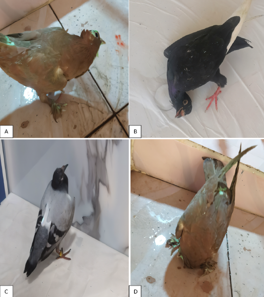
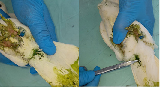
Field clinical examinations revealed a distinct set of symptoms indicating the severity of Newcastle disease infection in pigeons. Neurological signs were prominently observed, including torticollis, loss of balance, and partial paralysis of the wings or limbs as shown in Figure 2, suggesting direct involvement of the central nervous system by the virulent strain. Gastrointestinal manifestations were evident, with green watery diarrhea as shown in Figure 3, accompanied by a marked loss of appetite typical indicators of enteric involvement. The respiratory system also showed inflammatory responses such as labored breathing and abnormal respiratory sounds, reflecting significant damage to respiratory tissues. Additionally, general signs such as pronounced lethargy, wing drooping, feather breakage, and a noticeable increase in mortality rates were recorded. Collectively, these clinical findings strongly indicate the circulation of a highly virulent strain of Newcastle disease virus with substantial pathogenic potential.
Postmortem Findings
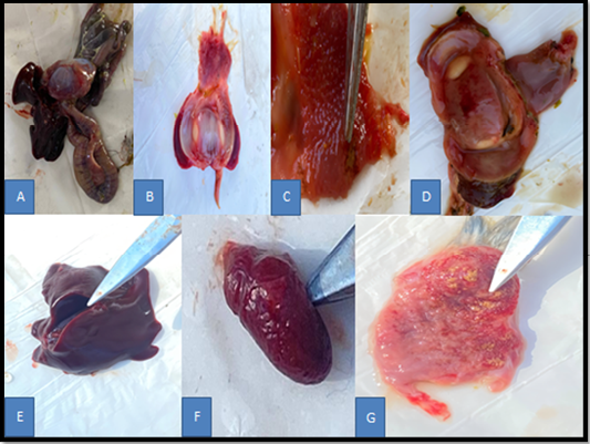
In pigeons affected by Newcastle disease reveal a series of characteristic lesions involving multiple organ systems. In the digestive tract, prominent enteritis is observed, characterized by thickening and congestion of the intestinal wall as shown in Figure 4A, along with petechial hemorrhages and, in some cases, mucosal ulcers resembling button ulcers. in the proventriculus show marked vascular congestion, petechial and linear hemorrhages as shown in Figure 4B, glandular hypertrophy, mucosal ulcers, and focal necrosis as shown in Figure 4C. In gizzard showing vascular congestion, petechial hemorrhages, and superficial ulcers as shown in Figure 4D. Also, enlargement and congested of the live as in the Figure 4E. In spleen show showing marked enlargement, severe congestion, and surface hemorrhagic foci as shown in Figure 4F. In Bursa of Fabricius show marked congestion and distinct hemorrhagic foci on its surface as shown in Figure 4G.
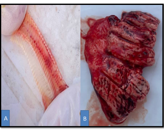
In the respiratory system, pathological changes include tracheal congestion, accumulation of thick mucus secretions as shown in Figure 5A. Also swelling of lung and pulmonary edema was evident (Figure 5B), and cloudiness of the air sacs, attributed to inflammation and infiltration of inflammatory cells.
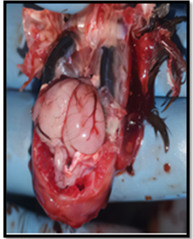
In severe cases, the central nervous system may also be affected, presenting with encephalitis, neuronal degeneration, and focal hemorrhages within the brain tissue as shown in Figure 6. These lesions collectively reflect the systemic nature of infection caused by virulent strains of Newcastle disease virus in pigeons.
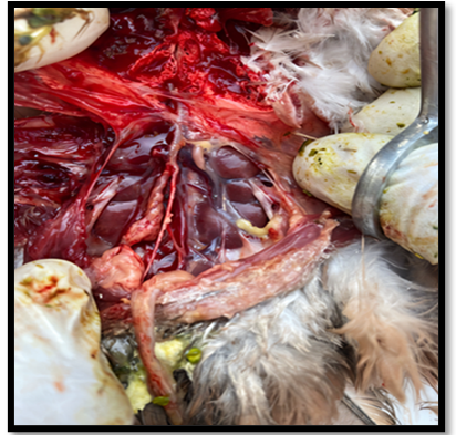
Also, the postmortem examination appears sever congestion in kidney of pigeon infected with ND AS shown in Figure 7.
Hemagglutination (HA) Test
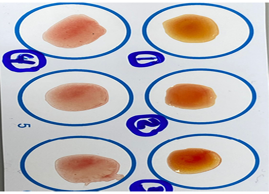
All tested samples showed positive hemagglutination (Figure 8), indicated by uniform reddish diffusion without central button formation. These results confirm the presence of an active virus capable of agglutinating red blood cells, supporting the diagnosis of NDV infection.
Hemagglutination Inhibition (HI) Test
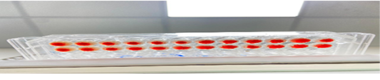
A hemagglutination inhibition (HI) test was performed to confirm the identity of the Newcastle Disease Virus (NDV) in the harvested allantoic fluid. Out of 28 collected pooled samples, 7 were positive for the HI test. The results demonstrated clear inhibition of hemagglutination in several wells, indicated by the absence of central button formation Figure 9.
Cytopathic Effect of NDV in Embryonated Chicken Eggs
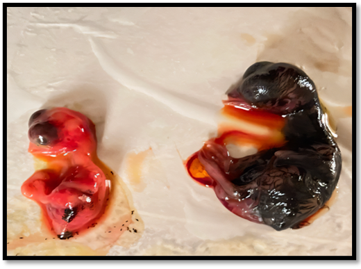
The cytopathic effects observed in embryonated chicken eggs post inoculating tissue analyzed collected from internal organs of pigeons suspected infected with NDV were marked growth retardation, in addition, visible hemorrhages were present on the body and in the brain region compared to the control embryo. as showed in Figure 10. The results showed that the pathotype of all seven HI-positive samples, based on the MDT test, was that of highly virulent isolates. The mean MDT for the seven isolates ranged from 40 to 56 hours.
The Results of Real Time PCR
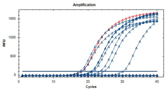
The Real-Time PCR amplification plots of fusion (F) gene for Newcastle disease virus using TaqMan probe (FAM) amplification from pigeon tissue sample. Only 7 samples show positive NDV plots with Vaccine sample red plot as positive control at threshold cycle numbers ranged (18-30 Cq) (Figure 11).
The results of Agarose Gel Electrophoresis
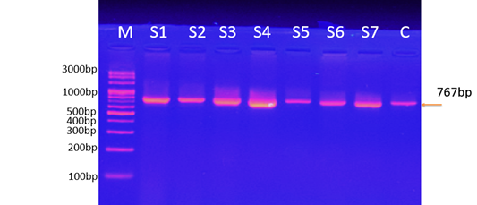
The results of agarose gel electrophoresis (Figure 12) showed that the expected size of the RT-PCR product (the fusion gene fragment used to detect for Newcastle disease virus from pigeon tissue samples). The marker M represent the 3000-100 bp ladder. The lane (S1-S7) showed selected positive Newcastle disease virus samples and the vaccine positive control samples with an expected 767 bp product.
DISCUSSION
Pigeons are among the many domestic and wild bird species infected with Newcastle disease virus (NDV), a highly contagious viral disease. In the last quarter of 2024, many pigeon breeders in Babylon Governorate, located in central Iraq, complained of various symptoms of infection were appearing in pigeon flocks. Therefore, the study aimed to confirm the primary diagnosis through molecular detection of the virus. In order to better understand the illness development and diagnostic characteristics of the virus in this avian group, pigeons displaying clinical symptoms were evaluated in this study. The clinical symptoms observed in pigeons in this study, were head twisting, greenish-white mucoid diarrhea, and neurological symptoms like tremors, were in line with earlier research ; whom referred to that infected pigeons with ND showed greenish-white diarrhea with nervous signs, including wings and legs paralysis, neck paresis, tremor of the head, rotation for one side, and incoordination. Postmortem examination revealed notable gross lesions, including congestion of the spleen and kidneys, hemorrhages in the intestinal and tracheal mucosa, hepatic necrosis, and brain congestion in pigeons with neurological signs. These lesions reflect the systemic and neurotropic characteristics of NDV and are consistent with findings by Marasigan et al. (2024). The results of current study agree with results obtain by previous study ; ; ; whom pointed to that in natural infection, the gross lesions of ND include hemorrhages and swelling in the brain, proventriculus, liver, spleen, and kidneys, and also, there is catarrhal enteritis and visceral congestion in addition to swelling and edematous of the Bursa of Fabricius. Because the clinical symptoms and gross lesions of ND in pigeons closely resemble those of other diseases such as salmonellosis, vitamin E deficiency, aflatoxinosis, mycoplasmosis, avian influenza, and other diseases, diagnosing the disease based on clinical signs is difficult . For that, to confirm the clinical diagnosis has been used some of laboratory tests such as rapid kits. The results of this test showed that only 28 pigeons were infected with ND out of a total of 100 pigeons collected from live bird markets in Babylon Governorate. The rapid kit proved to be a valuable field tool due to its ease of use and fast results, particularly suitable for on-site screening in suspected outbreak zones.
These findings align with the reports of ; , who have highlighted the high sensitivity and specificity of rapid tests for early NDV detection. Similarly, have emphasized that although rapid tests do not replace molecular methods in precision, they are essential for prompt field decisions when clinical signs are evident. To assess the virulence of the virus, inoculation into embryonated chicken eggs was performed to assay mean death time index. Embryos death occurred within less than 60 hours, indicating a highly virulent NDV strain. This observation is in line with previous findings ; , where it was found that highly virulent NDV strains cause rapid embryo mortality, serving as a biological marker of replication efficiency and viral severity. The agreement with previous studies strengthens the credibility of this method for evaluating virulence in pigeons. The dead chicken embryos showed clear pathological lesions, represented by bleeding throughout the body of the embryo, especially the head area these lesions were as observed previously . For further confirmatory analysis, the hemagglutination test and the hemagglutination inhibition test were performed. The results of these tests confirmed a total of 7 (25%) positive samples out of 28 and these results were also confirmed by real time RT-PCR which targeting the fusion (F) gene known to be associated with virulence. These findings align with previous several studies ; ; , where authors have emphasized the reliability of combining serological and molecular methods for accurate diagnosis of NDV. Several additional studies have ; confirmed the sensitivity of real-time PCR for detecting NDV strains in pigeons. Finally, gel electrophoresis results showed clear bands at expected sizes in the 7 positive samples and one vaccine sample, confirming the presence of NDV. These results are consistent with previous studies ; ; , where band sizes after RT-PCR serves as a reliable molecular indicator of NDV infection in pigeons.
CONCLUSIONS AND RECOMMENDATIONS
This study concluded that NDV is the main cause of the disease affecting pigeons, however, other pathogens can’t be ignored. We also conclude from this study that there is a close relationship between the results of virus isolation and molecular tests, both of which confirm the diagnosis of infection with the NDV. The rapid test kit is reliable for the initial screening and diagnosis of NDV in field conditions. However, further epidemiological studies and genetic analyses are still needed to better understand the antigenic properties of the virus and to develop effective strategies for controlling the disease in pigeon populations.
ACKNOWLEDGEMENTS
The authors would like to express their sincere gratitude to the College of Veterinary Medicine, Al-Qasim Green University, for providing the necessary laboratory facilities and technical support for the completion of this study. Special thanks are also extended to the staff of the Department of Pathology and Poultry Diseases for their assistance in sample collection and laboratory analysis. We are also grateful to the local authorities and live bird market vendors for facilitating access to pigeon samples.
NOVELTY STATEMENT
This study presents the first molecular evidence of virulent Avian Avulavirus-1 in Iraqi pigeons, emphasizing the diagnostic value of F gene detection and suggesting co-infection with other pathogens.
AUTHOR’S CONTRIBUTIONS
Both authors contributed to the preparation of this manuscript, its final review, and its approval for publication.
Conflict of Interest
The authors declare that there is no conflict of interest regarding the publication of this paper.
Figures
Figure 1:.
An example NDV-Ag rapid test results. Positive results are marked with a line at T and control marked with C indicate the successful execution of the test.
Figure 2:.
Photograghs showing pigeons naturally infected with Newcastle disease (ND), and showing neurological signs.
Figure 3:.
Photographs showing Newcastle disease-infected pigeon with vent staining due to green watery diarrhea.
Figure 4:.
Photographs showing gross lesions in visceral organ of pigeons infected with NDV. A: Thickening and congestion of the intestinal wall; B: Marked vascular congestion, petechial and linear hemorrhages in proventriculus; C: Glandular hypertrophy, mucosal ulcers, and focal necrosis in proventriculus; D: Vascular congestion, petechial hemorrhages, and superficial ulcers in gizzard; E: Enlarged and congested of the liver; F: Marked enlargement, severe congestion in the spleen; G: Marked congestion and distinct hemorrhagic foci in the Bursa of Fabricius.
Figure 5:.
A: Tracheal of a pigeon infected with ND showing severe mucosal congestion with petechial hemorrhage, and accumulation of thick mucus secretions; B: The lung of a pigeon infected with Newcastle disease shows severe congestion, swelling, and obvious hemorrhagies.
Figure 6:.
Photograph showing brain of a pigeon infected with Newcastle disease, indicating marked congestion of superficial blood vessels, sub-meningeal hemorrhages, and evident softening of the brain tissue.
Figure 7:.
The illustration of a clear enlargement of the kidneys in a pigeon infected with Newcastle disease, where the kidneys appear larger congestion.
Figure 8:.
Results of hemagglutination assay performed on allantoic fluid samples obtained from NDV infected pigeons. Wells 1, 3, and 5 show positive agglutination patterns, while wells 2, 4, and 6 are negative, indicating either absence of virus or inhibition by specific antibodies.
Figure 9:.
The photograph explains positive Hemagglutination hemagglutination inhibition for NDV isolates collected from pigeons.
Figure 10:.
The photograph showing the cytopathic effects observed in embryonated chicken eggs post inoculating with NDV which were marked with growth retardation. Additionally, visible hemorrhages were present on the body and in the brain region (embryo in left) compared to the control embryo (embryo in right).
Figure 11:.
The Real-Time PCR amplification plots of fusion (F) gene for Newcastle disease virus using TaqMan probe (FAM) from pigeon tissue sample. Only 7 samples showed positive NDV plots and Vaccine sample red plots as positive control at threshold cycle numbers ranged from 18 to -30 Cq.
Figure 12:.
The electrophoresis-based separation of the RT-PCR product analysis of the fusion (F) gene of Newcastle disease virus from pigeon tissue samples in agarose gel 1%. Amplified products was a DNA fragment with a size of 767 bp. RT-PCR products showed all 7 field isolates from pigeons. Where M: marker (3000-100 bp). The lane (S1-S7) showed selected positive NDV samples and C: vaccine positive control samples with expected 767 bp RT-PCR product.
References
Abbas AE, Abdelaziz SA, Khalifa KA, Alhassan AM, Ahmad SA (2022). Virological and serological studies of Newcastle disease virus in pigeons (Columba livia) in Khartoum State, Sudan. In: 2nd International Conference on Sustainable Ecological Agriculture, 2. Uluslararası Sürdürülebilir Ekolojik Tarım Kongresi, p. 458.
Abdisa T, Tagesu T (2017). Review on Newcastle disease of poultry and its public health importance. Journal of Veterinary Science and Technology, 8(3): 441. https://doi.org/10.4172/2157-7579.1000441[CrossRef]
Al-Shareef YAH, Abawi FHK (2024). Isolation and identification of Newcastle disease viruses from naturally infected chickens. Journal of Animal Health and Production, 12: 32–39. https://doi.org/10.17582/journal.jahp/2024/12.s1.32.39[Full text][CrossRef]
Al-Ziaydi AG, Al-Shammari AM, Hamzah MI (2020). Propagation of oncolytic newcastle disease virus in embryonated chicken eggs and its research applications in cell lines. Journal of Physics: Conference Series, 1664: 12129. https://doi.org/10.1088/1742-6596/1664/1/012129[CrossRef]
Almremdhy HA, Khamas EJ (2019). Cytotoxicity and antiviral activity of atorvastatin against Newcastle disease virus in chicken embryos. Biochemical and Cellular Archives, 19(2).
Al Shekaili T (2015). Epidemiological studies on avian influenza and other respiratory viruses in backyard poultry in Oman. The University of Liverpool (United Kingdom).
Alaarajy KHS, Almremdhy H (2025). Molecular diagnosis of natural pox virus infection in pigeons in Babylon province, central Iraq. Jornal of Animal Health and Production, 13(2): 488–495. https://doi.org/10.17582/journal.jahp/2025/13.7.488.495[Full text][CrossRef]
Ali HM, Rabee RHS (2024). Evaluation of oil vaccines of avian influenza H5 virus in layer by enzyme-linked immunosorbent assay. Journal of Animal Health and Production, 12: (Special Issue 1): 292–299. https://doi.org/10.17582/journal.jahp/2024/12.s1.292.299[Full text][CrossRef]
Bala, A. (2023). A historical overview of Newcastle disease and its global impact on poultry production. Veterinary Research Communications, 47(1): 133–140. https://doi.org/10.1007/s11259-022-10025-1[CrossRef]
Behboudi S (2023). Newcastle disease. CABI Compendium. https://doi.org/10.1079/cabicompendium.73358[CrossRef]
Chitty J (2018). Pigeons (Columba livia). Companion Animal Care and Welfare: The UFAW Companion Animal Handbook, 355–370. https://doi.org/10.1002/9781119333708[CrossRef]
Chowdhary M, Nashiruddullah N, Roychoudhury P, Bhat A, Ahmed JA, Kour K, Sood S (2020). Molecular and virulence characterization of newcastle disease virus in fowls and pigeons from Jammu, India. Indian Journal of Animal Research, 54(10): 1279–1284. https://doi.org/10.18805/ijar.b-3885[CrossRef]
Damena D, Fusaro A, Sombo M, Belaineh R, Heidari A, Kebede A, Kidane M, Chaka H (2016). Characterization of Newcastle disease virus isolates obtained from outbreak cases in commercial chickens and wild pigeons in Ethiopia. Springerplus, 5: 1–8. https://doi.org/10.1186/s40064-016-2114-8[CrossRef]
Dharmayanti NLPI, Nurjanah D, Nuradji H, Suyatno T, Indriani R (2023). Newcastle disease virus: the past and current situation in Indonesia. Journal of Veterinary Science, 25(1): e3. https://doi.org/10.4142/jvs.23022[CrossRef]
Ellakany HF, Elbestawy AR, Abd El-Hamid HS, Zedan RE, Gado AR, Taha AE, Soliman MA, Abd El-Hack ME, Swelum AA, Saadeldin IM (2019). Role of pigeons in the transmission of avian avulavirus (Newcastle disease-genotype VIId) to chickens. Animals, 9(6): 338. https://doi.org/10.3390/ani9060338[CrossRef]
Haddas R (2023). Newcastle disease virus. Infect Dis, 427. https://doi.org/10.1007/978-1-0716-2463-0_1093[CrossRef]
Haryanto A, Purwaningrum M, Verawati S, Irianingsih SH, Wijayanti N (2015). Pathotyping of local isolates Newcastle disease virus from field specimens by RT-PCR and restriction endonuclease analysis. Procedia Chemistry, 14: 85–90. https://doi.org/10.1016/j.proche.2015.03.013[CrossRef]
Hayes MC (2023). Molecular characterisation of pigeon paramyxovirus and other viruses identified by deep sequencing in pigeons and doves in South Africa. University of Pretoria (South Africa).
Hongzhuan Z, Ying T, Xia S, Jinsong G, Zhenhua Z, Beiyu J, Yanyan C, Lulu L, Jue Z, Bing Y (2020). Preparation of the inactivated Newcastle disease vaccine by plasma activated water and evaluation of its protection efficacy. Applied Microbiology and Biotechnology, 104: 107–117. https://doi.org/10.1007/s00253-019-10190-1[CrossRef]
Hossain I, Parvin R, Rahman MM, Begum JA, Chowdhury EH, Islam MR, Diel DG, Nooruzzaman M (2023). Comparative pathogenicity of a genotype XXI.1.2 pigeon Newcastle disease virus isolate in pigeons and chickens. Microbial Pathogenesis, 178: 106068. https://doi.org/10.1016/j.micpath.2023.106068[CrossRef]
Kraidi QA, Kareem Ramadhan MA, Seger WM, Najem HA (2024). Influence of pigeon paramyxovirus type-1 on clinico-pathological profiles in racing pigeons associated with recent outbreaks in Iraq. Research in Agricultural and Veterinary Sciences, 8(2). https://doi.org/10.62476/ravs8290[CrossRef]
Liang L, Wang D, Gao Z, Tang J, Li X, Ren P, Wang Y, Gao S, Wu X, Guo Y (2024). RAA-CRISPR/Cas12a-mediated rapid, sensitive, and onsite detection of Newcastle disease in pigeons. Veterinary Sciences, 11(10): 473. https://doi.org/10.3390/vetsci11100473[CrossRef]
Mao Q, Ma S, Schrickel PL, Zhao P, Wang J, Zhang Y, Li S, Wang C (2022). Review detection of Newcastle disease virus. Frontiers in Veterinary Science, 9:936251. https://doi.org/10.3389/fvets.2022.936251[CrossRef]
Marasigan CNBB, Lola MSEG, Umali DV (2024). Gross and microscopic pathology of pigeon paramyxovirus serotype 1 (PPMV-1) infection in racing pigeons (Columba livia domestica) from Luzon, Philippines. Philippine Journal of Veterinary Medicine, 61(1).
Marlier D, Vindevogel H (2006). Viral infections in pigeons. The Veterinary Journal, 172(1): 40–51. https://doi.org/10.1016/j.tvjl.2005.02.026[CrossRef]
Moustapha A, Talaki E, Akourki A, Ousseini M (2023). Newcastle disease virus in poultry: current status and control prospects. World’s Veterinary Journal, 2: 240–249. https://doi.org/10.54203/scil.2023.wvj26[CrossRef]
Muhammed QO, Rabee RHS (2024). Assessment of oil vaccination protection to Newcastle disease virus challenges in layer. Journal of Animal Health and Production, 12 (Special Issue 1): 351–356.
Nurzijah I, Elbohy OA, Kanyuka K, Daly JM, Dunham S (2022). Development of plant-based vaccines for prevention of avian influenza and Newcastle disease in poultry. Vaccines, 10(3): 478. https://doi.org/10.3390/vaccines10030478[CrossRef]
Parvez MNH, Islam MR, Akter MTD, Sarder MJU (2017). Clinico-histopathological observations of pigeons (Columba livia) suffering from Newcastle disease in Northern Bangladesh. Asian Journal of Medical and Biological Research, 3(1): 134–139. https://doi.org/10.3329/ajmbr.v3i1.32049[CrossRef]
Paul TK, Amin MR, Alam MA, Rahman MK, Sarker YA, Rizon MK (2015). Occurrence of pigeon diseases at Khulna Sadar, Bangladesh. Bangladesh Journal of Veterinary Medicine, 13(2): :21–25. https://doi.org/10.3329/bjvm.v13i2.26616[CrossRef]
Qosimah D, Murwani S, Sudjarwo E, Lesmana MA (2018). Effect of Newcastle disease virus level of infection on embryonic length, embryonic death, and protein profile changes. Veterinary World, 11(9): 1316. https://doi.org/10.14202/vetworld.2018.1316-1320[CrossRef]
Rivetz B, Weisman Y, Ritterband M, Fish F, Herzberg M (1985). Evaluation of a novel rapid kit for the visual detection of Newcastle disease virus antibodies. Avian Diseases, 29: 929–942. https://doi.org/10.2307/1590446[CrossRef]
Samal SK (2020). Paramyxoviruses as vaccine vectors. In: Viral Vectors in Veterinary Vaccine Development: A Textbook. Springer. pp. 113–139. https://doi.org/10.1007/978-3-030-51927-8_8[CrossRef]
Shaheen S, Anjum AD, Rizvi F (2005). Clinico-pathological observations of pigeons (Columba livia) suffering from Newcastle disease. Liver, 2: (7.14). https://doi.org/10.3329/ajmbr.v3i1.32049[CrossRef]
Xiang B, You R, Kang Y, Xie P, Zhu W, Sun M, Gao P, Li Y, Ren T (2019). Host immune responses of pigeons infected with Newcastle disease viruses isolated from pigeons. Microbial Pathogenesis, 127: 131–137. https://doi.org/10.1016/j.micpath.2018.11.049[CrossRef]
Xu D, Li C, Liu G, Chen Z, Jia R (2019). Generation and evaluation of a recombinant goose origin Newcastle disease virus expressing Cap protein of goose origin avastrovirus as a bivalent vaccine in goslings. Poultry Science, 98(10): 4426–4432. https://doi.org/10.3382/ps/pez255[CrossRef]