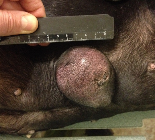
Ligfol and Fluorouracil were administered to the following patients. Patient 1. Doggess, Miniature Schnauzer breed, at the age of 10 years, 8.8 kg, not sterilized, ruttishness was every 6 months, last gestation and parturation were about 3 years ago. For the whole period of life, there were 4 gestations. Anamnesis morbi: localization of the primary M1 tumor on the right side, T2N0M0. Simple mastectomy was performed in the animal. The histological diagnosis was the following: carcinoma of the mammary gland, grade I malignancy, adjuvant chemotherapy (chemotherapy) was not performed. After 6 months, relapse was detected. The results of the clinical study were the following: localization of M2 tumor nodes (Figure 1) and M4 on the right side, T3N2/T2N0, distant metastases were not detected. The results of the hematological studies were the following: WBC 9.4 x 10⁹/L; Lymph 2.1 x 10⁹/L; Mon 0.3 x 10⁹/L; Gran 7.0 x 10⁹/L; Lymph % 22.4; Mon % 3.5; Gran % 74.1; Eos % 4.7; RBC 7.31 x 10 12/L; HGB 152 g/L; HCT % 51.5; PLT 516 x 109/L. The concomitant diseases were the following: cholecystitis and CKD.
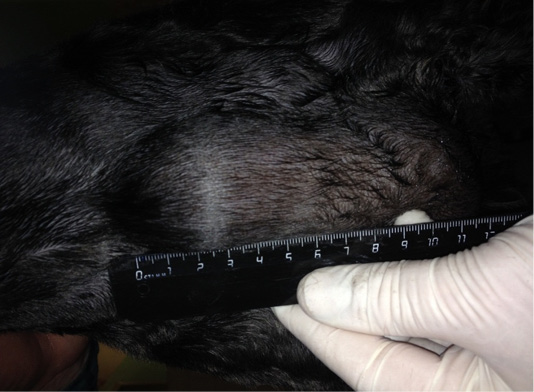
Figure 1: Neoplasm of the mammary gland in the M2 region on the right side. Doggess, miniature schnauzer breed, at the age of 10 years.
Patient 2. Doggess, standard dachshund breed, at the age of 10.5 years, with the weight of 7.5 kg, OGE was 2 years ago due to pyometra. Neither gestations, nor parturation episodes were during the whole period of life. Anamnesis morbi: primary tumor in M4 region on the right side, T2N0M0. Simple mastectomy was conducted in the animal. The histological diagnosis was the following: infiltrating adenocarcinoma of the mammary gland, grade I of malignancy, with a stromal reaction. Adjuvant CT was not performed in the animal. Relapse was noted after 12 months. The results of the clinical study were the following: localization of tumor nodes: M4 and M5 on the left side, T2N0/T2N1 (Figure 2); M1 and M5 on the right side T2N1/T2N2. Distant metastases were not detected during the primary admission. The hematological indicators were the following: WBC 7.6 x 10⁹/L; Lymph 2.0 x 10⁹/L; Mon 0.4 x 10⁹/L; Gran 5.2 x 10⁹/L; Lymph % 26.7; Mon % 4.9; Gran % 68.4; Eos % 0.4; RBC 8.32 x 10¹²/L; HGB 179 g/L; HCT % 61.5; PLT 343 x 109/L. The concomitant diseases were the following ones: mitral valve endocardiosis, HCM and CKD.
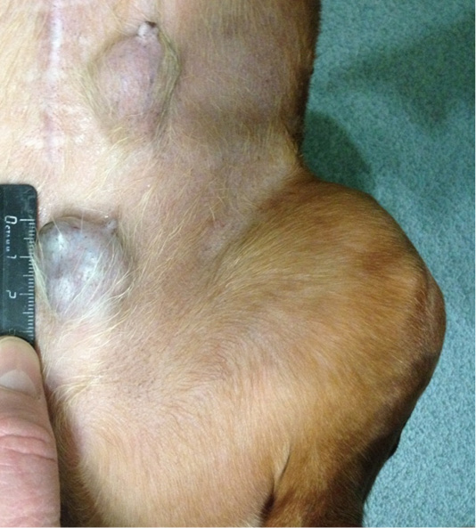
Figure 2: Localization of tumor nodules: M4 and M5 region on the left side. Doggess, breed dachshund standard, at the age of 10.5 years.
Patient 3. Doggess, breed jagdterrier, at the age of 12 years, with the weight of 10 kg, sterilized. There were no gestation and parturation episodes. Anamnesis morbi: primary M1 tumor on the right side, T2N0M0. Simple mastectomy was performed. The histological diagnosis was the following: carcinoma of the mammary gland mixed structure. No adjuvant chemotherapy was performed in the animal; relapse was detected after 7 months. The results of the clinical study were the following: localization of the tumor site at the suture site of the performed mastectomy (Figure 3), T3N2M0. The hematological indicators were the following: WBC 8.7 x 10⁹/L; Lymph 1.9 x 10⁹/L; Mon 0.4 x 10⁹/L; Gran 6.4 x 10⁹/L; Lymph % 21.8; Mon % 4.7; Gran % 73.5; Eos % 1.6; RBC 9.05 x 10¹²/L; HGB 193 g/L; HCT % 57.7; PLT 684 x 109/L. The concomitant diseases were the following ones: EHR, cholecystitis and CKD.
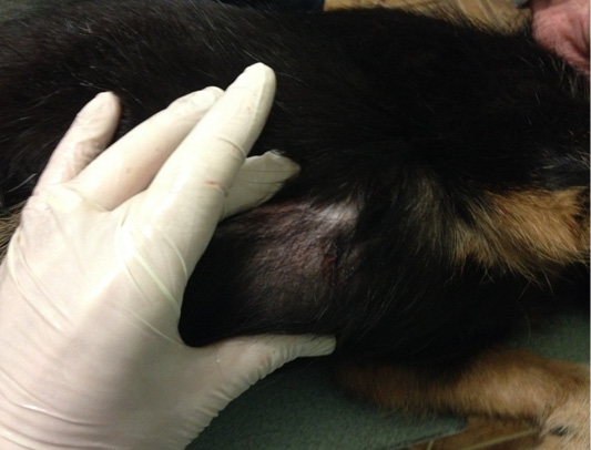
Figure 3: Mammary gland neoplasm in the M1 region on the right side. Doggess, breed jagdterrier, at the age of 12 years.
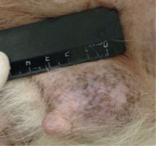
Figure 4: Mammary gland neoplasm in the M4 region on the left side. Doggess, American Bulldog, at the age of 11.5 years.
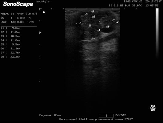
Patient 4. Doggess, American Bulldog, at the age of 11.5 years, with the weight of 37.2 kg, not sterilized, ruttishness was every 4–6 months. One episode of gestation and parturation was at the age of 4 years. Anamnesis morbi: primary tumor at M5 region on the left side, T3N1M0. Simple mastectomy was performed in the animal. The histological diagnosis was the following: infiltrative ductal carcinoma. Adjuvant CT was not performed. Relapse was detected after 6.5 months. The results of the clinical study were the following: localization of tumor sites in M4 and M5 regions on the right side, T2N0/T2N2; M4 on the left side–T2N0 (Figure 4). The ultrasound study revealed distant metastases in the liver (Figure 5). The hematological indicators were the following: WBC 9.6 x 10⁹/L; Lymph 1.5 x 10⁹/L; Mon 0.3 x 10⁹/L; Gran 7.8 x 10⁹/L; Lymph % 15.9; Mon % 3.6; Gran % 80.5; Eos % 1.9; RBC 6.84 x 10¹²/L; HGB 147 g/L; HCT % 52.7; PLT 735 x 109/L. The concomitant diseases were the following ones: DCM, CKD, and recurrent eczema.
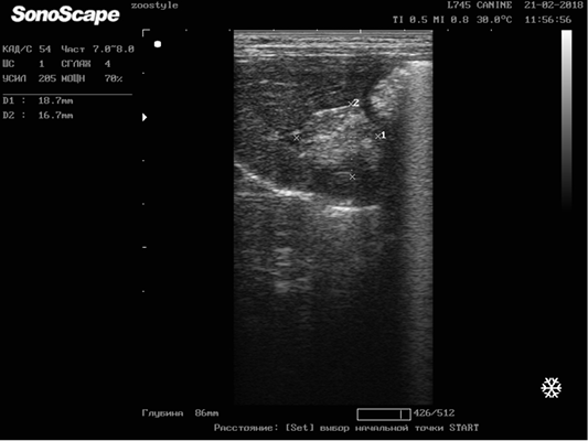
Figure 5: Metastasis of breast cancer in the liver parenchyma. Doggess, breed American Bulldog, at the age of 11.5 years.
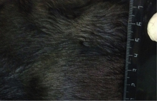
Figure 6: Localization of the tumor in M4 region on the left side. Doggess, Miniature Schnauzer, at the age of 10.5 years.
Patient 5. Doggess, miniature schnauzer, at the age of 10.5 years, 8.5 kg, not sterilized, ruttishness was every 6 months. There were 2 episodes of gestation and parturation, moreover, the last one occurred about 7 years ago. Anamnesis morbi: primary tumor in M5 region on the left side, T2N0M0. Patient underwent a simple mastecomy. Histological diagnosis of moderately differentiated non-infiltrating lobular mammary gland cancer. Adjuvant chemotherapy was not performed in the patient. Relapse was detected after 12 months. The results of the clinical study were the following: localization of the tumor in M4 region on the left side (Figure 6), T3N1M1. Distant metastases were detected on X-Ray in lungs–focuses up to 0.5 cm in diameter (Figure 7). The hematological indicators were the following: WBC 9.9 x 10⁹/L; Lymph 3.0 x 10⁹/L; Mon 0.4 x 10⁹/L; Gran 6.5 x 10⁹/L; Lymph % 30.3; Mon % 4.1; Gran % 65.6; Eos % 1.6; RBC 6.42 x 10¹²/L; HGB 134 g/L; HCT % 46.1; PLT 523 x 109/L. The concomitant diseases were the following ones: on ultrasonography – diffuse changes in the parenchyma of both kidneys and urolithiasis.
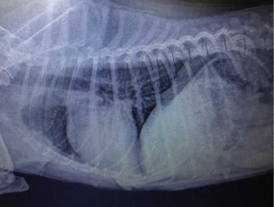
Figure 7: Metastasis of mammary gland cancer in the lungs. Doggess, Miniature Schnauzer, at the age of 10.5 years.
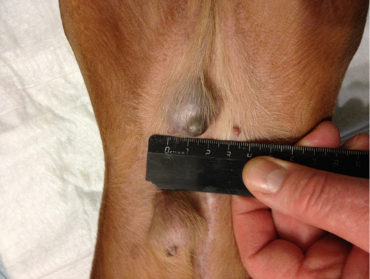
Figure 8: Localization of tumor nodules in M4 and M5 regions on the right side. Doggess, dachshund standard, at the age of 9 years.
Patient 6. Doggess, standard dachshund, at the age of 9 years, 7.8 kg, not sterilized, ruttishness was every 6 months. There were no episodes of gestation and parturation during the whole period of life. Anamnesis morbi: primary M3 tumor
Table 1: Dynamics of hematological indicators.