Research Article · Volume 12, Issue 3, 8 February 2024 · Open Access
Optimizing Bone Healing in Rabbit Models: A Comparative Study of Lidocaine Hydrochloride and Diclofenac: Histological Study
·
Affiliations & Notes ⌄
1Department of Physiology, Biochemistry and Pharmacology, College of Veterinary Medicine, University of Baghdad *Correspondence | Raffal A. Omar, Department of Physiology, Biochemistry and Pharmacology, College of Veterinary Medicine, University of Baghdad
This consistency strongly suggests that the effects of Lidocaine hydrochloride and Diclofenac on bone healing are enduring rather than transient.
The Lidocaine hydrochloride 2 mg/kg B.W. group consistently exhibited accelerated bone healing, increased bone density, and advanced tissue maturation.
In summary, this study illustrates that Lidocaine hydrochloride, particularly when administered at a 2 2 mg/kg B.W. dosage, exhibits significant potential in facilitating the bone healing process.
Recommendations stemming from this study encompass the necessity for further research to elucidate the mechanisms through which Lidocaine hydrochloride expedites bone healing and to explore its potential clinical applications.
Selected from the article's own text.
Abstract
The present study aimed to evaluate the effect of intraosseous injection rout of 2% lidocaine hydrochloride and 3.75% Diclofenac, on bone regeneration. Forty-five adult male rabbits of the local breed were employed to create a 3.5mm hole defect in the proximal third of the medial aspect of the tibia by an electric drill with dropping isotonic normal saline to prevent thermal necrosis. The experimental animals were divided randomly into three equal groups, each group include fifteen rabbits (n=15). control without any additive. group 1(lidocaine hcl). Which applied daily single dose of 2% lidocaine Hcl 2 mg/Kg. B. W. for five days post-operation (P. O.), while the group 2(diclofenac) applied 3.75% of 20mg/Kg. B. W. histopathological specimens were taken at the end of the 7th,14th, and 21th day p.o. The results revealed rapid bone regeneration improvement and development in group I compared to group II and control group. In conclusion, intra-osseous injection of 2% Lidocaine Hcl 2mg/kg BW (body weight) has a stimulatory effect on bone healing in which osteogenic tissue and trabecular bone were noticed clearly at the end of 1st week, which achieves histologically compared to Diclofenac and control groups.
Bone healing is a crucial process in the field of medical and surgical sciences, with various factors playing significant roles in either promoting or inhibiting this process. There is an increasing interest in medical research regarding the impact of certain substances on bone healing, including the use of lidocaine and diclofenac. These substances have diverse applications and effects on bone healing and tissue regeneration in general.
Anesthetic procedures and agents have a rich historical legacy in the field of surgery, with local anesthesia being particularly noteworthy . Consequently, as our understanding of anesthesia techniques has advanced, it has become feasible to perform a multitude of diagnostic and surgical procedures under local anesthesia . Local anesthesia means the loss of pain in a specific target area, briefly block pain signals in nerve fibers, halting pain transmission to the brain . Which is important to decrease the cost and side effects of general anesthesia, especially in large animal species. This depends on the local anesthetic agents’ ability to cross nerve sheaths and neural membranes . However, with the ever-increasing awareness of pain management that could be used in all veterinary species with a safety dose especially in dogs and rabbits ; .
Lidocaine hydrochloride, in various forms and concentrations, is widely utilized in veterinary medicine . Its versatility extends to applications beyond pain management, encompassing its potential influence on tissue regeneration processes, including bone healing. Besides lidocaine hcl established use for IO (intraosseous) injections and its capacity to regulate blood pressure and cardiac rhythm and its mainly excreted in the urine, with 90% as metabolites and 10% as the unchanged drug , also, its rapid onset, strong effectiveness, and low allergy risk , lidocaine’s role in veterinary surgery is notable for its rapid onset, ease of administration, and facilitating smooth postoperative recovery . Lidocaine, a local anesthetic, blocks nerve impulses by inhibiting sodium ion influx into nerve cells, resulting in a reversible loss of sensation Combining lidocaine with other drugs extends the duration of postoperative analgesia, achieving rapid sensory and motor block with minimal pain scores, which is also considered one of the advantages of lidocaine . This broad utility across a spectrum of procedures and administration routes . Underscores its significance in the context of tissue healing and regeneration, including its impact on bone tissue healing.
Diclofenac, a widely used NSAID, has potent analgesic and anti-inflammatory properties . Globally, injury and bone fracture patients often receive NSAID treatment. These drugs are effective in post-traumatic therapy due to their combined anti-inflammatory action and potent analgesic effects. However, it’s worth noting that some in vitro studies have suggested that NSAIDs, including diclofenac, may hinder bone fracture healing or the fixation of hydroxyapatite-coated implants . Diclofenac, specifically, has been associated with adverse effects on bone healing. Research indicates that diclofenac hampers the proliferation and triggers apoptosis in human osteoblast cells. This occurs due to the inhibition of prostaglandin E2 formation, consequently impeding pre-osteoblast differentiation and suppressing both bone formation and resorption . Furthermore, on a cellular level, diclofenac may have adverse effects on pre-osteoblast cell growth ; . Diclofenac is used for various medical conditions and possesses a broad spectrum of anti-inflammatory and analgesic properties ; . However, its use is associated with gastric damage, a potential side effect of NSAID use .
Numerous studies have been published regarding the cytotoxic effects of local anesthetics on various cell types, including osteoblastic cells . Additionally, intraosseous (IO) injection, which involves the direct administration of anesthetic agents into the bone, is experiencing a resurgence in popularity for regional anesthesia . There are numerous injection methods available for local anesthesia, with intraosseous injection being one of the historical techniques used since the early 1900s. Despite the advancements in plastic catheters facilitating vein access, there are situations where peripheral venous access is impractical. In such cases, intraosseous (IO) injection remains a valuable method, especially in dental and surgical procedures necessitating substantial anesthesia doses ; ; .
MATERIALs AND METHODS
In this study, 45 adult male rabbits of a local breed were utilized, and they underwent a one-week acclimation period in dedicated cages before the experiment commenced . These rabbits were chosen as a suitable model for bone healing research due to their relatively fast bone turnover and similarity to human bone physiology. It is essential to note that all animal procedures strictly adhered to ethical guidelines and were approved by the Institutional Animal Care and Use Committee (IACUC).
The rabbits were divided into three experimental groups as follows:
Control group
Fifteen rabbits in this group did not receive any drug intervention and were the control group to assess natural bone healing.
Lidocaine hydrochloride 2mg group
Another fifteen rabbits received intraosseous injections of Lidocaine at a dose of 2 mg/kg B.W. once daily for five days, following the protocol by .
Diclofenac group
The final group consisted of fifteen rabbits that underwent intraosseous injections of Diclofenac at a dose of 20 mg/kg B.W. once daily for five days, as outlined.
The study was conducted over 21 days, allowing for comprehensive observation of the bone healing process throughout the experiment.
Surgical procedure
Preoperative preparation
Prepare the proximal third of the medial aspect of Tibia, by clipping and shaving the hair, clean the area by tap water and medical soap, then disinfect the surgical site with 70% ethyl alcohol. after induction of general anesthesia and put the animal in lateral recompact and the medial aspect of the hind limb expose the surgeon, cover all the body with sterile drapes except the surgical site.
Anesthetics protocol
Induction of the general anesthesia done by intramuscular injection of 2% xylazine hydrochloride at the dose (17.5 mg/Kg. B. W.) After10 minutes, re-injection of 10% ketamine hydrochloride at the dose (25 mg/ Kg. B.W.) (Abd-Alreda, 2016).
Surgical technique
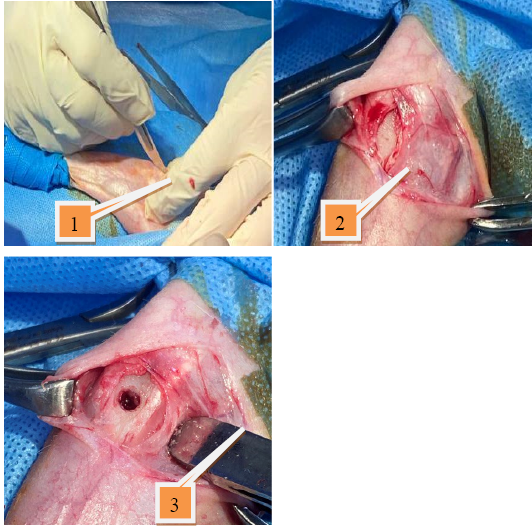
Create 2cm length skin incision by sharply dissect at the proximal and medial aspect of Tibia, separate all the soft tissue ,then remove the periosteum induced 3.5 mm hole defect with electrical drill with dropping normal sterile isotonic solution to prevent thermal necrosis of the bone , reposition the soft tissues and close the skin by simple interrupted suture pattern using 2/0 suture materials .the experimental animals divided to 4 groups as mentioned in the experimental design before.
Postoperative car
We are daily checking the site of operation.
Daily systemic antibiotics injection for three days p.o. Penicillin and streptomycin 10 Iu/Kg. B. W. and 5mg/Kg. B.W., respectively
Remove the suture materials seven days p.o.
RESULTS AND DISCUSSION
Histopathological results
First week: Control group
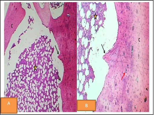
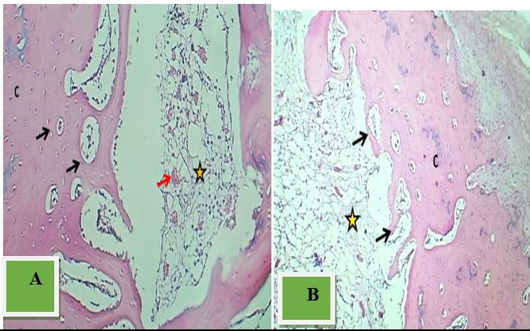
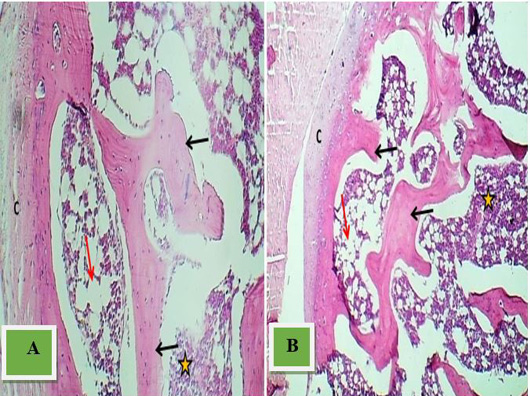
Histopathological evaluation revealed a normal appearance of the cortical bone with intact osteoid. The hall exhibited minimal new bone formation, primarily filled with marrow tissue (Figure 2).
Lidocaine Hcl group
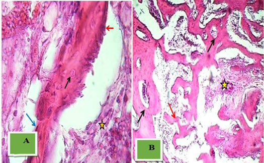
Histopathological findings depicted a thickened cortical bone with a densely populated marrow mass in the hall. The rim of the hall displayed a well-developed thick bone mass with numerous mature osteoid (Figure 3).
Diclofenac group
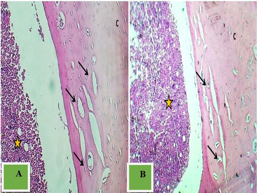
Histopathological analysis showed a significantly thinner and unremodeled cortical bone. The hall was filled with active osteogenic tissue, displaying active angiogenesis (numerous blood vessels), and marked the formation of an immature network of trabecular bone (Figure 4).
Second week
Control group
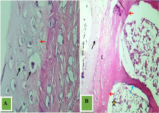
Histopathological examination displayed prominent osteogenesis within the periosteum surface of the cortical bone. The rim of the hall exhibited new bone formation, comprising some osteoid formation. The hall was filled with osteogenic tissue, blood vessels, and trabecular bone formation (Figure 5).
Lidocaine group
Histopathological examination demonstrated the hall filled with well-developed mature trabecular bone intermingled with a thick mass of active osteogenic tissue and numerous osteoids (Figure 6).
Diclofenac group
Histopathological analysis showed a commonly remodeled cortical bone with the formation of a thick bone layer, including a limited number of osteoids. The hall was primarily filled with marrow tissue (Figure 7).
Third week
Control group
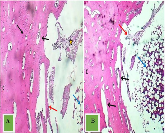
Histopathological examination revealed active endochondral ossification in the cortical bone, associated with a slightly thick layer of new bone formation. Numerous mature bone trabeculae filled the hall, along with osteogenic tissue and blood vessels (Figure 8).
Lidocaine group
Histopathological figures showed that the hall was filled with well-developed mature trabecular bone formation with a mass of red marrow tissue. The trabecular bone comprises mature osteocytes and osteoblast (Figure 9).
Diclofenac group
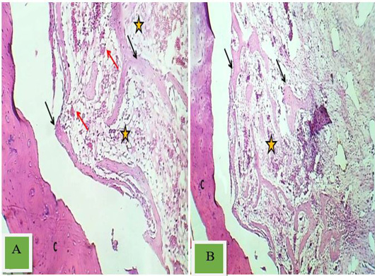
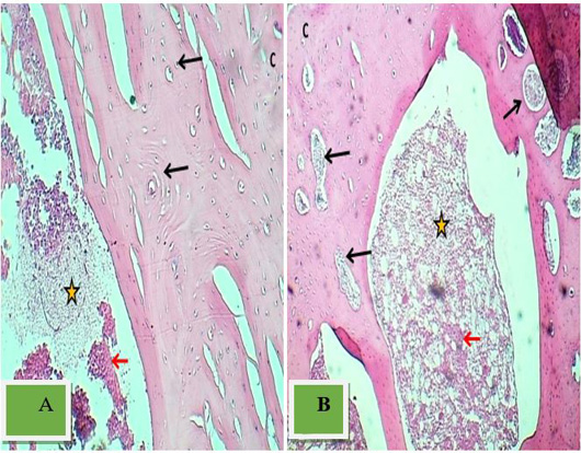
Histopathological findings indicated a commonly remodeled cortical bone with a thick and well-remodeled bone layer. Numerous mature osteoids were present, filling the hall with marrow and osteogenic tissue (Figure 10).
Persistent differences between the experimental groups remained stable over the 21-day study period. This consistency strongly suggests that the effects of Lidocaine hydrochloride and Diclofenac on bone healing are enduring rather than transient. The Lidocaine hydrochloride 2 mg/kg B.W group consistently exhibited accelerated bone healing, increased bone density, and advanced tissue maturation. In contrast, the Diclofenac group consistently demonstrated a slower progression of bone healing.
Acceleration of bone healing with lidocaine hydrochloride
During the initial seven days of the study, the histological observations suggest that Lidocaine hydrochloride can expedite the initiation of the bone healing process. Notably, the Lidocaine hydrochloride 2mg group displayed increased osteoblast activity and early woven bone formation, which aligns with previous research indicating that Lidocaine hydrochloride 2 mg/kg B.W. can stimulate osteoblastic differentiation and proliferation . These findings hold promise and may have significant clinical implications, particularly when accelerated bone repair is desired. Utilizing intraosseous lidocaine as a therapeutic agent for enhancing bone strength could be a viable consideration, particularly given the constant process of bone degradation and regeneration involving osteoclasts and osteoblasts .
Diclofenac-associated delay in bone healing
In contrast, the Diclofenac group experienced a noticeable delay in bone healing, with reduced osteoblast activity and less pronounced tissue remodeling ; ; . This delay aligns with concerns about NSAIDs, including Diclofenac, inhibiting bone healing, especially when COX-2 is absent or inhibited ; ; ; . Diclofenac’s anti-inflammatory properties may disrupt the crucial early inflammatory response necessary for bone repair. This raises questions about NSAID use in scenarios requiring optimal bone regeneration. Additionally, NSAIDs impair prostaglandin synthesis by inhibiting COX-1 and COX-2 in injured tissues and the central nervous system , with documented adverse effects on bone healing .
Consistency of results throughout the study
Persistent differences between the experimental groups remained stable over the 21-days study period. This consistency strongly suggests that the effects of Lidocaine hydrochloride and Diclofenac on bone healing are enduring rather than transient. The Lidocaine hydrochloride 2 mg/kg B.W. group consistently exhibited accelerated bone healing, increased bone density, and advanced tissue maturation. In contrast, the Diclofenac group consistently demonstrated a slower progression of bone healing.
CONCLUSIONS AND RECOMMENDATIONS
In summary, this study illustrates that Lidocaine hydrochloride, particularly when administered at a 2 2 mg/kg B.W. dosage, exhibits significant potential in facilitating the bone healing process. This is attributed to its capacity to enhance osteoblast activity and initiate early bone formation, offering the prospect of accelerated bone repair, particularly in situations demanding swift healing. Moreover, further comprehensive investigation is warranted into the therapeutic application of intraosseous lidocaine for fortifying skeletal structures.
Conversely, these findings raise concerns regarding the harmful impact of Diclofenac on bone healing, consistently prolonging the process, diminishing osteocyte activity, and obstructing tissue remodeling. This highlights the importance of exercising caution when contemplating the use of NSAIDs like Diclofenac in scenarios where optimal bone regeneration is of paramount importance.
Recommendations stemming from this study encompass the necessity for further research to elucidate the mechanisms through which Lidocaine hydrochloride expedites bone healing and to explore its potential clinical applications. Moreover, clinical studies are strongly recommended to evaluate the efficacy of intraosseous lidocaine in enhancing bone strength, particularly in cases necessitating expedited repair. Regarding Diclofenac, comprehensive investigations into its interference with bone healing mechanisms are imperative, and clinicians should meticulously assess the risks and benefits of NSAID use in patients requiring bone repair, all while considering alternative pain management strategies.
These findings may open the door to considering the therapeutic application of intraosseous lidocaine as a potential agent for enhancing bone strength. Given that bone undergoes a constant process of degradation and regeneration involving osteoclasts and osteoblasts, addressing conditions such as osteoporosis requires prompt solutions .
ACKNOWLEDGMENTS
The authors extend their heartfelt appreciation to the College of Veterinary Medicine at the University of Baghdad, including the department of physiology, biochemistry and Pharmacy and the department of Surgery and Obstetrics, for their invaluable assistance in conducting the surgical procedures, to make this research possible.
NOVELTY STATEMENT
This study makes a significant contribution to the field by shedding light on the contrasting effects of Lidocaine and Diclofenac on bone healing in a rabbit model. It underscores Lidocaine’s potential as a catalyst for bone repair and raises pertinent questions about the appropriateness of Diclofenac in orthopedic applications.
AUTHOR’S CONTRIBUTION
Each of the authors played a pivotal role in shaping the design, execution, and analysis of this research.
Ethical statement
Before starting this study, the local animal care committee granted ethical approval and use at the College of Veterinary Medicine, University of Baghdad (number P.G 2035 on 25/9/2023).
Conflict of interest
The authors have declared no conflict of interest.
Figures
Figure 1:.
The surgical stages,1: Skin incision, 2: Expose the medial aspect of the proximal end of the Tibia, 3: Induce a 3.5 mm hole.
Figure 2:.
Histopathological of bone Section at the end of 1stwk p. o. control group revealed little bone formation (black arrow) and bone marrow (yellow star). H & E stain A40x. B.100x
Figure 3:.
Section at the end of 1stwk p. o -2 mg/kg B.W lidocaine hcl shows normal cortical bone (C), active osteogenic tissue (Red arrow), and thick formed bone layer (Black arrows), Red marrow (blue arrows). H & E stain A
Figure 4:.
the Section at the end of 1stwk p. o Diclofenac shows thin cortical bone (C), osteogenic tissue (Asterisk), and a network of new trabecular bone formation (Black arrows). H & E stain A. 40x. and B.100x
Figure 5:.
Section at the end of 2nd wk p. o -control) shows normal cortical bone (C), new bone formation (Black arrow), and osteogenic tissue (Asterisk). H & E stain.40x.and B.100x
Figure 6:.
Section at the end of 2ndwk p. o -2 mg/kg B.W lidocaine hcl) shows well- developed mature trabecular bone (Black arrows), active osteogenic tissue (Asterisk), and heavy formation of trabecular bone (Red arrows). H & E.
Figure 7:.
Section at the end of 2ndt wk p. o -diclofenac shows a thick remodeled bone layer (Arrows), Red marrow (Asterisk), and normal cortical bone (C). H & E stain A. 40x.and B.100x.
Figure 8:.
Section at the end of 3rd p. o control shows cortical bone with active endochondral ossification (Black arrow), layer of new bone formation (Red arrows), trabecula (Blue arrows), and osteogenic tissue (Asterisk). H & E stain. A. 40x. and B.100x.
Figure 9:.
Section at the end of 3rd p.o. 2 mg/kg B.W lidocaine hcl shows red marrow (Asterisk) with little adipocytes (Red arrow) and thick mature trabecular bone (Black arrows). H & E stain. A. 40x. and B.100x
Figure 10:.
Section at the end of 3rd p. o -Diclofenac shows thick remodeled bone layer (Black arrows), marrow tissue (Red arrow) within osteogenic tissue (Asterisk), and normal cortical bone (C). H & E stain. A. 40x. and B.100x
References
Abd Al-Reda AM (2016). Influence of ketorolac on bone repair in rabbits. MSc. thesis Department of Surgery and Obstetrics Veterinary Medicine College University of Baghdad, pp. 19.
Ahmed MA (2011). Attenuation of the cardiovascular response during rigid bronchoscope a comparative study using intravenous lidocaine and sublingual glyceryltrinitrate. J. Fac. Med. Baghdad, 53(2): 115–120. https://doi.org/10.32007/jfacmedbagdad.532848[CrossRef]
Al-Atrakji MQY, Al-ZohyriAM, Al-Janabi AS (2012). Comparative study of the effects of some NSAIDs on ovulation in female mice. J. Fac. Med. Bagdad, 54(2): 158-162. Available from: https://iqjmc.uobaghdad.edu.iq/index.php/19JFacMedBaghdad36/article/view/748, https://doi.org/10.32007/jfacmedbagdad.542748[CrossRef]
Alfaro RA, Davis DD (2020). Diclofenac. In: Stat Pearls. Stat Pearls Publishing, pp. 22-45. PMID: 32491802.
Ali AF (2013). Evaluation of midazolam and ketamine preceding by xylazine as general anesthesia in rabbits. Iraqi J. Vet. Med., 37(2): 144–148. https://doi.org/10.30539/iraqijvm.v37i2.274[CrossRef]
Bayir Y, Odabasoglu F, Cakir A, Aslan A, Suleyman H, Halici M, Kazaz C (2006). The inhibition of gastric mucosal lesion, oxidative stress and neutrophil-infiltration in rats by the lichen constituent diffractaic acid. Phytomedicine, 13(8): 584-590. https://doi.org/10.1016/j.phymed.2005.07.002[CrossRef]
Beaussier M, Delbos A, Maurice-Szamburski A, Ecoffey C, Mercadal L (2018). Perioperative use of intravenous lidocaine. Drugs, 78(12): 1229-1246. https://doi.org/10.1007/s40265-018-0955-x[CrossRef]
Bindu S, Mazumder S, Bandyopadhyay U (2020). Non-steroidal anti-inflammatory drugs (NSAIDs) and organ damage: A current perspective. Biochem. Pharmacol., 180: 114147. https://doi.org/10.1016/j.bcp.2020.114147[CrossRef]
Brune K, Patrignani P (2015). New insights into the use of currently available non-steroidal anti-inflammatory drugs. J. Pain Res., pp. 105-118. https://doi.org/10.2147/JPR.S75160[CrossRef]
Cousins MJ, Bridenbaugh PO, Carr DB, Horlocker TT (1998). Neural blockade in clinical anesthesia and management of pain. 4th edition. Philadelphia: Lippincott-Williams and Wilkins; pp. 1308.
Covino BG (1972). Local anesthesia. N. Eng. J. Med., 286(18): 975-983. https://doi.org/10.1056/NEJM197205042861805[CrossRef]
Ege B, Calisir M, Al-Haideri Y, Ege M, Gungormus M (2018). Comparison of local anesthetic efficiency of tramadol hydrochloride and lidocaine hydrochloride. J. Oral Maxillofacial Surg., 76(4): 744-751. https://doi.org/10.1016/j.joms.2017.11.011[CrossRef]
El-Boghdadly K, Pawa A, Chin KJ (2018). Local anesthetic systemic toxicity: current perspectives. Local and Regional Anesthesia, pp. 35-44. https://doi.org/10.2147/LRA.S154512[CrossRef]
Fracon RN, Teofilo JM, Stain RB, Lamano T (2008). Prostaglandins and bone: Potential risks and benefits related to the use of nonsteroidal anti-inflammatory drugs in clinical dentistry. J. Oral Sci., 50(3): 247-252. https://doi.org/10.2334/josnusd.50.247[CrossRef]
García-Martínez O, De Luna-Bertos E, Ramos-Torrecillas J, Manzano-Moreno FJ, Ruiz C (2015). Repercussions of NSAIDS drugs on bone tissue: The osteoblast. Life Sci., 123: 72-77. https://doi.org/10.1016/j.lfs.2015.01.009[CrossRef]
Gerstenfeld LC, Einhorn TA (2004). COX inhibitors and their effects on bone healing (Review). Expert Opin. Drug Saf., 3(2): 131-136. https://doi.org/10.1517/14740338.3.2.131[CrossRef]
Golzari SE, Soleimanpour H, Mahmoodpoor A, Safari S, Ala A (2014). Lidocaine and pain management in the emergency department: A review article. Anesthesiol. Pain Med., 4(1): e15444. https://doi.org/10.5812/aapm.15444[CrossRef]
Gurge BCV, Ribeiro FV, da Silva MAD, Júnior HN, Sallum W, Sallum EA, de Toledo S, Casati MZ (2005). Selective COX-2 inhibitor reduces bone healing in bone defects. Braz. Oral Res., 19(4): 312-316. https://doi.org/10.1590/S1806-83242005000400014[CrossRef]
Hadjicharalambous C, Alpantaki K, Chatzinikolaidou M (2021). Effects of NSAIDs on pre-osteoblast viability and osteogenic differentiation. Exp. Therapeut. Med., 22: 740. https://doi.org/10.3892/etm.2021.10172[CrossRef]
Haider HS, Mahdi FA (2013). The combination effect of lidocaine, ketamine and atracurium in intravenous regional anesthesia. Al-Kindy Coll. Med. J., 9(2): 61–63. Retrieved from https://jkmc.uobaghdad.edu.iq/index.php/MEDICAL/article/view/531
Herbenick MA, Sprott D, Stills H, Lawless M (2008). Effects of a cyclooxygenase 2 inhibitor on fracture healing in a rat model. Am. J. Orthop., 37(7): E133-E137.
Hoskins SL, Nascimento Jr P, Lima RM, Espana-Tenorio JM, Kramer GC (2012). Pharmacokinetics of intraosseous and central venous drug delivery during cardiopulmonary resuscitation. Resuscitation, 83(1): 107-112. https://doi.org/10.1016/j.resuscitation.2011.07.041[CrossRef]
Khalil L (2019). Administration of I.V. lidocaine before induction of general anesthesia prolongsuxamethonium action in caesarian section surgeries. clinical assessment. Al-Kindy Coll. Med. J., 13(2): 97–100. https://doi.org/10.47723/kcmj.v13i2.103[CrossRef]
Kozica L, Kapić D, Šahinović M, Kapur E, Lujinović A, Aličelebić S, Ćosović E (2018). Reactive changes of the sciatic nerve connective tissue sheaths following paraneural lidocaine application. J. Neurosurg., Homo sporticus. 23(2).
Krischak GD, Augat P, Blakytny R, Claes L, Kinzl L, Beck A (2007). The non-steroidal anti-inflammatory drug diclofenac reduces appearance of osteoblasts in bone defect healing in rats. Arch. Orthopaed. Trauma Surg., 127: 453-458. https://doi.org/10.1007/s00402-007-0288-9[CrossRef]
Leunig M, Yuan F, Gerweck LE, Berk DA, Jain RK (1995). Quantitative analysis of angiogenesis and growth of bone: Effect of indomethacin exposure in a combined in vitro-in vivo approach. Res. Exp. Med., 195: 275-288. https://doi.org/10.1007/BF02576798[CrossRef]
Lillis T, Veis A, Sakellaridis N, Tsirlis A, Dailiana Z (2019). Effect of clopidogrel in bone healing-experimental study in rabbits. World J. Orthoped., 10(12): 434–445. https://doi.org/10.5312/wjo.v10.i12.434[CrossRef]
Mithila SN (2022). Role of voltage-gated sodium channel modulators in peripheral nervous system disorders. Doctoral dissertation, Brac University.
Nazht HH, Adnan SN, Omar RA (2020). Repair tibial chronic defect by using 810±10 nm continuous diode laser in rabbits. J. Vet. Med. Health, 5(1): 127.
Omar RA (2009). Efficiency of some analgesics mixed with general anaesthesia and their influence on bone healing in rabbits. Ministry of higher education and scientific research. Ph.D. thesis, Department of pharmacology and physiology, Veterinary Medicine College, University of Baghdad.
Papich MG (2008). An update on nonsteroidal anti-inflammatory drugs (NSAIDs) in small animals. Veterinary Clinics of North America: Small Anim. Pract., 38(6): 1243-1266. https://doi.org/10.1016/j.cvsm.2008.09.002[CrossRef]
Passavanti MB, Piccinno G, Alfieri A, Di Franco S, Sansone P, Mangoni G, Fiore M (2020). Local infiltration of tramadol as an effective strategy to reduce post-operative pain: A systematic review protocol and meta-analysis. Syst. Rev., 9: 1-6. https://doi.org/10.1186/s13643-020-01419-1[CrossRef]
Pentyala S, Hughes E, Mysore P, Mishra S, Miller J, Rahman A, Urbanczyk K (2012). Effect of lidocaine on bone matrix formation by osteoblasts. https://doi.org/10.7243/2049-9752-1-1[CrossRef]
Perez-Castro R, Patel S, Garavito-Aguilar ZV, Rosenberg A, Recio-Pinto E, Zhang J, Xu F (2009). Cytotoxicity of local anesthetics in human neuronal cells. Anesth. Analgesia, 108(3): 997-1007. https://doi.org/10.1213/ane.0b013e31819385e1[CrossRef]
Pugh JA, Tyler J, Churchill TA, Fox RJ, Aronyk KE (2007). Intraosseous infusion into the skull: potential application for the management of hydrocephalus. J. Neurosurg., 106(2): 120-125. https://doi.org/10.3171/ped.2007.106.2.120[CrossRef]
Sato S, Kim T, Arai T, Maruyama S, Tajima M, Utsumi N (1988). Comparison between the effects of dexamethasone and indomethacin on bone wound healing. Jpn. J. Pharmacol., 42(1): 71-78. https://doi.org/10.1254/jjp.42.71[CrossRef]
Simon AM, O’Connor JP (2007). Dose and time-dependent effects of cyclooxygenase-2 inhibition on fracture-healing. J. Bone Joint Surg. Am., 89: 500-511. https://doi.org/10.2106/JBJS.F.00127[CrossRef]
Srivastava, V. K., Agrawal, S., Kumar, S., Khan, S., Sharma, S., Kumar, R. (2018). Comparative evaluation of dexmedetomidine and propofol along with scalp block on haemodynamic and postoperative recovery for chronic subdural haematoma evacuation under monitored anaesthesia care. Turkish journal of anaesthesiology and reanimation, 46(1), 51.
Taylor A, McLeod G (2020). Basic pharmacology of local anaesthetics. BJA Educ., 20(2): 34-41. https://doi.org/10.1016/j.bjae.2019.10.002[CrossRef]
Tranquilli WJ, Grimm KA (2015). Introduction: Use, definitions, history, concepts, classification, and considerations for anesthesia and analgesia. Vet. Anesth. Analg. Fifth Ed. Lumb Jones, pp. 1-10. https://doi.org/10.1002/9781119421375.ch1[CrossRef]
Vuolteenaho K, Moilanen T, Moilanen E (2008). Non-steroidal anti-inflammatory drugs, cyclooxygenase-2 and the bone healing process. Basic Clin. Pharmacol. Toxicol., 102(1): 10-14. https://doi.org/10.1111/j.1742-7843.2007.00149.x[CrossRef]
Xian CJ, Zhou XF (2009). Treating skeletal pain: Limitations of conventional anti-inflammatory drugs and anti-neurotrophic factor as a possible alternative. Nat. Clin. Pract. Rheumatol., 5(2): 92-98. https://doi.org/10.1038/ncprheum0982[CrossRef]
Xie Y, Pan M, Gao Y, Zhang L, Ge W, Tang P. 2019. Dose-dependent roles of aspirin and other non-steroidal anti-inflammatory drugs in abnormal bone remodeling and skeletal regeneration. Cell Biosci., 9: 103. https://doi.org/10.1186/s13578-019-0369-9[CrossRef]