
The current study aimed to investigate the antidiabetic effects of polyunsaturated Omega-3-fatty in streptozocin (65 mg/kg body weight) induced diabetic Wistar rats. Animal were assorted to five groups: negative control group A and positive control group B and experimental groups C, D and E. Experimental groups C, D and E received Omega-3-fatty acid supplemented food in 0.3g, 0.4g and 0.5g/kg bodyweight dosage for 12 weeks, respectively. These Omega-3-fatty acids treated rats showed significant decrease in blood glucose level and rise in serum insulin as compared to positive control group (P < 0.001). At the same time, they showed significantly increased expression of insulin gene along with transcription factor PDX1 in group A (P < 0.001). It was concluded that the effect of appropriate dosage of O3FAs on the transactivation of insulin gene by expressing PDX1 can be a new therapeutic strategy in managing T2DM in future.
Short Communication · Volume 56, Issue 1, 23 December 2023 · Open Access
Omega-3-Fatty Acids: A Supportive Remedial Therapy for Type 2 Diabetes Mellitus
·
Affiliations & Notes ⌄
- 1Department of Physiology, Faculty of Basic Medical Sciences, Isra University Hyderabad, Pakistan. * Corresponding author: abrooqazi@gmail.com
Article Info ⌄
- Journal:
- Pakistan Journal of Zoology
- Published:
- 23 December 2023

From the article
- PCR-based expression (up and down regulation) of insulin gene and PDX1 in control and experimental group of animals were calculated.
- A significantly favorable impact of O3FAs was found on insulin gene expression, its related transcription factor PDX1 and other secondary outcome variable like blood fasting sugar at different doses.
- After O3FAs administration, group C showed normal expression of PDX1 in three animals; in group D, six animal showed upregulation and group E had its expression in eight animals, after O3FAs administration.
- It is concluded that O3FAs can delay the occurrence of insulin resistance and enhance the improvement in glycemic status in response to high-fat diet and so monitor the expression and secretion of adipocytokines in the animal model.
Selected from the article's own text.
Abstract
Materials and methods
The animals and procedures used for the experiments involved in the research project were accepted by Research Ethics Committee of Isra University, Hyderabad and Parasitology Department, Sindh Agricultural University, Tandojam (Letter # IU/RR-10/D(MandDR)/BASAR-27/2016/1573). Seventy five male albino Wistar rats were used in this research work. For a week, animals were made familiar to environments in the laboratory. Rats were kept in separate stainless steel cages at 24°-25°C with 12-h light-dark cycle in the Animal House of Parasitology Department of Sindh Agricultural University (SAU). Food and water were available ad libitum. All rats were weighed accurately to 200-250g before experimental procedures began and they were randomly categorized into control and experimental groups based on treatment protocol.
To induce diabetes in an experimental model, 60 male albino rats of Wistar strains were injected intraperitoneally with 65mg/kg body weight of freshly prepared streptozocin (STZ) in 0.9 % normal saline after six to eight hour fasting.
Group A (control group) were given normal diet for 90 days (N=15).
Group B (Diabetic control group) were treated with STZ (65mg/kg body weight) for confirmation of diabetogenic effect in an animal model (N=15).
Group C: Diabetic-induced of animals group were treated with 0.3 g/kg body weight of O3FAs for 90 days (N=15).
Group D: Diabetic-induced group of animals were treated with 0.4 g/kg body weight of O3FAs for 90 days (N=15).
Group E: Diabetic-induced group of animals were treated with O3FAs in a dose of 0.5 g/kg for 90 days (N=15).
On day1 and day 03, after fasting for 6-8 h, fasting blood glucose level was determined by using Breuer Glucometer to confirm diabetes and to check fasting glucose in control and experimental groups. Animals with blood glucose level 250mg/dl or above were selected for this study.
On the finalization of the experiment, all the animals were anesthetized with ketamine (80 mg/kg bw) to collect blood samples via retro-orbital puncture technique. Later, all animals were euthanized by the procedure of cervical dislocation after withdrawing the blood samples. Blood was collected in EDTA and plain red top bottles without anticoagulant (BD, Vactainer, USA). The pancreas of animals were removed, rinsed with normal saline and stored in RNA Later solution at −80 °C for RNA isolation and quantitative real-time reverse transcription-polymerase chain reaction (RT-PCR) analysis.
Extracted RNA from pancreatic tissue (30 mg) was quantified and qualified using NanoDrop 1000 spectrophotometer (Thermo scientific, USA). Real time PCR and cDNA synthesis were carried out via SuperScriptTM III platinum SYBR green one-step- qRT PCR kit with ROX according to manufacturer’s protocol. Reactions were placed in a preheated real-time instrument programmed. Applied Biosystems 7500 was used for mRNA expression for real-time PCR. 18sRNA was used as an internal control for qPCR analysis. Primer sequences used for current research were taken from previous study done by Claudia-Soto et al. (2014) (Table I).
Table I. Primer sequences and length for realtime PCR.
| Gene | Primer Sequence | Size |
| Insulin | F: 5’-CCA GTT GGT AGA GGG AGC AG-3 R: 5’-CAC CTT TGT GGT CCT CAG CT-3’ | 20 m |
| PDX1 | F: 5’- GGG ACC GCT CAA GTT TGT AA 3’ R: 5’-GGC TTA ACC TAA ACG CCA CA- 3’ | 20 m |
| 18sRNA | F: 5’- GTA ACC CGT TGA ACC CCA TT-3’ R: 5’- CCA TCC AAT CGC TAG TAG CG-3’ | 20 m |
Results were expressed as mean ± standard deviation (S.D) with statistically significant p value ≤ 0.05. One-way ANOVA was utilized to find out the significance of differences in the mean values of the parameters measured among different groups of rats. The post-Hoc Fischer`s LSD test for multiple comparisons was carried out to analyze the variables with significant p value and F ratio. The graphs were made using GraphPad Prism version 8 (GraphPad Software Inc., CA, USA).
Results and discussion
PCR-based expression (up and down regulation) of insulin gene and PDX1 in control and experimental group of animals were calculated.
A significantly favorable impact of O3FAs was found on insulin gene expression, its related transcription factor PDX1 and other secondary outcome variable like blood fasting sugar at different doses.
In group A (n=15), insulin gene was normally expressed in all animals while in group B (n=15), it was downregulated in all animals after STZ administration. After O3FAs administration, group C showed normal expression in four out of 15 animals; in group D, six out of 15 animal showed upregulation and group E had its expression in ten out of 15 animals, after O3FAs administration (Fig. 1).
In group A (n=15), PDX1 was normally expressed in all animals while in group B (n=15), it was downregulated in all animals after STZ administration. After O3FAs administration, group C showed normal expression of PDX1 in three animals; in group D, six animal showed upregulation and group E had its expression in eight animals, after O3FAs administration.
Glucose was noted as 78 mg/dl in control (group A) rats in contrast to the experimental groups B (450 mg/dl), C (351 mg/dl), D (244 mg/dl) and E (150 mg/dl). Blood glucose levels significantly improved with the supplementation of O3FAs in group C, D and E as compared to rats of positive control group B. Difference was statistically highly significant (P=0.001) (Table II).
Table II. Glucose (mg/dl) level post-treatment in control and experimental groups of rats (n= 75).
| Animals grouping | Mean | SD | F ratio | p value |
| Group A (Control) | 78 | 13.33 | 367.4 | 0.001 |
| Group B (STZ –Induced) | 450 | 39.2 | ||
| Group C (0.3g – O3FAs) | 351 | 33.3 | ||
| Group D (0.4g – O3FAs) | 244 | 30.4 | ||
| Group E (0.5g – O3FAs) | 150 | 28.7 |
STZ, Streptozocin; O3FAs, Omega 3 fatty acids.
Many researchers have evaluated the ameliorating effect of O3FAs on glycemic status ; . Findings related to glycemic status of the current research work are consistent with a randomized double blind placebo control study on T2DM patients conducted by to see the effects of O3FAs on glycemic indices. A study by Chacinska et al. (2019) reported that O3FAs can improve the glucose metabolism and decreases inflammation in rodents. In this study, Wistar rats were selected and were grouped into three classes as standard diet-control, high-fat diet and high fat diet with fish oil to examine the impact of fish oil administration on expression levels of adipocytokines, ceramide and diacylglycerol expression. Therefore, the finding of this study are almost in line with the current study, which shows that O3FAs up-regulated genes of Homeodomain family important for development of pancreas functionality. PDX1 expressed in pancreatic islets has an important action in transactivation of insulin gene.
Conclusion
It is concluded that O3FAs can delay the occurrence of insulin resistance and enhance the improvement in glycemic status in response to high-fat diet and so monitor the expression and secretion of adipocytokines in the animal model. The effect of appropriate dosage of O3FAs on the transactivation of insulin gene by expressing PDX1 may provide new insight into the novel development for therapeutic strategies in the management of T2DM in future.
Acknowledgment
The authors acknowledge the Prof. Dr. A.G. Arijo and his team, the Department of Parasitology, SAU, Tandojam for their kind cooperation and support in completion of this work.
Funding
No funding was provided for this project.
Ethical statement
The research project were approved by Research Ethics Committee of Isra university, Hyderabad, and Parasitology Department, Sindh Agricultural University, Tandojam (Letter # IU/RR-10/D(M&DR)/BASAR-27/2016/1573)
Statement of conflict of interest
The authors have declared no conflict of interest.
Figures
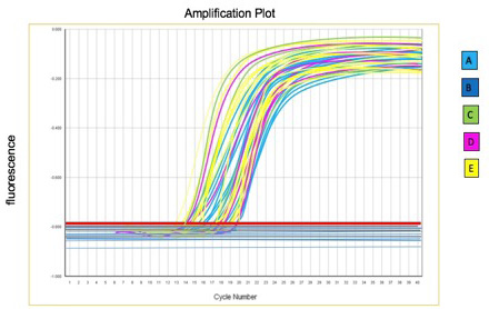
PCR-based expression of insulin gene in control and experimental group of animals.
References
- Abdissa, D., 2021. Nutr. Diet Suppl., 13: 53. https://doi.org/10.2147/NDS.S298870[CrossRef]
- Chacińska, M., Zabielski, P., Książek, M., Szałaj, P., Jarząbek, K., Kojta, I., Chabowski, A. and Błachnio-Zabielska, A.U., 2019. Nutrients, 11: 835. https://doi.org/10.3390/nu11040835[CrossRef]
- Chen, J., Ning, C., Mu, J., Li, D., Ma, Y. and Meng, X., 2021. Mol. Cell Biochem., 476: 1-14.
- Claudia-Soto, Jess, J., Julia, P., Imelda, G., Ana, E., Esther, U., Perez, l. and Raya, L., 2 2014. Int. J. Lat. Res. Sci. Technol., 3: 221-227.
- De-Souza, D.R., da Silva Pieri, B.L., Comim, V.H., de Oliveira Marques, S., Luciano, T.F., Rodrigues, M.S. and De Souza, C.T., 2020. J. Diabetes Complicat., 34: 107553. https://doi.org/10.1016/j.jdiacomp.2020.107553[CrossRef]
- Hashiesh, H.M., Meeran, M.F., Sharma, C., Sadek, B., Kaabi, J.A. and Ojha, S.K., 2020. Nutrients, 12: 2963. https://doi.org/10.3390/nu12102963[CrossRef]
- Moin, A.S.M. and Butler, A.E., 2019. Curr. Diabetes Rep., 19: 1-12. https://doi.org/10.1007/s11892-019-1194-6[CrossRef]
- Numan, A., Khawaja, K.I., Qureshi, A.B., Yousaf, M.S., Rabbani, I., Zaneb, H. and Rehman, H., 2020. Pakistan J. Zool., 52: 1217-1220. https://doi.org/10.17582/journal.pjz/20160213056633[Full text][CrossRef]
- Qazi, A.F. and Shaikh, D.M., 2019. J. Pharm. Res. Int., 31: 1-7.
- Sarbolouki, S., Javanbakht, M.H., Derakhshanian, H., Hosseinzadeh, P., Zareei, M., Hashemi, S.B., Dorosty, A.R., Eshraghian, M.R., and Djalali, M., 2013. Singapore med. J., 54: 387-390. https://doi.org/10.11622/smedj.2013139[CrossRef]
- Tamarai, K., Bhatti, J.S. and Reddy, P.H., 2019. Biochim. Biophys. Acta Mol. Basis, 1865: 2276-2284. https://doi.org/10.1016/j.bbadis.2019.05.004[CrossRef]
- Wang, J.F., Zhang, H.M., Li, Y.Y., Xia, S., Wei, Y., Yang, L., Wang, D., Ye, J.J., Li, H.X., Yuan, J. and Pan, R.R., 2019. Lipids Hlth. Dis., 18: 1-9. https://doi.org/10.1186/s12944-019-1048-x[CrossRef]
- Zhu, M., Liu, X., Liu, W., Lu, Y., Cheng, J. and Chen, Y., 2021. Aging (Albany N. Y.), 13: 7691. https://doi.org/10.18632/aging.202593[CrossRef]
Services
Article usage statistics
Loading…