
The present study was aimed to examine the morphological, behavioral changes and histopathological changes in the liver of Nile tilapia, Oreochromis niloticus after acute exposure to methylmercury. The toxicity of methylmercury of Nile tilapia was studied in a static renewal bioassay for 48 hours and 96 hours (96 hours L.C-50 0.22 mg/L). The study was conducted on 40 fish that weredivided into 4 groups A, B, C and D. Group A was considered as control group.Group B, C and D were exposed to sublethal concentrations of methylmercury 0.044 mg/L, 0.055 mg/ L and 0.073 mg/L in tap water respectively. The fish exhibited tail and fin disruption, bulging of eyes, scales erosion, mucous secretion, hemorrhage, color change, slow down swimming, lake of balance, restlessness, swimming side-wise, retardation of opercular movement and quick sudden movements. The fish was anesthetized and liver were removed after 48 and 96 hours for histopathologicalstudies. The histopathological changes observed in liver were degeneration of hemopoietic tissue, destruction of hepatocytes, degeneration of bile duct, inflammatory infiltration, vacuolization, pyknosis, presence of MMCs and necrosis in hepatocytes. It can be concludedthat methylmercury has harmful effects on histology of liver of fish. Therefore, the present work suggests that the exposure to the methylmercury should be prevented to avoid injurious health hazard risks to fish.
Research Article · Volume 5, Issue 1, 19 October 2019 · Open Access
Examination of Morphological, Behavioral and Histopathological Effects on Oreochromis niloticus after Acute Exposure to Methylmercury
· · · ·
Affiliations & Notes ⌄
- 1Department of Zoology, Islamia College, Cooper Road, Affiliated with Lahore College for Woman University, Lahore, Pakistan
- 2Department of Institute of Education and Research, University of Punjab, Lahore, Pakistan. , Email: Aqsapervaiz7687@gmail.com
Article Info ⌄
- Journal:
- Journal of Innovative Sciences
- Published:
- 19 October 2019
Abstract
1. Introduction
The methylmercury known as a notorious compound not only regarding to human intoxication but also to aquatic life i.e. fish. As it concentrated in fish tissues and muscles and cause histopathological changes . Consumption of methylmercury diets in controlled experiments on fish have shown that it had a number of effects on them like hormonal, reproductive, behavioral and neurochemical changes . Hatching success and heart rate is directly linked with the waterborne methylmercury concentration in Walleye larvae . Fin regeneration is interloped with methylmercury concentration in fish . 7β-estradiol level decreased in female whereas testosterone level depleted in male in Fathead Minnows after exposure to methylmercury . Experiment on Hoplias malabaricus which has consumed defile prey for more than 70 days results in morphological changes in liver and kidney . Changes in liver histology was observed in Atherinella brasiliensis when it introduced in lake contaminated with methylmercury . Wang et al. (2011) studied changing in respiration rate as well as water pumping after methylmercury exposure in Oreochromis niloticus. Changing in the liver and gills histology was observed in Cirrhinus mrigala after exposure to mercuric chloride as well as lead acetate one month by Chavan and Muley (2014). In natural stream water gills histology was observed in Gambusia holbrooki by Jagoe et al. (1996) after giving methylmercury’s nominal concentrations. Histopathological changes in liver and gills of Oryzias latipes was observed by Liao et al. (2007) after methylmercury’s bioaccumulation. Development of the blind fin fold and the tail fin was seen in Zebra fish embryos by Yang et al. (2010) after methylmercury exposure. Olfactory epithelium, gills, kidneys as well as liver histopathological changes were observed after 96 hr experiment with methylmercury chloride in Salvelinus alpinus by Riberio et al. (2002). The aim of study is to examine the harmful effects of methyl mercury on fish morphology, behavior along with its histopathological effects on the liver of Oreochromis niloticus after acute exposure.
2. Materials and Methods
The current study was conducted to evaluate the toxicity of methylmercury on fish liver. The 96 hours LC-50 of mercury (Hgcl2) for tilapia was 0.22 mg/L . The Nile tilapia (Oreochromis niloticus) was used as experimental animal for the present project and weight before experiment as given in Table 1. The fish were randomly distributed in four groups A, B, C and D. Each group contained ten fish. The fish remained starved during the experiment. For acute phase, Group A was considering as control group where the experimental groups, B, C and D were exposed to sub-lethal concentrations of 0.044 mg/L, 0.055 mg/L and 0.073 mg/L of methylmercury for 96 hours. These concentrations were selected after dummy experiments in order to check at which sub lethal concentration minimum or no death can occur. During these four days’ experiment, mortality rate at 24 hours, 48 hours, 72 hours and 96 hours was also observed keenly and behavior of fish along with morphological changes were also observed at 96 hours. Liver histopathology had also observed after 48 and 96 hours. The experimental and control fish was anaesthetized and dissected at the end of 48 and 96 hours and liver took out to examine. Just before its fixation, liver was rinsed in 0.85 % saline solution for three times to remove blood and debris.
Table 1: Average body weight + S.E.M of different groups of fish, Oreochromis niloticus.
| Fish Group | Average Body Weight + S.E.M gms. (n=10) |
| A | 52.8±4.04 |
| B | 51.2 ±5.04 |
| C | 50.4±4.04 |
| D | 48.1±2.39 |
| E | 40.0±2.39 |
| F | 42.0±4.44 |
In histological technique, microscopic examination of tissues was prepared in order to study their anatomy. Fixation and tissue processing are the two main steps for this. In 1st step, physical and chemical state of components of cells and tissue are fixed so that they will resist in further treatment with various reagents with minimum loss of framework. Bouin’s fluid was used as fixative in present study.
Tissue Processing comprises of several steps, dehydration is the 1st step in which water was removed from the tissues by treating it with the 70 and 90 % alcohol respectively after that Xylene was used as clearing agent and paraffin wax was used to remove clearing agent. Tissues were immersed in paraffin wax at 56-58oC for overnight in oven and its volume must be more than tissues. Four hours is enough for impregnation. In embedding, square blocks of tissues were cut into pieces with sharp scalpel. Then all blocks were marked with paper and kept in cool place (refrigerator). After trimming of tissues, sectioning occurred and sections was made of 10-5 cm length. After this, five minutes bath with xylene was given to remove wax and hydration was taking place by giving tissues bath with different alcohol concentration. Then the last step is staining and mounting. Haematoxylin stains the nucleus while Eosin stains cytoplasm. Candabalsam was used as mounting agent for the present study to prevent contamination from dust and air. Then carefully and quickly, a clean cover slip was applied.
3. Results and Discussion
3.1 Behavioral and morphological changes
Abnormal behavior such as restlessness, incessant jumping, gulping of air, sudden quick movement, increase in opercular movement and swimming on the back were observed at 96 hours when the media started to act on test species. The bulging of eyes, tail and fin distortion, scale erosion, excess mucous secretion and hemorrhage were seen in affected fish at 96 hours. Normal color and behavior were observed in the control group. The behavioral and morphological changes observed in treated fish groups are represented in Table 6 and 7, respectively.
Table 2: LC-50, Mortality data for Oreochromis niloticus, following treatment with different concentrations of Methylmercury for 24 hour.
| Fish group | Concentration of Methylmercury mg/L | Fish Exposed | No. of dead fish | No.of fish survived |
| A | 0 | 10 | 0 | 10 |
| B | 0.044 | 10 | 0 | 10 |
| C | 0.055 | 10 | 0 | 10 |
| D | 0.073 | 10 | 0 | 10 |
Table 3: LC-50, Mortality data for Oreochromis niloticus, following treatment with different concentrations of Methylmercury for 48 hours.
| Fish group | Concentration of Methylmercury mg/L | Fish Exposed | No. of dead fish | No. of fish survived |
| A | 0 | 10 | 0 | 10 |
| B | 0.044 | 10 | 0 | 10 |
| C | 0.055 | 10 | 0 | 10 |
| D | 0.073 | 10 | 1 | 9 |
Table 4: LC-50, Mortality data for Oreochromis niloticus, following treatment with different concentrations of Methylmercury for 72 hours.
| Fish group | Concentration of Methylmercury mg/L | Fish Exposed | No. of dead fish | No. of fish survived |
| A | 0 | 10 | 0 | 10 |
| B | 0.044 | 10 | 0 | 10 |
| C | 0.055 | 10 | 1 | 9 |
| D | 0.073 | 10 | 1 | 8 |
3.2 Histopathological studies of liver
Group A (control group) Liver of the control fish presented normal appearance. Hepatocytes were compact and were arranged in cords, sinusoids were distinct. Blood vessels are filled with RBCs. The liver with normal histology is shown in Figure 1.
Histopathology of experimental groups different pathological changes in the liver were observed in experimental groups. These changes increased in their severity with respect to increased dose and exposure time period. The pathological changes observed in each group after 48 and 96 hours are as follows.
Group B Fish exposed to 0.044 mg/L of methylmercury exhibited some distinct changes. After 48 hours histology of liver showed sinusoids, destruction of hepatocytes and pyknotic nuclei (Figure 2). After 96 hours the changes observed were the cellular necrosis, appearance of the melano-macrophages centers, vacuolar degeneration in necrotic cells, disorganization of hepatic cords and degeneration of bile duct. The nucleus become pyknotic and karyolosis occur (Figure 5).
Group C was treated with 0.055 mg/L of methylmercury. The liver exhibits relatively severe form of destruction. After 48 hours, the disorganization of hepatic cords, tissue degeneration, vacuolization, degeneration of bile ducts and pyknosis were more severe than group B (Figure 3). After 96 hours, the liver showed degeneration of hemopoietic tissue, degeneration of hepatic cords, degeneration of bile duct, inflammation of bile duct, cellular necrosis, Melano Macrophage Centers (MMCs) centers and pyknotic nuclei (Figure 6).
Group D was exposed to 0.073 mg/L methylmercury. After 48 hours degeneration of hemopoietic tissue, degeneration of bile duct, disorganization of hepatic cords vacuolization, necrosis and pyknotic nuclei were observed (Figure 4). Pathological condition of the liver was most severe after 96 hours. Degeneration of hematopoietic tissue was dispersed in many areas of liver. Cellular necrosis in the hemopoietic tissue was also observed. Cellular necrosis, pyknotic nuclei and vacuolization, degeneration of hemopoietic tissue and Melano Macrophage Centers (MMCs) were the other changes noticed after 48 hours (Figure 7). Histopathological changes in liver of Nile tilapia after 48 and 96 hours observed from experimental studies is showed in Tables 8 and 9, respectively. Mortality of the fish after 24 hours, 48 hours, 72 hours and 96 hours was also recorded during the whole experiment and the data of these mortalities is summarized in Tables 2, 3, 4 and 5, respectively.
Liver is the organ for transformation of toxin materials . The teleost liver is one of the most touchy organ to the biochemical changes by pollutants ; . Excessive mucus secretion is first protective defense against toxic pollutants . Mucus secretion was observed in Rainbow trout, Salmo gairdneri Richardson and also in Sword fish by Vazquez et al. (2013). Increased gill opercular movements observed under alarmed situations to the increased physiological activities ; . Opercular movements were observed in other fishes also by the methylmercury chloride exposure like Zebra fish (Danio rerio) . Gulping of air was noticed to avoid contact of toxic medium in treated fish . Gulping air and disrupted shoaling behavior in fish were seen after sub-lethal exposure . Gulping of air was also observed in trout (Salvelinus fontinalis) by Kim et al. (1976). Abrupt swimming is avoidance response to avoid the toxicant. The sluggish movements are also due to action of chemical on nervous system . Abrupt swimming due to methylmercury was observed in Zebra fish (Danio rerio) .
Table 5: LC-50, mortality data for Oreochromis niloticus, following treatment with different concentrations of Methylmercury for 96 hours.
| Fish group | Concentration of Methylmercury mg/L | Fish Exposed | No. of dead fish | No. of fish survived |
| A | 0 | 10 | 0 | 10 |
| B | 0.044 | 10 | 1 | 9 |
| C | 0.055 | 10 | 1 | 8 |
| D | 0.073 | 10 | 1 | 7 |
Histopathological results of the present study indicated that liver of Nile tilapia was the primary target tissue affected by methylmercury. Many lesions in liver were recorded as hemorrhage, necrosis, depletion of hemopoietic tissue, vacuoles, disorganization of hepatic chords and MMCs. The different concentration of methylmercury used as well as exposure for different time periods showed different degree of histopathological changes. The main change was necrosis that is observed in the liver of treated groups. Vacuolation and pyknotic nuclei are significant of the early stages of necrosis ; . Cell death in fish is related with the presence of pyknotic nucleus . Pyknotic nuclei corresponds with cell hypo functionality ; . The melano-macrophage centers (MMCs) are corresponds with the removal of particles by its phagocytotic activity .
Table 6: Behavioral changes in Oreochromis niloticus during exposure time for methylmercury.
| Behavioral Changes | Concentration of Methylmercury | |||
| 0.00mg/L | 0.044mg/L | 0.055mg/L | 0.073mg/L | |
| Lake of Balance | - | - | + | ++ |
| Rate of Swimming | - | ++ | +++ | +++ |
| Rate of Opercular Activity | - | + | ++ | ++ |
| Swimming Sidewise | - | - | + | ++ |
| Restlessness | - | + | ++ | ++ |
| Sudden quick movements | - | - | + | ++ |
Foot note: No change (-); Mild change (+); Moderate change (++); Severe change (+++).
Table 7: Morphological changes in Oreochromis niloticus during exposure time for methylmercury.
| Morphological changes | Concentration of Methylmercury | |||
| 0.00mg/L | 0.044mg/L | 0.055mg/L | 0.073mg/L | |
| Fins Disruption | - | - | + | ++ |
| Bulging of eyes | - | + | ++ | +++ |
| Tail disruption | - | + | ++ | +++ |
| Scale erosion | - | + | ++ | ++ |
| Mucous Secretion | - | + | ++ | +++ |
| Hemorrhage | - | + | ++ | ++ |
| Color Change | - | - | - | - |
Foot note: No Change (-); Mild Change (+); Moderate Change (++); Severe Change (+++).
The histopathological changes in Poecilia reticulata (Guppy) were observed by aqueous methyl mercury chloride exposure. Fish were exposed to concentrations of 0, 1.0, 1.8, 3.2, 5.6, or 10 μg/L for 1 and 3 months and histopathological changes such as necrosis, MMCs and degeneration of hemopoietic tissues were observed . The histopathological effects of methylmercury were observed in Tiger fish (Hoplias malabaricus) after feed upon contaminated prey fish for more than 70 days. The liver of exposed individuals presented increased number of melano- macrophage centers (MMCs), necrotic areas, degeneration of hemopoietic tissue
Table 8: Histopathological changes in liver of Oreochromis niloticus after 48 hours exposure to methylmercury.
| Histopathological changes in liver | Concentrations | |||
| 0.0 mg/L | 0.044mg/L | 0.055mg/L | 0.073mg/L | |
| Pyknotic nuclei in the hemopoietic tissue | - | + | ++ | ++ |
| Necrosis | - | - | ++ | ++ |
| Disorganization of hepatic cords | - | + | ++ | ++ |
| Vacuolar degeneration | - | - | + | ++ |
| Melano-macrophages centers (MMCs) | - | - | - | + |
| Degeneration of hemopoietic tissue | - | - | - | + |
Foot note: No Change (-); Mild Change (+); Moderate Change (++); Severe Change (+++).
Table 9: Histopathological changes in liver of Oreochromis niloticus after 96 hours exposure to methylmercury.
| Histopathological changes in liver | Concentrations | |||
| 0.0 mg/L | 0.044mg/L | 0.055mg/L | 0.073mg/L | |
| Pyknotic nuclei in the hemopoietic tissue | - | + | ++ | +++ |
| Necrosis | - | + | ++ | +++ |
| Disorganization of hepatic cords | - | + | ++ | +++ |
| Vacuolar degeneration | - | + | ++ | +++ |
| Melano-macrophages centers (MMCs) | - | + | ++ | +++ |
| Degeneration of hemopoietic tissue | - | ++ | ++ | +++ |
Foot note: No change (-); Mild change (+); Moderate change (++); Severe chang.
and disorganization of hepatic chords and vessels . Histopathological changes like presence of pyknotic nuclei, melano- macrophage centers (MMCs) and degeneration of hemopoietic tissues were reported by Ribeiro et al. (2002) in Arctic Charr (Salvelinus alpinus) exposed to a single dose of methylmercury. Medaka (Oryzias latipes) when exposed to methylmercury chloride showed histopathological changes in liver. These changes are vacuolar degeneration, presence of MMCs and pyknotic nuclei . Brazilian silversides (Atherinella brasiliensis) when introduced in the Brazilian lake affected with methylmercury show changes in liver histology. Vacuolar degeneration, Hemopoietic tissue degeneration, necrosis, presence of MMCs and pyknotic nuclei were seen . Yellow Mystus (Hemibagrus filamentus) when exposed to methylmercury show histopathological changes like necrosis, degeneration of hemopoietic tissue, vacuoles, presence of pyknotic nuclei, sinusoids, swelling and MMCs centers (Senarat et al., 2015). All these histopathological changes are also observed in present experimental work on tilapia (Oreochromis niloticus) exposed to methylmercury chloride for 96 hours.
4. Conclusion
It is concluded that sublethal concentrations of methylmercury lead to abnormal behavior in fish along with morphological changes and histopathological results indicate that exposure to sub-lethal concentrations of methylmercury in fish caused destructive changes in the liver leading to the malfunctioning of it that ultimately cause fish death and would eventually cause a change in the population structure of fish and if it is survived, it is also harmful for humans that love to eat tilapia. As methylmercury is quickly uptake by fish muscles. So we should prevent the usage of this chemical as it is not good for fish as well as for human. Its usage should be minimize and ban in the Pakistan. And this is also conveyed to Public that methylmercury should not be used.
Acknowledgements
I offer my sincerest gratitude to my supervisor, Mrs. Rukhshanda Afridi for guiding me in the best possible way. I am especially grateful to my coworkers Zahra Pervaiz, Rabia Masood and Hira Pervaiz. I own everything to my parents, Mr. and Mrs. Pervaiz Akhtar. Without them all, I can’t be able to complete my work.
Author’s Contribution
Aqsa Pervaiz planned, perform experiment and wrote manuscript. Rukhshanda Afridi provide guidance in performance and helped in critical analysis and review of data. Zahra Pervaiz helped in interpretation of results and Rabia Masood helped in data collection and Hira Pervaiz helped in writing the manuscript.
Figures
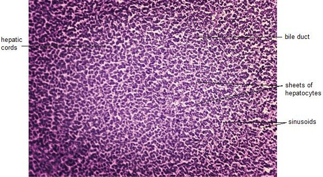
Photomicrograph of T.S. of liver of group A (control group) showing compact hepatocytes, bile duct, hepatic cords and sinusoids, H and E, (40x).
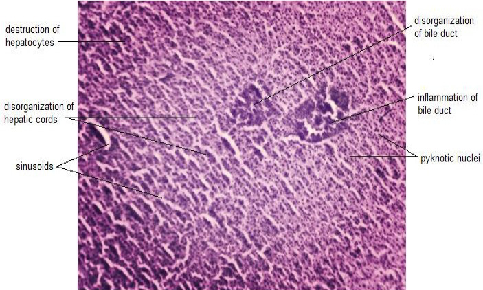
Photomicrograph of T.S. of liver of group-B (experimental) after 48 hours, showing pyknotic nuclei, disorganization of hepatic cords, inflammation of bile duct, disorganization of bile duct, destruction of hepatocytes and sinusoids, H and E, (40x).
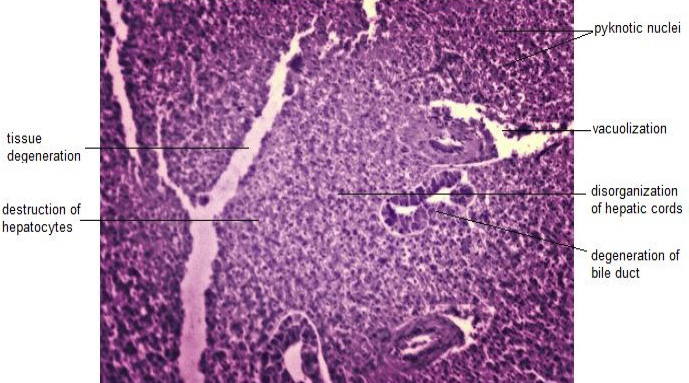
Photomicrograph of T.S. of liver of group-C (experimental) after 48 hours, showing pyknotic nuclei, tissue degeneration, disorganization of hepatic vessel, degeneration of bile duct and vacuolization, H and E, (40x).
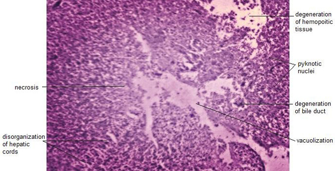
Photomicrograph of T.S. of liver of group-D (experimental) after 48 hours, showing necrosis, pyknotic nuclei, degeneration of hemopoietic tissue, degeneration of bile duct, disorganization of hepatic cords and vacuolization, H and E, (40x).
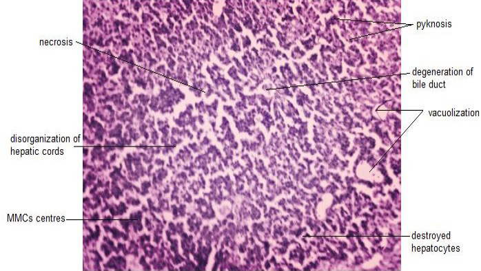
Photomicrograph of T.S. of liver of group-B (experimental) after 96 hours, showing necrosis, pyknosis, destroyed hepatocytes, degeneration of bile duct, MMCs, disorganization of hepatic cords and vacuolization, H and E, (40x).
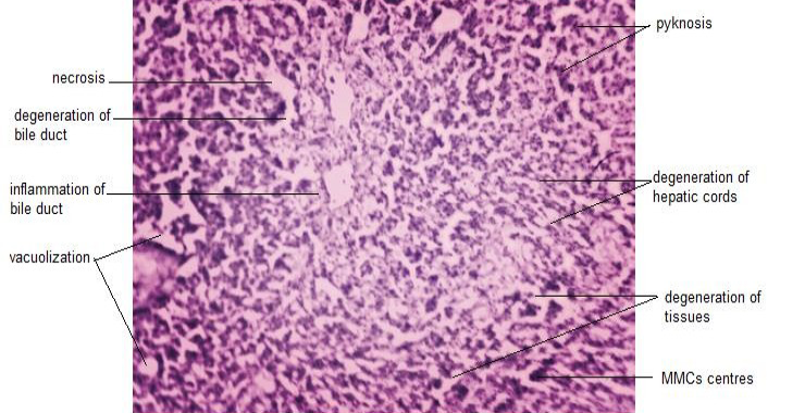
Photomicrograph of T.S. of liver of group-C (experimental) after 96 hours, showing necrosis, pyknosis, degeneration of tissue, degeneration of bile duct, degeneration of hepatic cords, inflammation of bile duct and vacuolization, H and E, (40x).
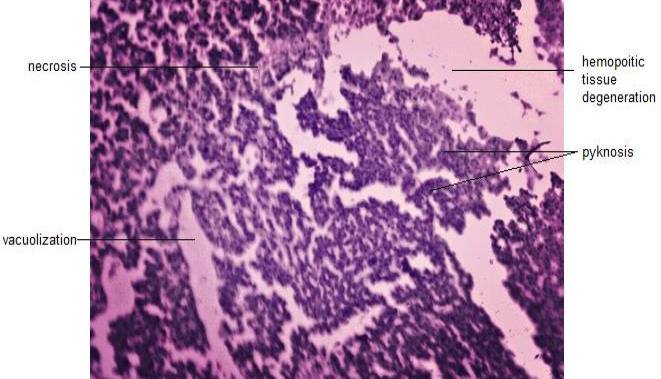
Photomicrograph of T.S. of liver of group-D (experimental) after 96 hours, showing necrosis, pyknosis, hemopoietic tissue degeneration and vacuolization, H and E, (40x).
References
- Alkahem, H.F., 1995. Acute and sub lethal exposure of cat fish, Clarias gariepinus to cadmium chloride: survival, behavioural and physiological responses. Pakistan Journal of Zoology, 27(1): 33-37.
- Bairuty, G.A.A., Shaw, B.J., Handy, R.D. and Henry, T.B., 2013. Histopathological effects of waterborne copper nanoparticles and copper sulphate on the organs of rainbow trout (Oncorhynchus mykiss). Aquatic Toxicology, 126: 104-15. https://doi.org/10.1016/j.aquatox.2012.10.005[CrossRef]
- Chavan, V.R. and Muley, D.V., 2014. Effect of heavy metals on liver and gill of fish Cirrhinus mrigala. Int. J. Curr. Microbiol. Appl. Sci., 3(5): 277-288.
- Chavan, V.R. and Muley, D.V., 2015. Effect of heavy metals on liver and gill of fish Cirrhinus mrigala. International Journal of Current Microbiology and Applied Sciences, 3(5): 277-288.
- Drevnick, P.E., Roberts, A.P., Otter, R.R., Schmidt, H.C.R., Klaper and Oris, J.T., 2008. Mercury toxicity in livers of northern pike (Esox lucius) from Isle Royal, USA. Comparative Biochemistry and Physiology- Part C: Toxicology and Pharmacology, 147: 331-338. https://doi.org/10.1016/j.cbpc.2007.12.003[CrossRef]
- Dyk, J.V., Pieterse, G. and Vuren, J.V., 2007. Histological changes in the liver of Oreochromis mossambicus (Cichlidae) after exposure to cadmium and zinc. Ecotoxicology and Environment Safety, 66: 432-440. https://doi.org/10.1016/j.ecoenv.2005.10.012[CrossRef]
- Fernandez, W.S., Dias, J.D., Oliveira Ribeiro, C.A.O. and Azevedo, J.R., 2011. Liver damages and nuclear abnormalities in erythrocytes of Atherinella brasiliensis from two beaches in Southeast of Brazil. Brazilian Journal of Oceanography, 59(2). https://doi.org/10.1590/S1679-87592011000200005[CrossRef]
- Handy, R.D. and Maunder, R.J., 2009. The biological roles of mucus: importance for osmoregulation and osmoregulatory disorders of fish health. Cambridge Society for Experimental Biology, Vol. 1. pp. 203-235.
- Hao, L., Wang, Z. and Xing, B., 2009. Effect of sub-acute exposure to TiO2 nanoparticles on oxidative stress and histopathological changes in Juvenile Carp (Cyprinus carpio). Journal of Environmental Sciences, 21(10): 1459-1466. https://doi.org/10.1016/S1001-0742(08)62440-7[CrossRef]
- Heath, A.G., 1995. Water pollution and fish physiology. 2nd ed. Florida: CRC Press, pp. 384.
- Ishikawa, N.M., Ranzani-Paiva, M.T.T. and Lombardi, J.V., 2007. Acute toxicity of mercury (hgcl2) to nile tilapia, oreochromis niloticus. B. Inst. Pesca, Sao Paulo, 33(1): 99-104.
- Jagoe, C.H., Faivre, A. and Newman, M.C., 1996. Morphological and morphometric changes in the gills of mosquito fish (Gambusia holbrooki) after exposure to mercury (II). Aquatic Toxicology, 34: 163-183. https://doi.org/10.1016/0166-445X(95)00033-Z[CrossRef]
- Katja, S., Georg, B.O.S., Stephan, P. and Christian, E.W.S., 2005. Impact of PCB mixture (Aroclor 1254) and TBT and a mixture of both on swimming behaviour, body growth and enzymatic biotransformation activities (GST) of young carp, Cyprinus carpio. Aquatic. Toxicology, 71: 49– 59. https://doi.org/10.1016/j.aquatox.2004.10.012[CrossRef]
- Kim, J.M.M., Olson, G.F., Holcomb, G.W. and Hunt, E.P., 1976. Long term effects of methylmercuric chloride on three generations of brook trout (Salvelinus fontinalis). Journal of Fisheries Research Board of Canada, Vol. 33. pp. 2726- 2739. https://doi.org/10.1139/f76-324[CrossRef]
- Latif, M.A., Bodaly, R.A. Johnston, T.A. and Fudge, R.J.P., 2001. Effects of environmental and maternally derived methylmercury on the embryonic and larval stages of walleye (Stizostedion vitreum). Environmental Pollution, 111(1): 139-148. https://doi.org/10.1016/S0269-7491(99)00330-9[CrossRef]
- Liao, C.Y., Zhou, Q.F., Fu, J.J. and Jiang, G., 2007. Interaction of methylmercury and selenium on the bioaccumulation and histopathology in Medaka (Oryzias latipes). Environmental Toxicology, 22(1): 69-77. https://doi.org/10.1002/tox.20236[CrossRef]
- Lock, R.A. and Overbeeke, V.A.P., 1981. Effects of mercuric chloride and methylmercuric chloride on mucus secretion in rainbow trout, Salmo gairdneri Richardson. Comparative Biochemistry and Physiology- Part C, 69(1): 67-73. https://doi.org/10.1016/0306-4492(81)90103-9[CrossRef]
- Mela, M., Randi, M.A., Ventura, D.F., Carvalho, C.E., Pelletier, E. and Oliveira Ribeiro, C.A., 2007. Effects of dietary methylmercury on liver and kidney histology in the neotropical fish Hoplias malabaricus. Ecotoxicology and Environment Safety, 68(3): 426-435. https://doi.org/10.1016/j.ecoenv.2006.11.013[CrossRef]
- Meyers, T.R. and Hendricks, J.D., 1982. A summary of tissue lesions in aquatic animals induced by controlled exposures to environmental contaminants, chemotherapeutic agents, and potential carcinogens. Marine Fish. Rev., 44: 1-17.
- Nikiforova, A.I. and Golichenkov, V.A., 2012. Characteristics of the reparative regeneration of fins in the polypterid fish (Polypteridae, Actinopterygii). Ontogenez, 43(2): 136-142. https://doi.org/10.1134/S1062360412020063[CrossRef]
- Omoniyi, I.A. and Sodunke, S.A., 2002. Effects of lethal and sub-lethal concentrations of Tobacco (Nicotiana tobaccum), Leaf dust extraction on weight and hematological changes in Clarias gariepinus (Burchell). Applied Sciences and Environment Management, 6: 37-41. https://doi.org/10.4314/jasem.v6i2.17174[CrossRef]
- Rabitto, I.S., Costa, J.R.M.A., Assis, H.C.S., Randi, M.A.F., Akaishi, F.M., Pelletier, E., Oliveira and Ribeiro, C.A., 2005. Dietary Pb (II) and TBT (tributyltin) exposures to neotropical fish Hoplias malabaricus: Histopathological and biochemical findings. Ecotoxicology and Environment Safety, 60: 147-156. https://doi.org/10.1016/j.ecoenv.2004.03.002[CrossRef]
- Rahman, M.Z., Hossain, Z., Mollah, M.F.A. and Ahmed, G.U., 2002. Effect of diazinum 60 EC on Anabas testudineus, Channa punctatus and Barbodes gonionotus. The ICLARM Quarterly, 25: 8-12.
- Ribeiro, C.A., de-O., Belger, L., Pelletier, E. and Rouleauc, C., 2002. Histopathological evidence of inorganic mercury and methyl mercury toxicity in the arctic charr (Salvelinus alpinus). Environmental Research, 90: 217-225. https://doi.org/10.1016/S0013-9351(02)00025-7[CrossRef]
- Roberts, R.J., 1981. Patologia de los peces. Mundi-Prensa. pp. 366.
- Scheuhammer, A.M., Meyer, M.W., Sandheinrich, M.B. and Murray, M.W., 2007. Effects of environmental methylmercury on the health of wild birds, mammals, and fish. Ambio, 36(1): 12-8. https://doi.org/10.1579/0044-7447(2007)36[12:EOEMOT]2.0.CO;2[CrossRef]
- Senarat, S., Kettratad, J., Poolprasert, P., Yenchum, W. and Jiraungkoorskul, W., 2015. Histopathological finding of liver and kidney tissues of the yellow mystus, Hemibagrus filamentus from the Tapee River, Thailand. Songklanakarin. Journal of science and technology, 37(1): 1-5.
- Udotong, J.I.R., 2015. Histopathological Changes in Liver and Muscle of Tilapia Fish from QIRE Exposed to Concentrations of Heavy Metals. International Journal of Biology Biomolecular Agricultural Food and Biotechnological Engineering, 9(6).
- Ural, M.S. and Simsek, K.S., 2006. Acute toxicity of dichlorvos on fingerling of European catfish, Silurus glanis., Bulletin of Environmental Contamination and Toxicology, 76: 871-876.
- Vazquez, M., Calatayud, M., Velez, D. and Devesa, V., 2013. Intestinal transport of methylmercury and inorganic mercury in various models of Caco-2 and HT29-MTX cells. Toxicology, 311(3): 147-153. https://doi.org/10.1016/j.tox.2013.06.002[CrossRef]
- Vazquez, M., Calatayud, M., Velez, D. and Devesa, V., 2015. Intestinal transport of methylmercury and inorganic mercury in various models of Caco-2 and HT29-MTX cells. Toxicology, 311(3): 147-153. https://doi.org/10.1016/j.tox.2013.06.002[CrossRef]
- Wang, R., Wong, M.H. and Wang, W.X., 2011. Coupling of methylmercury uptake with respiration and water pumping in freshwater tilapia, Oreochromis niloticus. Environmental Toxicology and Chemistry, 30(9): 2142-2147. https://doi.org/10.1002/etc.604[CrossRef]
- Welingtom, S., Dias, F.J.F., Oliveira, R.C.A. and Azevedo, J.S., 2011. Liver damages and nuclear abnormalities in erythrocytes of Atherinella brasiliensis from two beaches in Southeast of Brazil. Brazilian Journal of Oceanography, 59(2). https://doi.org/10.1590/S1679-87592011000200005[CrossRef]
- Wester, P.W. and Canton, H.H., 1992. Histopathological effects in Poecilia reticulata (guppy) exposed to methyl mercury chloride. Toxicology Pathology, 20(1): 81-92. https://doi.org/10.1177/019262339202000110[CrossRef]
- Xiaojuan, X., Daniel, W., Michael, J.C., Ryan, C., Crystal, L., Stefan, G. and Lillian, A.S., 2012. Comparison of Neurobehavioral Effects of Methylmercury Exposure in Older and Younger Adult zebra fish (Danio rerio). Neuro Toxicology, 33: 1212-1218. https://doi.org/10.1016/j.neuro.2012.06.011[CrossRef]
- Yang, L., Kemadjou, J.R., Zinsmeister, C., Bauer, M., Legradi, J., Muller, F., Pankratz, M., Jakel, J. and Strahle, U., 2007. Transcriptional profiling reveals barcode-like toxicogenomic responses in the zebra fish embryo. Genome Biology, 8(10): 227. https://doi.org/10.1186/gb-2007-8-10-r227[CrossRef]
- Younis, E.M., Abdel-Warith, A.A. and Al-Asgah, N.A., 2012. Hematological and enzymatic responses of Nile tilapia, Oreochromis niloticus during short and long term sub-lethal exposure to zinc. Afr. Jornal of Biotechnology, 11: 4442-4446. https://doi.org/10.5897/AJB11.3987[CrossRef]
- Zamorano, F.X.M., Klingler, C.A.R., Head, J. and Carvan, M.J., 2016. Parental whole life cycle exposure to dietary methylmercury in Zebra fish (Danio rerio) affects the behavior of offspring. Environmental Science and Technology, 50(9): 4808-4816. https://doi.org/10.1021/acs.est.6b00223[CrossRef]
Cited by 1 article
In journals published by ResearchersLinks.
Services
Article usage statistics
Loading…