Research Article · Volume 14, Issue 11, 10 October 2026 · Open Access
Multi-Organ Histopathology and Systemic Safety Profile of a High-Dose Recombinant Nipah Vaccine Candidate Formulated with SWE Adjuvant in Syrian Hamsters (Mesocricetus auratus)
· · · · · · · · · · · · · ·
Affiliations & Notes ⌄
1Faculty of Veterinary Medicine, Universitas Airlangga, Surabaya, Indonesia
2Professor Nidom Foundation, Surabaya, Indonesia
3Riset AIRC Indonesia, Surabaya, Indonesia
4Department of Pharmacy, Faculty of Health Science, Universitas PGRI Adi Buana Surabaya
5Email: nidomca@fkh.unair.ac.id or reviany@pnfinstitute.org
6Faculty of Medicine, Universitas Airlangga, Surabaya, Indonesia. *Correspondence | Reviany V. Nidom, Chairul A. Nidom, Faculty of Veterinary Medicine, Universitas Airlangga, Surabaya, Indonesia
Repeated-dose intramuscular administration of a high-dose recombinant Nipah vaccine candidate formulated with SWE adjuvant demonstrated a favorable systemic safety profile in Syrian hamsters.
Evaluation under the Patterned Physiological Criteria framework provided preliminary safety metrics supporting host tolerance across clearance tissues.
Selected from the article's own text.
Abstract
Nipah virus (NiV) causes severe zoonotic disease with high fatality rates. Preclinical toxicity screening of recombinant attachment glycoprotein (G) seed vaccine candidates under repeated high-dose exposure is essential to confirm host tolerance limits. This study evaluated the repeated-dose systemic toxicity and multi-organ histopathology safety profile of a high-dose recombinant NiV seed vaccine candidate formulated with Squalene Oil-in-Water Emulsion (SWE) adjuvant in Syrian hamster (Mesocricetus auratus), using the Patterned Physiological Criteria framework evaluates synchronized multi-organ clearance responses against non-target tissue structural preservation. Twenty-four specific-pathogen-free Syrian hamsters were randomly allocated into four experimental groups (n=6/group, sex-balanced): P1 received high-dose vaccine antigen (200 μ”g/mL” , 0.05” mL” ); P2 received high-dose vaccine formulated with SWE adjuvant (1:1 v/v); P3 received distilled water; and P4 received SWE placebo. Formulations were administered via intramuscular injection at weeks 0 and 4. Daily clinical monitoring and body weights were maintained until week 8, whereupon total necropsy and semi-quantitative INHAND microscopic organ scoring (Grades 0–4) were executed. Inter-organ score relationship were evaluated using Spearman’s rank correlation. Zero mortality (0/24) and no clinical distress were recorded across all cohorts over 56-day. Gross necropsy revealed normal visceral integrity without hemorrhages or gross structural abnormalities. Microscopic evaluation showed that repeated high-dose exposure induced only mild cellular adaptations (Grades ≤2) in the liver, lungs, and kidneys, as well as splenic white pulp hyperplasia (Grade 1). Non-target vital systems (heart, brain, intestine, and gonads) exhibited Grade 0 across all animals. Inter-organ analysis showed strong synchronized positive linear correlations (p<0.01), anchored by the lung-spleen axis (r_s=0.944) and liver-lung axis (r_s=0.919). Recombinant NiV seed vaccine candidate formulated with SWE adjuvant demonstrated a favorable systemic safety profile in Syrian hamsters. Microscopic variations were interpreted as potential adaptive cellular responses during macromolecular processing. Further preclinical evaluation of immunogenicity and protective efficacy is recommended.
Nipah virus (NiV) represents a highly lethal zoonotic Henipavirus that poses a severe threat to global health security through recurring spillovers events and pandemic potential ; . Viral attachment glycoprotein (G) binding to host ephrin-B2/B3 receptors drives widespread microvascular disruption, microthrombosis, and hyperinflammatory host responses, precipitating fatal acute respiratory distress syndrome (ARDS) and necrotizing encephalitis ; ; . Among emerging vaccine technologies, recombinant subunit platforms targeting the attachment (G) glycoprotein offer key advantages over mRNA and viral vector platforms, including an absence of pre-existing anti-vector immunity, high thermal stability, an established non-replicating safety profile, and consistent industrial-scale manufacturing scalability ; Gao et tal., 2022; ; . Industrial-scale manufacturing mandates rigorous safety screening of the master vaccine seed lot under repeated worst-case high-dose scenarios (200 μ”g/mL” ) to define biological tolerance thresholds prior to human clinical trials ; ; .
Formulating non-replicating antigens with advanced Squalene Oil-in-Water Emulsion (SWE) adjuvants enhances immune longevity, yet systemic biodistribution of submicron oil droplets introduces potential risks of transient reactogenicity, acute-phase hepatic clearance burdens, and transient microvascular neutrophil margination in pulmonary capillary beds ; ; ; .Evaluating these biological responses requires an appropriate animal model, with Syrian hamsters (Mesocricetus auratus) serve as a translational model of choice due to their microvascular endothelial sensitivity and ephrin-B2/B3 receptor distribution mirroring human tissue targets, allowing safety to be established in the primary small animal species used for Henipavirus efficacy testing ; ; ; .
Nonclinical safety guidelines (ICH S6, WHO guidelines) and standard diagnostic frameworks such as International Harmonization of Nomenclature and Diagnostic Criteria (INHAND) mandate multi-organ histopathology evaluation ; . However, traditional interpretive approaches often evaluate organ pathology as isolated endpoints, creating ambiguity when distinguishing expected, coordinated clearance responses from uncoupled systemic toxicity ; . To address this interpretive ambiguity, the “Patterned Physiological Criteria” as an exploratory analytical framework designed to complement established guidelines by mapping co-directional organ clearance trajectories . Operationally, this proposed exploratory framework defines host safety through two quantitative criteria: (1) statistically significant positive rank correlations exceeding an exploratory threshold of r_s>0.70 representing strong positive rank concordance based on established statistical conventions ; among responsive clearance organs (liver, lungs, kidneys, spleen) exhibiting low-grade, reversible cellular adaptations (≤ Grade 2), and (2) unvaried Grade 0 structural preservation in non-target vital systems (brain, heart, intestine, gonads). This study evaluated the repeated-dose systemic toxicity and multi-organ histopathology safety profile of a high-dose SWE-adjuvanted recombinant NiV seed vaccine candidate in juvenile Syrian hamsters under this exploratory framework, establishing a safety baseline for further preclinical development.
Materials and methods
Ethical statement
All experimental procedures involving animals were conducted in strict accordance with the national guidelines for animal care and use. Institutional ethical approval was granted by the Animal Care and Use Committee (IACUC) under approval number Approval No. 030623/IACUC/VII/2023. In vivo experimental pro cedures, immunization protocols, daily clinical monitoring, and necropsy operations were executed within the certified animal Biosafety Level 3 (BSL-3) facility at Professor Nidom Foundation (PNF), Surabaya, Indonesia.
Hamsters were housed in standard polycarbonate cages (42×27×18” cm”) lined with autoclaved paper bedding and equipped with wooden chew blocks for environmental enrichment. Animals were housed 3 per cage (segregated by sex) under controlled environmental conditions (temperature 22±3^∘ “C”, relative humidity 55±10%, 12-hour light/dark cycle) with a standard commercial pellet diet and water provided ad libitum. Tissue processing, paraffin embedding, microtomy, Hematoxylin-Eosin (HE) staining, digital micro-imaging, and semi-quantitative INHAND microscopic scoring were performed at the Animal Diagnostic Laboratory (PADIA-Lab), PNF, Surabaya, Indonesia.
Experimental design
Twenty-four specificpathogen-free (SPF) juvenile Syrian hamsters (Mesocricetus auratus), aged 3–4 weeks withbody weights ranging between 20–30 grams were obtained from certified breeding stocks. Sample size calaculation was determined using Mead’s Resource Equation to adhere to the Reduction principle of the 3Rs framework:
E=N-T
Where N represents the total number of experimental units (N=24) and T represents the total degrees of freedom for the treatment factor combinations. Accounting for a 4×2 factorial design (4 treatment conditions × 2 sexes; T=8), the error degrees of freedom is:
E=N-T=24-8=16
An E value of 16 satisfies the ideal regulatory threshold (10≤E≤20), validating statistical power without animal redundancy. Animals were randomly assigned into four treatment groups n=6/group (3 males and 3 females):P1 (High-Dose Vaccine): Received 0,05 mL intramuscular inoculation of a high-dose (200 μ”g/mL”) recombinant Nipah virus attachment G-protein seed preparation; P2 (High-Dose Vaccine + SWE Adjuvant): Received 0,05 mL intramuscular inoculation of the high-dose vaccine antigen (200 μ”g/mL” ) formulated with Squalene Oil-in-Water Emulsion (SWE) adjuvant at a 1:1 volume ratio; P3 (Negative Control): Received 0,05 mL intramuscular injection of sterile distilled water; P4 (Adjuvant Control): Received 0,05 mL intramuscular injection of placebo SWE vehicle, composed of a squalene-in-water emulsion (squalene, polysorbate 80, and sorbitan trioleate in a isotonic citrate buffer system) formulated identically to the adjuvant component of P2 but without viral antigen
All test formulations were administered by intramuscular injection into the cranial thigh muscle (musculus quadriceps femoris), an injection volume of 0,05 mL per site avoids muscle tearing, local tissue tension, and formulation leakage, while injection into the quadriceps femoris eliminates the risk of sciatic nerve damage associated with posterior thigh routes. However, localized mechanical tissue tension induced by this volume in 20–30 g juvenile muscle mass is acknowledged as a potential minor confounding factor independent of vaccine reactogenicity. Prime immunization was administered on Day 0, followed by a booster immunization on Day 28, simulating a worst-case systemic exposure scenario. To optimize animal usage in accordance with the Reduction principle (3Rs) during this initial safety screening, a post-treatment recovery cohort was excluded. The study design focused resources on characterizing peak tissue reactogenicity at Day 56 (28 days post-booster) prior to committing to full-scale recovery evaluations.
Husbandry and clinical observation
Hamsters were acclimatized for 7 days under controlled temperature (22±3^∘ “C” ), relative humidity (55±10%), and a 12-hour light/dark cycle. Standard commercial pellet diet and fresh water were provided ad libitum. Animals were observed daily at 09.00 AM by blinded observers for qualitative clinical signs of distress or systemic toxicity, including lethargy, piloerection, respiratory distress, motor deficits, and injection site reactogenicitythroughout the 56-day observation period. Body weight was recorded at baseline and prior to terminal necropsy at Week 8 to evaluate overall health maintenance and screen for treatment-related weight depression.
On Day 56, animals were euthanized following deep intramuscular anesthesia using a combination of ketamine (80” mg/kg body weight) and xylazine (5” mg/kg” body weight), followed by cervical dislocation. Full gross necropsy was performed immediately to inspect visceral organs for hemorrhages, lesions, or gross anatomical changes . Target organs (liver, lungs, kidneys, spleen, heart, brain, intestine, and gonads) were harvested and immersion-fixed in 10% Neutral Buffered Formalin (NBF) for 48 hours to ensure thorough tissue penetration prior to paraffin embedding.
Tissue processing and histopathology
Fixed tissue specimens were processed using an automated tissue processor. Dehydration was performed through ascending grade ethanol steps (70%, 80%, 95%, and 100% for 1 hour each). Clearing was achieved by immersing tissues in xylene across three separate jars for 1 hour each. Impregnation was completed using molten pure paraffin wax across three separate containers for 1 hour each.
Processed tissues were embedded into paraffin blocks using a standard embedding center. Thin sectioning was performend using a Leica RM2125RTS microtome to obtain tissue ribbons with a thickness of 4-5 μ”m”. Hematoxylin-Eosin (HE) staining was initiated with deparaffinization in xylene for three stages (5 minutes each). Rehydration followed via descending ethanol grades (100%, 95%, 80%, and 70% for 2 minutes each) before rinsing in running tap water for 5 minutes. Cell nuclei were stained with Mayer’s hematoxylin for 10 minutes, rinsed in running tap water for 5 minutes, and cytoplasm was counterstained using 1% alcoholic eosin Y for 2 minutes . Final dehydration used 95% and 100% ethanol (1 minute each) followed by final clearing in xylene (5 minutes) ; . Section mounting was completed using glass cover slips and a xylene-based liquid mounting medium.
Microscopic evaluation was conducted by blinded pathologists using a Nikon® Eclipse Ci microscope integrated with digital software. Diagnostic nomenclature and lesion identification were standardized using INHAND criteria, tissue alterations were scored using an adapted semi-quantitative 5-point grading scheme (Grades 0–4) . Scoring prioritized primary morphological changes per organ evaluated by percentage tissue area involvement. The consolidated multi-organ histopathology scoring matrix is detailed in Table 1.
Statistical analysis
Statistical computations were executed using IBM SPSS Statistics (Version 28.0). Data distributional traits were tested using the Shapiro-Wilk test, while variance homogeneity was assessed via Levene’s test. Because histopathology data consist of ordinal semi-quantitative grades (Grades 0–4) exhibiting non-normal distributions (p<0.05), non-parametric inferential methods were applied.
To evaluate potential sex-related differences, the Scheirer-Ray-Hare extensions of the Kruskal-Wallis test was conducted, which confirmed no significant main effects of Sex (p>0.05) and no Treatment × Sex interactions (p>0.05) across any organ system. Main treatment effects across experimental cohorts (n=6/”group”) were evaluated using the Kruskal-Wallis test (df=3,α=0.05). Post-hoc pairwise comparisons between treatment groups were conducted using the Dunn-Bonferroni test, with significance levels adjusted for the 6 pairwise comparisons within each organ family (α=0.05/6=0.0083).
To evaluate systemic response trajectories across the complete experimental exposure spectrum (from unexposed control to high-dose exposure), Spearman’s rank correlation coefficient (rs) were computed across the combined cohort (n=24). Organs exhibiting unvaried Grade 0 across all cohorts were documented descriptively to confirm structural safety. Analytical roles of each statistical test are summarized in Table 2.
Results
Clinical safety and necropsy
Intramuscular administration of the high-dose recombinant Nipah seed vaccine alone (P1) or formulated with SWE adjuvant (P2) produced zero mortality (0/24) across all experimental cohorts throughout the 56-day trial. Daily clinical monitoring confirmed normal behavioral patterns without signs of systemic distress, lethargy, piloerection, respiratory abnormalities, or motor deficits. Injected sites exhibited no swelling, erythema, or local reactogenicity.
All experimental cohorts exhibited steady, age-appropriate body weight gain over the 56-day observation period. Mean initial body weights ranged from 24.1±2.0” g” to 24.8±1.9” g” , and final body weights ranged from 110.1±8.2” g” to 114.3±6.9” g” . Systematic post-mortem examination revealed normal gross morphology across all visceral organs, with no evidence of congestion, petechial hemorrhage, or gross structural abnormalities. Gross necropsy findings are summarized in Table 3.
Histopathology of active organs
Semi-quantitative microscopic scoring revealed statistically significant treatment effects (p<0.05) across the liver, lungs, kidneys, and spleen. Non-parametric Scheirer-Ray-Hare analysis confirmed no main effects of Sex (p>0.05) and no Treatment ×Sex interactions (p>0.05). Consolidated median scores and post-hoc significance letterings across combined cohorts (n=6/”group”) are summarized in Table 4.
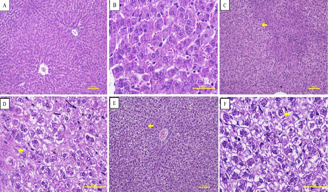
Microscopic evaluation of liver tissue from control hamsters (P3 and P4) verified intact lobular architecture and normal hepatic cords (Figure 1A, B). Active treatment groups (P1 and P2) exhibited centrilobular focal vacuolar degeneration of hepatocyte (Grade 1–2) without evidence of ballooning necrosis or lobular structural disruption (Figure 1C–F). These low-grade changes ae morphologically characteristic of reversible cellular adaptation during macromolecular processing.
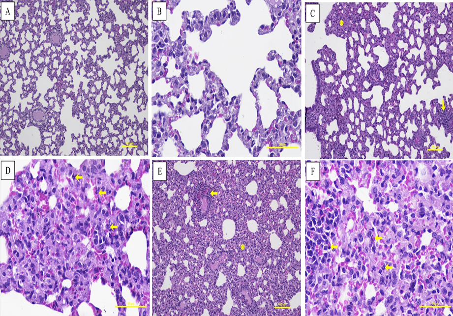
Pulmonary sections from control cohorts presented thin interalveolar septa and patent alveolar spaces (Figure 2A, B). Inoculated animals (P1 and P2) showed mild interalveolar
Table 1: Consolidated scoring matrix for multi-organ histopathology evaluation.
Organ Target
Primary Diagnositc Lesion Evaluated
Score Definition and Tissue Grading Criteria (Grade 0–4)
4: Severe mucosal disruption (>50% mucosal area) including ulceration.
Erben et al. (2014); Gibson-Corley et al. (2013)
Testis
Seminiferous tubular vacuolation
0: Normal physiological maturation;
1: Minimal tubular vacuolation (<10% area evaluated);
2: Mild tubular vacuolation/alteration (10 – 25% area evaluated);
3: Moderate tubular degeneration (26 – 50% area evaluated);
4: Severe tubular alteration/atrophy (>50% area evaluated).
Creasy et al. (2012); Gibson-Corley et al. (2013)
Ovary
Follicular atresia
0: Normal physiological maturation;
1: Minimal follicular alteration, atresia (<10% area evaluated);
2: Mild follicular alteration, atresia (10 – 25% area evaluated);
3: Moderate follicular alteration, atresia (26 – 50% area evaluated);
4: Diffuse follicular loss, atresia (>50% area evaluated).
Dixon et al. (2014); Gibson-Corley et al. (2013)
Table 2: Summary of statistical tests and analytical roles.
Statistical Test Method
Specific Data Target
Analytical Role and Decision Treshold
Shapiro-Wilk Test
Histopathological scores (n=24)
Assesses distributional normality (p<0.05 indicates non-normal distribution guiding non-parametric test selection)
Levene’s Test
Group score variances (n=6/"group" )
Evaluates homogeneity of variance (p<0.05 indicates non-homogeneous variance)
Scheirer-Ray-Hare Test
Factorial scores (4" Treatments"×2" Sexes" )
Evaluates main effects of Treatment, Sex, and Treatment × Sex interaction (α=0.05).
Kruskal-Wallis Test
Between-group score comparisons (df=3)
Computes overall treatment effect significance across cohorts (n=6/"group",p<0.05).
Dunn-Bonferroni Test
Post-hoc pairwise comparisons (6 pairs/organ)
Identifies specific group differences and assigns significance letters based on organ-wise adjusted p-values (α=0.0083).
Spearman’s Rho (rs)
Inter-organ score pairs (n=24)
Maps co-directional multi-organ clearance trajectories across the experimental exposure continuum (p<0.01)
Table 3: Necropsy and gross pathology checklist across experimental cohorts.
Evaluation Parameter
P1 (High-Dose Vaccine)
P2 (Vaccine + SWE)
P3 (Distilled Water)
P4 (SWE Placebo)
Mortality Rate
0%
0%
0%
0%
Clinical Distress Signs
Absent
Absent
Absent
Absent
Body Weight Progression
Normal
Normal
Normal
Normal
Injection Site Reactogenicity
None
None
None
None
Hepatic Gross Appearance
Normal size/color
Normal size/color
Normal size/color
Normal size/color
Pulmonary Gross Appearance
Fully inflated/pink
Fully inflated/pink
Fully inflated/pink
Fully inflated/pink
Renal Gross Appearance
Intact capsule
Intact capsule
Intact capsule
Intact capsule
Splenic Gross Appearance
Normal gross appearance
Normal gross appearance
Normal gross appearance
Normal gross appearance
Visceral Hemorrhages
Absent
Absent
Absent
Absent
Table 4: Histopathology median grades (min–max) of active organs.
Organ Target
Experimental Cohort
Male
Female
p-value
Liver
P1
1 (1-2)^a
1 (1-2)^a
p_"Treatment" =〖0.00033〗^*
p_"Sex" =0.60635
p_"Interaction" =0.85029
P2
1 (1-2)^a
1 (0-1)^ab
P3
0 (0-0)^b
0 (0-0)^b
P4
0 (0-0)^b
0 (0-0)^b
Lung
P1
2 (1-2)^a
2 (1-2)^a
p_"Treatment" =〖0.00013〗^*
p_"Sex" =0.85033
p_"Interaction" =0.99101
P2
1 (1-2)^a
2 (1-2)^a
P3
0 (0-0)^b
0 (0-0)^b
P4
0 (0-0)^b
0 (0-0)^b
Kidney
P1
1 (1-1)^a
1 (1-2)^a
p_"Treatment" =〖0.00050〗^*
p_"Sex" =0.86761
p_"Interaction" =0.99376
P2
1 (0-1)^ab
1 (0-1)^ab
P3
0 (0-0)^b
0 (0-0)^b
P4
0 (0-0)^b
0 (0-0)^b
Spleen
P1
1 (1-1)^a
1 (1-1)^a
p_"Treatment" =〖0.00004〗^*
p_"Sex" =1.000
p_"Interaction" =1.000
P2
1 (1-1)^a
1 (1-1)^a
P3
0 (0-0)^b
0 (0-0)^b
P4
0 (0-0)^b
0 (0-0)^b
Note: Values represent Median (Min–Max). Main treatment effects (p_”Treatment”), sex main effects (p_”Sex”), and interaction effects (p_”Interaction”) were calculated using the Scheirer-Ray-Hare extension of the Kruskal-Wallis test (α=0.05). p_”Sex” = 1.000 and p_”Interaction” = 1.000 for splenic white pulp hyperplasia resulted mathematically from completely identical median score distributions between male and female cohorts across all treatment groups. Asterisks (*) indicate significant main treatment effects (p<0.05). Different superscript letters (a,b,ab) within the same column for an individual organ indicate statistically significant differences determined by the Dunn-Bonferroni post-hoc test (p<0.05).
Table 5: Spearman’s rho correlation matrix of responsive organs.
Organ Target
Liver
Lung
Kidney
Spleen
Liver
--
Lung
0.919∗∗
--
Kidney
0.749∗∗
0.814∗∗
--
Spleen
0.886∗∗
0.944∗∗
0.834∗∗
--
Note: Values represent Spearman’s rank correlation coefficient (rs). The superscript symbol ** indicates that the correlation is highly significant at the p<0.01 level. Non-target vital organs exhibiting Grade 0 across all subjects (heart, brain, intestine, and gonads) possessed zero variance and evaluated descriptively.
septal thickening associated with focal neutrophilic PMN infiltration (Figure 2C–F), indiciating localized acute inflammatory reactogenicity. Alveolar lumen patency was fully maintained across all fields without pulmonary edema or intra-alveolar hemorrhage.
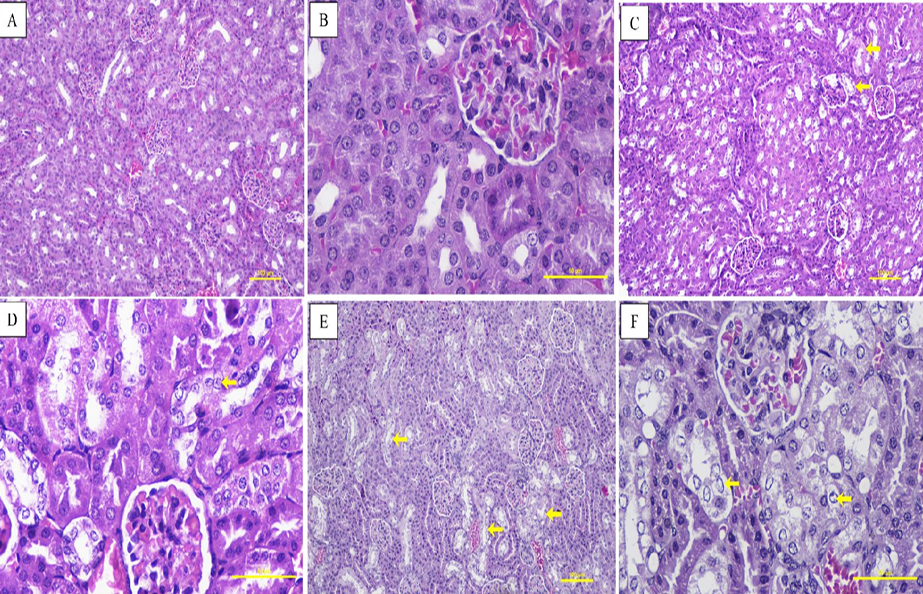
Renal architecture in control animals presented intact glomeruli and cortical tubules (Figure 3A, B). Active cohorts (P1 and P2) exhibited focal vacuolar degeneration restricted to the proximal tubular epithelium (Figure 3C–F). Glomerular tufts, Bowman’s spaces, and tubular basement membranes remained intact across all groups without proteinaceous cast formation.
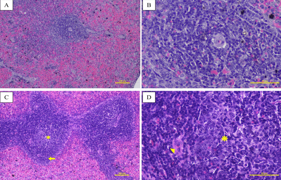
Splenic tissue from control hamsters showed resting lymphoid follicles (Figure 4A, B). Active treatment cohorts (P1 and P2) displayed a uniform shift to Grade 1, characterized by reactive white pulp follicular B-cell hyperplasia and germinal center expansion (Figure 4C, D). Expanded follicles were bounded by intact mantle zones without white pulp lymphoid depletion or red pulp congestion, reflecting reactogenicity following antigen/adjuvant administration.
Histopathology of non-target organs
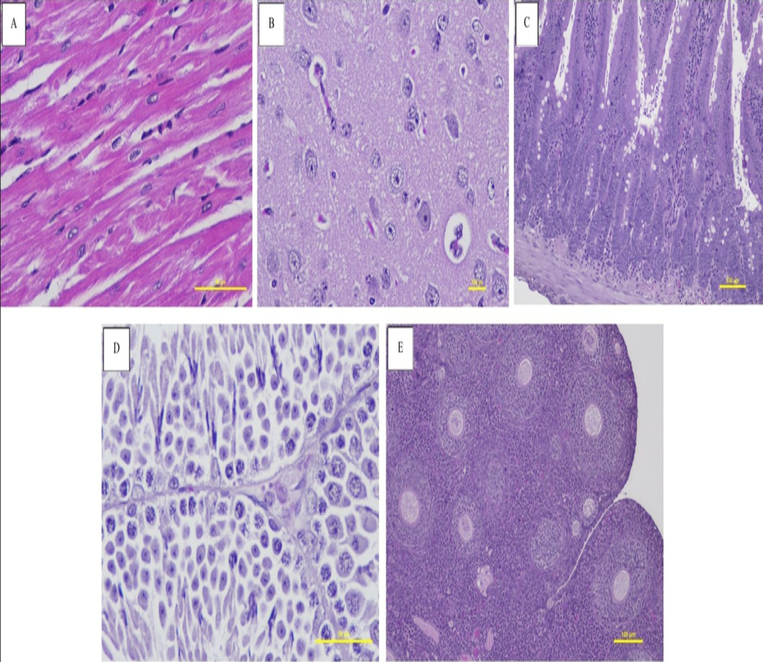
Microscopic evaluation of life-sustaining non-target organs (heart, brain, intestine, and gonads) demonstrated complete structural preservation. All subjects across all cohorts achieved unvaried Grade 0.
Myocardial tissue retained normal branching myofiber syncytia without necrosis, hyaline degeneration, or inflammatory cell infiltration (Figure 5A). Cerebral cortical neuropil maintained intact cellular architecture without evidence of microglial gliosis, neurononal necrosis, or perivascular cuffing (Figure 5B). Intestinal mucosal sections showed intact villi architecture and normal lamina propria without mucosal erosion or ulceration (Figure 5C). Gonadal tissues displayed physiological maturation evidenced by active spermatogenesis in seminiferous tubules (Figure 5D), and normal follicular development in ovaries (Figure 5E).
To evaluate systemic response trajectories across the experimental exposure spectrum (from unexposed baseline to high-dose exposure), Spearman’s rank correlation analysis was performed across all active responsive organs (n=24). Statistically significant positive rank correlations (p<0.01) were recorded among all active organ pairs (Table 5).
The lung-spleen axis recorded the highest rank correlation coefficient (r_s=0.944), followed closely by the liver-lung axis (r_s=0.919), liver–spleen axis (r_s=0.886), spleen–kidney axis (r_s=0.834), lung–kidney axis (r_s=0.814), and liver–kidney axis (r_s=0.749) These global rank correlations reflect cohort-wide, treatment-induced score distributions driven primarily by group-level separation across exposure conditions (Grade 0 in unexposed controls versus Grade 1-2 in active cohorts), rather than demstrating direct intra-individual causal mechanisms or synchronized homeostatic clearance.
Discussion
Preclinical safety evaluation of biological master seed lots under repeated worst-case high-dose exposure scenarios is essential to define host tolerance limits prior to clinical trials ; . In this study, intramuscular administration of a high-dose recombinant
Nipah seed vaccine formulated with SWE adjuvant demonstrated high host tolerance in Syrian hamsters, contrasting with wild-type henipavirus infections that cause lethal microvascular disruption, fatal respiratory distress, and necrotizing encephalitis ; .
Microscopic examination showed mild, localized cellular changes in the liver, lungs, and kidneys (Grades ≤2). Hepatic cellular swelling changes may represent an adaptive metabolic response during macromolecular clearance ; . Elevated metabolic demands on hepatocytes during high-dose antigen biotransformation provide a plausible explanation for transient intracellular fluid accumulationwithout structural ballooning necrosis or lobular disruption ; . Interalveolar septal thickening and focal PMN neutrophilic infiltration in lungs reflects localized acute inflammatory reactogenicity to circulating squalene microemulsion droplets ; . This localized inflammatory reactogenicity occurred without compromising alveolar gas exchange or inducing pulmonary edema ; . Renal proximal tubular vacuolar degeneration represents an established physiological response during macromolecular filtration and tubular reabsorption ; , occurring without glomerular damage or proteinaceous cast formation ; .
Splenic white pulp follicular B-cell hyperplasia (Grade 1) is consistent with immune reactogenicity following administration of adjuvanted protein antigen . Squalene oil-in-water emulsions prolong antigen presentation, optimizing systemic immune engagement within regional lymphoid tissues . Controlled germinal center expansion bounded by intact mantle zones signifies an organized reactive immune response rather than immunopathology .
Non-target vital systems (heart, brain, intestine, gonads) maintained Grade 0 across all cohorts, demonstrating host structural preservation at the tested dose and 56-day observation window. The absence of microglial gliosis or perivascular cuffing indicates that recombinant attachment glycoproteins (G) macromolecules did not alter cerebral neurophil architecture under physiological conditions ; . Likewise, intact myocardial syncytia, intestinal mucosal villi, and gonadal architectures confirm structural tolerance across non-target tissues ; ; ; ; .
Application of the proposed exploratory Patterned Physiological Criteria framework provided an integrative analytical approach to evaluate multi-organ host responses across exposure levels. While the strong inter-organ rank correlation (r_s=0.749-0.944,p<0.01) are driven primarily by group-level score separation between unexposed control cohorts and active treatment groups rather than establishing direct physiological causation, the concurrent manifestation of low-grade cellular adaptations across clearance tissues (liver, lungs, kidneys, spleen)—without isolated severe pathology in any single organ—is consistent with broad host tolerance across clearance pathways. Lung-spleen axis (r_s=0.944) aligns with established pulmonary-splenic immune axes, where splenic B-cell activation regulates endothelial cell adhesion and leukocyte recruitment within pulmonary capillaries ; ; . The liver-lung axis (r_s=0.919) demonstrates coordinated metabolic clearance between hepatic parenchymal cells and pulmonary intravascular macrophages ; . Complementary excretory balancing across liver-kidney (r_s=0.749), lung-kidney (r_s=0.814), and spleen-kidney (r_s=0.834). While the global correlations are driven primarily by treatment group assignments across the cohort, the concurrent low-grade alterations across filtration and immune organs–without isolated severe pathology in any single organ–support the conclusion that microscopic changes represent an organized systemic clearance process rather than uncoupled, idiosyncratic toxicity ; .
This multi-organ evaluation directly satisfies regulatory requirements established by BPOM Regulation No. 10 of 2022, WHO guidelines for nonclinical evaluation of vaccines, US-FDA guidance on starting dose safety, and EMA vaccine standards. Specifically, the study meets key regulatory criteria by validating repeated worst-case dosing safety, 56-day post-immunization observation stability, sex-balanced cohort tolerance, and standardized INHAND multi-organ histopathology screening (WHO, 2005; FDA, 2005; EMA, 2010; .
Study limitations and recommendation
Several methodological constraints warrant consideration when interpreting these safety findings, including the absence of a post-treatment recovery cohort to verify lesion reversibility, serial euthanasia time-points to characterize acute reactogenicity kinetics, dose-ranging groups to establish NOAEL, immunogenicity correlates (neutralizing antibody titers, IgG subclasses, cellular responses), protective challenge data, comparative adjuvant arms, injection site histopathology, quantitative organ weights, longitudinal body weight trajectories, biochemical/hematological parameters, and non-human primate validation, while the exclusive use of juvenile hamsters (3–4 weeks old) with limited group sizes (n=6/group, n=3/sex) constrains extrapolation to adult populations and statistical power for detecting subtle sex-specific responses.
Future investigations should incorporate dose-ranging toxicity studies with dedicated recovery cohorts to establish NOAEL and lesion reversibility kinetics, execute serial time-point harvests to characterize the temporal evolution of tissue responses, expand cohort sizes to n=10–12/group to enhance statistical power for sex-specific analyses, perform detailed histopathological evaluation of local injection sites with quantitative organ weight assessments, conduct integrated immunogenicity and protective efficacy studies in both juvenile and adult hamster models, benchmark SWE adjuvant against established adjuvants (alum, MF59), and ultimately validate systemic safety and protective efficacy in non-human primate models prior to human clinical trial progression, with concurrent serum biochemistry and hematological monitoring to complement histopathological evaluations.
Conclusion
Repeated-dose intramuscular administration of a high-dose recombinant Nipah vaccine candidate formulated with SWE adjuvant demonstrated a favorable systemic safety profile in Syrian hamsters. Histopathological alterations were limited to mild, low-grade cellular changes in the liver, lungs, and kidneys, while the brain, heart, intestine, and gonads showed no treatment-related lesions. Splenic white pulp hyperplasia was consistent with a reactive immune response following formulation exposure. Evaluation under the Patterned Physiological Criteria framework provided preliminary safety metrics supporting host tolerance across clearance tissues. These findings provide preclinical safety data to support continued development of the candidate vaccine formulation .
Acknowledgements
We would like to thank BRIN Research and Innovation for Advanced Indonesia (RIIM). This research was supported by the RIIM-3, LPDP and BRIN (Grant no. 12/II.7/HK/2023). We would also like to thank Professor Nidom Foundation for providing the Animal BSL3 facilities.
NOVELTY STATEMENT
This study provides the first comprehensive multi-organ histopathological baseline safety profile of a high-dose recombinant Nipah virus attachment (G) glycoprotein seed vaccine candidate formulated with a squalene oil-in-water emulsion (SWE) adjuvant in Syrian hamsters. Beyond establishing organ-level tolerance under a worst-case repeated-dose regimen, this work introduces an integrative “Patterned Physiological Criteria” exploratory analytical framework to differentiate expected, co-directional physiological clearance adaptations from uncoupled systemic toxicity across eight visceral organs. Standardized under modern INHAND diagnostic criteria, these findings demonstrate that the SWE-adjuvanted formulation maintains broad systemic safety without inducing irreversible tissue lesions or sex-specific toxicological variations, establishing a foundational preclinical benchmark for future Henipavirus vaccine platforms.
Author’s Contribution
Conceptualization: RVN, SI, and CAN. Methodology: RVN, SI, and CAN. Software: ZIA, ANN, EAR and IYR. Validation: RVN, KPS, KR, DL, JR, and LRY. Formal analysis: ANN, IYR, EAR. Investigation: EDP, TWM, and RG. Resources: RVN, SI, and ANN. Data curation: SI, EDP, and TWM. Writing original draft: ZIA. Writing review and editing: ZIA and EDP. Visualization: ZIA, EDP, TWM, and RG. Supervision: RVN, SI and CAN. Project administration: ZIA, SI, and TWM.
Generative AI and AI assisted technology statement
The authors declare that generative AI was used solely for language refinement and grammatical improvement. The authors take full responsibility for the content of the manuscript.
Conflict of interest
The authors declare that they have no known competing financial interests or personal relationships that could have appeared to influence the work reported in this paper.
Tabel S1: Consolidated table of abbreviations.
Abbreviation
Full Technical Definition
ARDS
Acute Respiratory Distress Syndrome
ATP
Adenosine Triphosphate
BPOM
Badan Pengawas Obat dan Makanan (Indonesian FDA)
BSL-3
Biosafety Level-3
EMA
European Medicines Agency
G-protein
Attachment Glycoprotein
HE
Hematoxylin and Eosin
IM
Intramuscular
INHAND
International Harmonization of Nomenclature and Diagnostic Criteria
Na+/K+ ATPase
Sodium-Potassium Adenosine Triphosphatase Pump
NBF
Neutral Buffered Formalin
NiV
Nipah Virus
PALS
Periarteriolar Lymphoid Sheath
PMN
Polymorphonuclear Leukocyte
SPF
Specific-Pathogen-Free
SWE
Squalene Oil-in-Water Emulsion
US-FDA
United States Food and Drug Administration
WHO
World Health Organization
Figures
Figure 1:.
Microscopic features of Syrian hamster hepatic tissue (HE staining). (A) Grade 0 (100×) and (B) 400× magnification from group P3 showing normal lobular cords; (C) Grade 1 (100×) and (D) 400× magnification from P2 group; (E) Grades 2 (100×) and (F) 400× magnification from P1 group. Arrows indicate focal vacuolar degeneration (Scale bars: 100× = 200 μm; 400× = 50 μm).
Figure 2:.
Microscopic features of Syrian hamster pulmonary tissue (HE staining). (A) Grade 0 (100×) and (B) 400× magnification from control group P4; (C) Grade 1 (100×) and (D) 400× magnification from P2 group; (E) Grades 2 (100×) and (F) 400× magnification from P1 group. Arrows denote PMN neutrophil infiltration; asterisks (*) mark interalveolar septal thickening (Scale bars: 100× = 200 μm; 400× = 50 μm).
Figure 3:.
Microscopic features of Syrian hamster renal tissue (HE staining). (A) Grade 0 (100×) and (B) 400× magnification from control group P3; (C) Grade 1 (100×) and (D) 400× magnification from P2 group; (E) Grades 2 (100×) and (F) 400× magnification from P1 group. Arrows indicate proximal tubular epithelial vacuolar degeneration (Scale bars: 100× = 200 μm; 400× = 50 μm).
Figure 4:.
Microscopic features of Syrian hamster splenic tissue (HE staining). (A) Grade 0 (100×) and (B) 400× magnification from control group P4; (C) Grade 1 (100×) and (D) 400× magnification from P2 group. Arrows mark the intact mantle zone; asterisks (*) denote germinal center proliferation; arrowheads locate central arterioles (Scale bars: 100× = 200 μm; 400× = 50 μm).
Figure 5:.
Representative microscopic panels of non-target vital organs demonstrating Grade 0 structural preservationGrade. (A) Normal cardiac myocardium; (B) Intact cerebral cortex neuropil; (C) Normal intestinal mucosal villi; (D) Testicular seminiferous tubules showing active spermatogenesis; (E) Ovarian parenchyma demonstrating normal follicular maturation (Scale bars: 100× = 200 μm; 400× = 50 μm).
References
Akoglu, H. (2018). User’s guide to correlation coefficients. Turk. J. Emergency Med., 18(3), 91–93. https://doi.org/10.1016/j.tjem.2018.08.001[CrossRef]
Al-Obaidi MMJ, Muthanna A, Desa MNM (2024). Nipah virus neurotropism: Insights into blood-brain barrier disruption. J. Integr. Neurosci., 23(5): 90. https://doi.org/10.31083/j.jin2305090[CrossRef]
Banks WA (2016). From blood–brain barrier to blood–brain interface: New opportunities for CNS drug delivery. Nat. Rev. Drug Discov., 15(4): 275–292. https://doi.org/10.1038/nrd.2015.21[CrossRef]
Berridge BR, Mowat V, Nagai H, Nyska A, Okazaki Y, Clements PJ, Rinke M, Snyder PW, Boyle MC, Wells MY (2016). Nonproliferative and proliferative lesions of the cardiovascular system of the rat and mouse. Toxicol. Pathol., 44(3): 286–350. https://doi.org/10.1293/tox.29.3S-1[CrossRef]
Bossart KN, Zhu Z, Middleton D, Klippel J, Crameri G, Bingham J, McEachern JA, Green D, Hancock TJ, Chan Y-P, Hickey AC, Dimitrov DS, Wang L-F, Broder CC (2009). A neutralizing human monoclonal antibody protects against lethal disease in a new ferret model of acute Nipah virus infection. PLoS Pathog., 5(10): e1000642. https://doi.org/10.1371/journal.ppat.1000642[CrossRef]
BPOM (2022). Pedoman uji toksisitas praklinik secara in vivo. peraturan badan pengawas obat dan makanan republik indonesia nomor 10 tahun. Jakarta, Indonesia.
Bradley A, Bowsher C, Bube A, Chamanza R, Forest T, Kovi RC, Krinke G, Little PB, Narama I, Rao DB, Sharma AK, Shibutani M, Sills R (2020). Proliferative and nonproliferative lesions of the rat and mouse central and peripheral nervous systems. Toxicol. Pathol., 48(1): 112–141. https://doi.org/10.1177/0192623320951154[CrossRef]
Brown B, Gravier T, Fricke I, Al-Sheboul SA, Carp TN, Leow CY, Imarogbe C, Arabpour J (2023). Immunopathogenesis of Nipah virus infection and associated immune responses. Immuno, 3(2): 160–181. https://doi.org/10.3390/immuno3020011[CrossRef]
Calabro S, Tortoli M, Baudner BC, Pacitto A, Cortese M, O’Hagan DT, De Gregorio E, Seubert A, Wack A (2011). Vaccine adjuvants alum and MF59 induce rapid recruitment of neutrophils and monocytes that participate in antigen transport to draining lymph nodes. Vaccine, 29(9): 1812–1823. https://doi.org/10.1016/j.vaccine.2010.12.090[CrossRef]
Cantor J (2025). Rodent models of lung disease: A road map for translational research. Int. J. Mol. Sci., 26(17): 8386. https://doi.org/10.3390/ijms26178386[CrossRef]
Cheville NF (2009). Ultrastructural pathology: the comparative cellular basis of disease. 2nd edition. Wiley-Blackwell, Ames, Iowa. https://doi.org/10.1002/9780813810379[CrossRef]
Choque-Guevara R, Perez-Zuniga R, Garcia-Lozano M, Torres-Ramos Y, Flores-Romo L (2022). Systemic cellular mobilization and endothelial adhesion kinetics following adjuvanted subunit vaccination. Front. Immunol., 13: 884120.
Creasy D, Bube A, de Rijk E, Kandori H, Kuwahara M, Masson R, Nolte T, Reams R, Regan, K, Rehm S, Rogerson P, Whitney K (2012). Proliferative and nonproliferative lesions of the rat and mouse male reproductive system. Toxicol. Pathol., 40(6_suppl): 8S–120S. https://doi.org/10.1177/0192623312454337[CrossRef]
Cullen JM, Stalker MJ (2016). Liver and biliary system. in: jubb, kennedy and palmer’s pathology of domestic animals, 6th edition, volume 2, elsevier, St. Louis, MO, pp. 258–352. https://doi.org/10.1016/B978-0-7020-5318-4.00008-5[CrossRef]
Daneman R, Prat A (2015). The blood–brain barrier. Cold Spring Harb. Perspect. Biol., 7(1): a020412. https://doi.org/10.1101/cshperspect.a020412[CrossRef]
Didierlaurent AM, Collignon C, Bourguignon P, Wouters S, Fierens K, Fochesato M, et al. (2014). Enhancement of adaptive immunity by the human vaccine adjuvant AS01 depends on activated dendritic cells. J. Immunol., 193(4): 1920–1930. https://doi.org/10.4049/jimmunol.1400948[CrossRef]
Dixon D, Alison R, Bach U, Colman K, Foley GL, Harleman JH, Haworth R, Herbert R, Heuser A, Long G, Mirsky M, Regan K, Van Esch E, Westwood FR, Vidal J, Yoshida M (2014). Nonproliferative and proliferative lesions of the rat and mouse female reproductive system. J. Toxicol. Pathol., 27(3_suppl): 1S–107S. https://doi.org/10.1293/tox.27.1S[CrossRef]
Elmore SA (2006). Enhanced histopathology of the spleen. Toxicol. Pathol., 34(5): 648–655. https://doi.org/10.1080/01926230600865523[CrossRef]
EMA (2010). Guideline on non-clinical evaluation of vaccines. european medicines agency (EMEA/CHMP/VWP/164653/2005), Amsterdam, Netherlands.
Erben U, Loddenkemper C, Doerfel K, Spieckermann S, Haller D, Heimesaat MM, Zeitz M, Siegmund B, Kühl AA (2014). A guide to histomorphological evaluation of intestinal inflammation in mouse models. Int. J. Clin. Exp. Pathol., 7(8): 4557–4576.
Facciolà A, Visalli G, Laganà A, Di Pietro A (2022). An overview of vaccine adjuvants: Current evidence and future perspectives. Vaccines, 10(5): 819. https://doi.org/10.3390/vaccines10050819[CrossRef]
FDA (2005). Guidance for Industry: Estimating the maximum safe starting dose in initial clinical trials for therapeutics in adult healthy volunteers. US Food and Drug Administration,. Rockville, MD.,
Frazier KS, Seely JC, Hard GC, Betton G, Burnett R, Nakatsuji S, Nishikawa A, Durchfeld-Meyer B, Bube A (2012). Proliferative and nonproliferative lesions of the rat and mouse urinary system. Toxicol. Pathol., 40(4_suppl): 14S–86S. https://doi.org/10.1177/0192623312438736[CrossRef]
Gao Z, Li T, Han J, Feng S, Li L, Jiang Y, Xu Z, Hao P, Chen J, Hao J, Xu P, Tian M, Jin N, Huang W, Li C (2022). Assessment of the immunogenicity and protection of a Nipah virus soluble G vaccine candidate in mice and pigs. Front. Microbiol., 13: 1031523. https://doi.org/10.3389/fmicb.2022.1031523[CrossRef]
Geisbert TW, Daddario-DiCaprio KM, Hickey AC, Smith MA, Chan YP, Wang LF, Mattapallil JJ, Geisbert JB, Bossart KN, Broder CC (2010). Development of an acute and highly pathogenic nonhuman primate model of Nipah virus infection. PLoS ONE, 5(5): e10690. https://doi.org/10.1371/journal.pone.0010690[CrossRef]
Giacomini KM, Huang SM, Tweedie DJ, Benet LZ, Brouwer KLR, Chu X, Dahlin A, Evers R, Fischer V, Hillgren KM, Hoffmaster KA, Ishikawa T, Keppler D, Kim RB, Lee CA, Niemi M, Polli JW, Sugiyama Y, Zhang L (2010). Membrane transporters in drug development. Nat. Rev. Drug Discov., 9(3): 215–236. https://doi.org/10.1038/nrd3028[CrossRef]
Gibson-Corley KN, Olivier AK, Meyerholz DK (2013). Principles for Valid Histopathologic Scoring in Research. Veterinary Pathology, 50(6), 1007–1015. https://doi.org/10.1177/0300985813485099[CrossRef]
Greaves P (2012). Histopathology of preclinical toxicity studies: interpretation and relevance in drug safety studies. 4th edition. Academic. Press, Boston, MA.,
Green MD, Al-Humadi NH (2016). Preclinical toxicology of vaccines. In: A Comprehensive Guide to toxicology in nonclinical drug development, 2nd edition, Elsevier, Boston, MA, pp. 709–735. https://doi.org/10.1016/B978-0-12-803620-4.00027-X[CrossRef]
Gupta S, Wisialowski T, DaSilva JK (2025). Association between gastrointestinal effects in rodent toxicity studies and decreased locomotor activity in safety pharmacology studies. J. Pharmacol. Toxicol. Methods, 135: 107797. https://doi.org/10.1016/j.vascn.2025.107797[CrossRef]
Hur W, Seong GH, Choi HS (2025). Next generation drug clearance insights: Real-time tracking in hepatobiliary and renal systems. Light Sci. Appl., 14(1): 98. https://doi.org/10.1038/s41377-025-01782-5[CrossRef]
Jang E, Lee JK, Inn KS, Chung EK, Lee KT, Lee JH (2020). Renal dysfunction and tubulopathy induced by high-dose tenofovir disoproxil fumarate in C57BL/6 mice. Healthcare, 8(4): 417. https://doi.org/10.3390/healthcare8040417[CrossRef]
Kim S, Kang H, Skrip L, Sahastrabuddhe S, Islam A, Jung SM, Vesga JF, Endo A, Edmunds WJ, Abbas K ) (2025). Progress and challenges in Nipah vaccine development and licensure for epidemic preparedness. Expert Rev. Vaccines, 24(1): 183–193. https://doi.org/10.1080/14760584.2025.2476523[CrossRef]
Klingelhöfer D, Braun M, Naser CA, Brüggmann D, Groneberg DA (2025). Emerging nipah virus with pandemic potential: Is the scientific community learning from former pandemics? Rev. Med. Virol., 35(2): e70028.https://doi.org/10.1002/rmv.70028[CrossRef]
Klucker MF, Dalençon F, Probeck P, Haensler J (2012). AF03, an alternative squalene emulsion-based vaccine adjuvant prepared by a phase inversion temperature method. J. Pharm. Sci., 101(12): 4490–4500. https://doi.org/10.1002/jps.23311[CrossRef]
Kumar V, Abbas AK, Aster JC (2020). Robbins and cotran pathologic basis of disease. 10th edition. Elsevier, Philadelphia, PA.
Kurts C, von Vietinghoff S, Krebs CF, Panzer U (2025). Kidney immunology from pathophysiology to clinical translation. Nat. Rev. Immunol., 25(6): 460–476. https://doi.org/10.1038/s41577-025-01131-y[CrossRef]
Li J, Hsu K, Hoang T, Xia Z, Sui Y, Berzofsky J (2024). Sex-biased immunogenicity of a mucosal subunit vaccine against SARS-CoV-2 in mice. J. Immunol., 212(1_suppl): 0702. https://doi.org/10.4049/jimmunol.212.supp.0702.6184[CrossRef]
Li Y, Li R, Wang M, Liu Y, Yin Y, Zai X, Song X, Chen Y, Xu J, Chen W (2020). Fc-based recombinant henipavirus vaccines elicit broad neutralizing antibody responses in mice. Viruses, 12(4): 480. https://doi.org/10.3390/v12040480[CrossRef]
Lv C, He J, Zhang Q, Wang T (2025). Vaccines and animal models of Nipah virus: Current situation and future prospects. Antiviral Res., 221: 105780. https://doi.org/10.3390/vaccines13060608[CrossRef]
Madhukalya R, Yadav U, Parray HA, Raj N, Lupitha SS, Kumar V, Saroj A, Agarwal V, Kumar D, Das S, Kumar R (2025). Nipah virus: Pathogenesis, genome, diagnosis, and treatment. Appl. Microbiol. Biotechnol., 109(1): 158. https://doi.org/10.1007/s00253-025-13474-6[CrossRef]
Mead R (1990). The Design of experiments: statistical principles for practical applications. cambridge university press, Cambridge, UK.
Meyerholz DK, Beck AP (2020). Histopathologic evaluation and scoring of viral lung infection. Methods Mol. Biol., 2099: 205–220. https://doi.org/10.1007/978-1-0716-0211-9_16[CrossRef]
Miao J, Chard LS, Wang Z, Wang Y (2019). Syrian hamster as an animal model for the study on infectious diseases. Front. Immunol., 10: 2329. https://doi.org/10.3389/fimmu.2019.02329[CrossRef]
Mishra G, Prajapat V, Nayak D (2024). Advancements in nipah virus treatment: Analysis of current progress in vaccines, antivirals, and therapeutics. Immunology, 171(2): 155–169. https://doi.org/10.1111/imm.13695[CrossRef]
Mody KT, Zhang B, Li X, Fletcher NL, Akhter DT, Jarrett S, Zhang J, Yu C, Thurecht KJ, Mahony TJ, Mitter N (2021). Characterization of the biodistribution of a silica vesicle nanovaccine carrying a protective antigen with in vivo imaging. Front. Bioeng. Biotechnol., 8: 606652. https://doi.org/10.3389/fbioe.2020.606652[CrossRef]
Mukaka, M. M. (2012). A guide to appropriate use of correlation coefficient in medical research. Malawi Medical Journal, 24(3), 69–71.
Ott G, Radhakrishnan R, Fang JH, Hora M (2000). The adjuvant MF59: A 10-year perspective. Methods Mol. Med., 42: 211–228.
Pandiri AR, Kerlin RL, Mann PC, Everds NE, Sharma AK, Myers LP, Steinbach, TJ (2017). Is it adverse, nonadverse, adaptive, or artifact? Toxicol. Pathol., 45(1): 238–247. https://doi.org/10.1177/0192623316672352[CrossRef]
Pigeaud DD, Geisbert TW, Woolsey C (2023). Animal models for Henipavirus research. Viruses, 15(10): 1980. https://doi.org/10.3390/v15101980[CrossRef]
Pratiwi ED, Mawaddah TW, Sadjuri AR, Nugroho DT, Hidayat A, Nidom AN, Ayyuba ZI, Wahyuningsih ES, Santoso KP, Plumeriastuti H, Soeharsono, Indrasari S, Nidom RV, Wijayadikusumah AR, Nidom CA (2026). Liver safety assessment of an indonesian hexavalent vaccine candidate through histopathology and ALT/AST evaluation in rats and rabbits. Vaccines, 14(1): 94. https://doi.org/10.3390/vaccines14010094[CrossRef]
Renne R, Brix A, Harkema J, Kittel B, Lewis D, March T, Nagano K, Pino M, Rittinghausen S, Rosenbruch M, Tellier P, Wohrmann T (2009). Proliferative and nonproliferative lesions of the rat and mouse respiratory tract. Toxicol. Pathol., 37(7_suppl): 5S–125S. https://doi.org/10.1177/0192623309353423[CrossRef]
Sakurai Ogawa T, Matsumoto J, Kihira T, Fukushima S, Miyata I, Shimizu H, Itamura S, Ouchi K, Hamada A, Tani K, Okabe N, Yamaguchi T (2019). Regulatory aspects of quality and safety for live recombinant viral vaccines against infectious diseases in Japan. Vaccine, 37(43): 6573–6579. https://doi.org/10.1016/j.vaccine.2019.08.031[CrossRef]
Sasaki E, Momose H, Hiradate Y, Mizukami T, Hamaguchi I (2018). Establishment of a novel safety assessment method for vaccine adjuvant development. Vaccine, 36(46): 7112–7118. https://doi.org/10.1016/j.vaccine.2018.10.009[CrossRef]
Satapathy T, Sahu P, Satapathy A, Bhardwaj SK, Satapathy A, Yadav N, et al. (2025). Nipah virus (NiV) at the human-animal-environment interface: Emerging insights into spillover dynamics and pandemic risk. J. Drug Deliv. Ther., 15(11): 124–133. https://doi.org/10.22270/jddt.v15i11.7457[CrossRef]
Schnellmann RG (2022). Toxic responses of the kidney. In: Casarett &and Doull’s Essentials of Toxicology, 4th Edition, McGraw-Hill, New York, NY, pp. 291–308.
Seubert A, Monaci E, Pizza M, O’Hagan DT, Wack A (2008). The adjuvants aluminum hydroxide and MF59 induce monocyte and granulocyte chemoattractants and enhance monocyte differentiation toward dendritic cells. J. Immunol., 180(8): 5402–5412. https://doi.org/10.4049/jimmunol.180.8.5402[CrossRef]
Slauson DO, Cooper BJ (2002). Mechanisms of Disease: A textbook of comparative general pathology. 3rd edition. Mosby, St. Louis, MO.
Soleimani S (2022). A review of the establishment of the seed lot system in the production of biological products and its importance. Arch. Razi Inst., 77(4): 1251–1260.
Suckow MA, Stevens KA, Wilson RP (2012). The laboratory rabbit, guinea pig, hamster, and other rodents. academic. Press, Boston, MA.,
Thoolen B, Maronpot RR, Harada T, Nyska A, Rouse C, Nolte T, Malarkey DE, Kaufmann W, Küttler K, Deschl U, Nakae D, Gregson R, Vinlove MP, Brix AE, Singh B, Belpoggi F, Ward JM (2010). Proliferative and nonproliferative lesions of the rat and mouse hepatobiliary system. Toxicol. Pathol., 38(7_suppl): 5S–81S. https://doi.org/10.1177/0192623310386499[CrossRef]
Vervaeke P, Borgos SE, Sanders NN, Combes F (2022). Regulatory guidelines and preclinical tools to study the biodistribution of RNA therapeutics. Adv. Drug Deliv. Rev., 184: 114236. https://doi.org/10.1016/j.addr.2022.114236[CrossRef]
Vuppu S, Chavda VP, Mishra T, Sharma N, Kamaraj S (2025). Regulatory guidelines for vaccine adjuvants. In: Emerging pathways of vaccine adjuvants, Wiley-blackwell, Hoboken, NJ, pp. 215–240. https://doi.org/10.1002/9781394237654.ch8[CrossRef]
Watanabe S, Yoshikawa T, Kaku Y, Kurosu T, Fukushi S, Sugimoto S, Nishisaka Y, Fuji H, Marsh G, Maeda K, Ebihara H, Morikawa S, Shimojima M, Saijo M (2023). Construction of a recombinant vaccine expressing Nipah virus glycoprotein using the replicative and highly attenuated vaccinia virus strain LC16m8. PLoS Negl. Trop. Dis., 17(12): e0011851. https://doi.org/10.1371/journal.pntd.0011851[CrossRef]
WHO (2005). Guidelines on nonclinical evaluation of vaccines. world health organization technical report series no. 927, Geneva, Switzerland.
Wong KT, Ong KC (2011). Pathology of acute henipavirus infection in humans and animals. Patholog. Res. Int., 567248. https://doi.org/10.4061/2011/567248[CrossRef]
Wong KT, Ong KC (2002). Nipah virus infection: Pathology and pathogenesis of an emerging paramyxoviral zoonosis. Am. J. Pathol., 161(6): 2153–2167. https://doi.org/10.1016/S0002-9440(10)64493-8[CrossRef]
Yang Z, Cong B, Wang B, Liu S, Liu Y, Ren X, Cao X (2026). Single-cell spatiotemporal mapping reveals a spleen-to-lung neutrophil axis in antiviral defense. Immun. Inflamm., 2(1): 13. https://doi.org/10.1007/s44466-026-00030-8[CrossRef]
Zachary JF, McGavin MD (2017). Pathologic basis of veterinary disease. 6th edition. elsevier, St. Louis, MO.
Zhao T, Cai Y, Jiang Y, He X, Wei Y, Yu Y, Tian X (2023). Vaccine adjuvants: Mechanisms and platforms. Signal Transduct. Target. Ther., 8(1): 283. https://doi.org/10.1080/21645515.2023.2273155[CrossRef]