Research Article · Volume 14, Issue 11, 10 October 2026 · Open Access
Hydrolysis Time Differentially Affects Antioxidant and Anti-Inflammatory Activities of Goose Egg (Anser anser) Ovomucoid Peptides
· · · ·
Affiliations & Notes ⌄
1Department of Animal Products Technology, Faculty of Animal Science, Universitas Gadjah Mada, Yogyakarta 55281, Indonesia
2Department of Animal Production, Faculty of Animal Science, Universitas Gadjah Mada, Yogyakarta 55281, Indonesia. *Correspondence | Mohammad Zainal Abidin, Department of Animal Products Technology, Faculty of Animal Science, Universitas Gadjah Mada, Sleman 55281, Indonesia
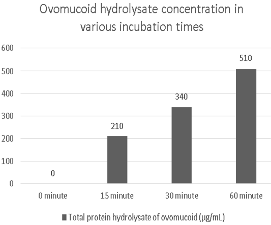
Alcalase effectively broke down goose egg ovomucoid, as indicated by the progressive increase in free amino groups from 210 to 510 μg/mL and the disappearance of ovomucoid protein bands during hydrolysis.
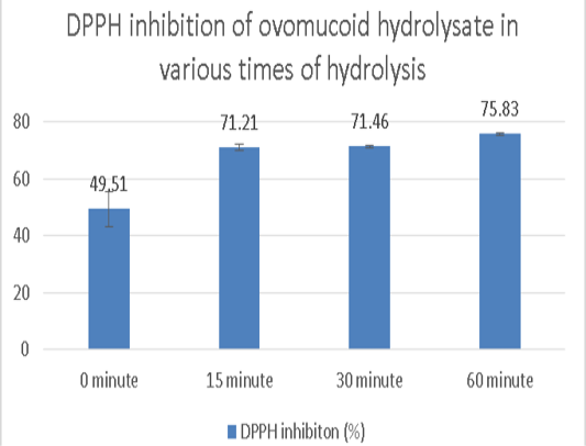
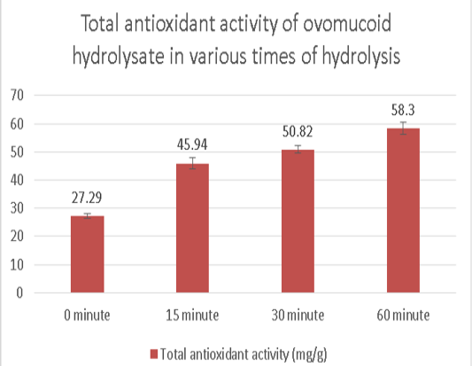
Extending the hydrolysis time significantly enhanced antioxidant activity, with the 60 min hydrolysate exhibiting the highest DPPH scavenging activity (75.83%), ABTS scavenging activity (95.21%), reducing power (2.74 mg AAE/g), and total antioxidant capacity (58.30 mg AAE/g).
These results demonstrate that prolonged hydrolysis promotes the release of antioxidant bioactive peptides from goose egg ovomucoid.
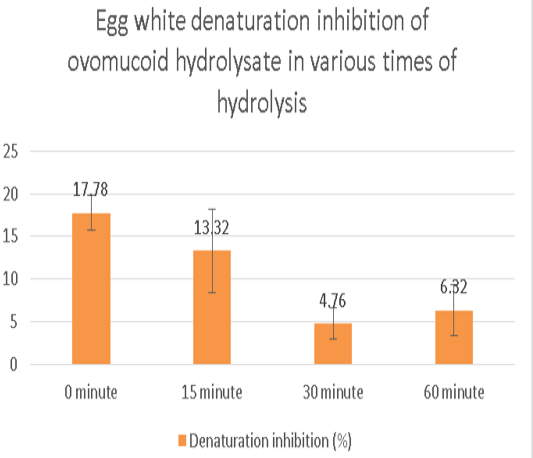
Conversely, anti-inflammatory activity decreased following hydrolysis, with native ovomucoid showing the greatest inhibition of protein denaturation (17.78%), while the highest activity among hydrolysates was observed after 15 min of hydrolysis (13.32%).
Selected from the article's own text.
Abstract
Ovomucoid is a major egg white protein (about 11%) with a unique glycosylated structure, ovomucoid holds significant potential as a precursor for bioactive peptides. Using enzymatic hydrolysis could unlock novel, natural ingredients for the functional food and nutraceutical sectors, which urgently need of effective antioxidants and anti-inflammatory agents. The objective of this study was to evaluate the antioxidant and anti-inflammatory activities of ovomucoid hydrolysates, and to determine the effect of varying enzymatic hydrolysis times on the enhancement of these bioactive properties. Ovomucoid was extracted from egg white using ethanol. Its purity was confirmed by SDS-PAGE. The protein was then enzymatically hydrolyzed with alcalase for different durations (0, 15, 30, and 60 minutes). The antioxidant activity of the hydrolysates was assessed using DPPH, ABTS, Reducing Power Assay (RPA), and Total Antioxidant Capacity (TAC) methods. Anti-inflammatory activity was determined via an egg albumin denaturation inhibition assay. The results showed that increasing the ovomucoid hydrolysis time to 60 minutes resulted in significantly higher antioxidant activity compared to the 0, 15, and 30 minute hydrolysates (P < 0.05). Significant differences in anti-inflammatory activity were also observed among the various hydrolysis times (P < 0.05). The optimal inhibitory capacity was achieved under non-hydrolyzed conditions 0 minutes and at 15 minutes of hydrolysis, whereas extending the time to 30 and 60 minutes did not yield further improvement and instead showed a tendency to decline. Enzymatic hydrolysis duration is a critical factor in maximizing the release of bioactive peptides, as longer hydrolysis time significantly increases the antioxidant capacity of ovomucoid hydrolysates. Overall, ovomucoid hydrolysates could be a source of antioxidants for functional foods. However, they did not demonstrate significant anti-inflammatory activity or inflammatory inhibition capability.
Eggs are an important source of animal protein, rich in vitamins, minerals, essential fatty acids, phospholipids, sphingomyelin, lutein, and zeaxanthin. Furthermore, they contain various bioactive components, such as antioxidants, anti-inflammatory agents, and choline (Sanlier and Ustun., 2021). A goose egg typically weighs about 162 grams, but in Asia, especially in Indonesia, ranges from approximately 120-130 g and it contains albumen (57.2%), yolk (30.6%), and eggshell (12.2%) (Kokoszyński., 2017; Karabulut., 2021). The bulk composition of fresh raw eggs includes water (76.1%), protein (12.6%), fat (9.5%), ash (1.1%), and carbohydrates (0.7%) . Currently, very limited literature exists regarding the specific protein composition of goose egg white. However, as a general reference, chicken egg white proteins are composed of ovalbumin (54%), ovotransferrin (12%), ovomucoid (11%), lysozyme (3.5%), and ovomucin (3.5%), all of which are closely associated with albumen viscosity. Minor proteins include avidin (0.05%), cystatin (0.05%), ovomacroglobulin (ovostatin) (0.5%), ovoflavoprotein (0.8%), ovoglycoprotein (1.0%), and ovoinhibitor (1.5%) ; . Ovomucoid is the most heavily glycosylated protein in egg whites, with a molecular weight of 20.1 kDa, typically visualized in SDS-PAGE gels in the range of 30-40 kDa ; . As a glycoprotein, ovomucoid contains a high proportion of carbohydrates (approximately 25-30% of the overall mass), making it heat-stable and resistant to clumping together .
As a major component of egg white, chicken egg white ovomucoid has been found to exhibit functional properties, including antioxidant and anti-inflammatory activities ; ; . To maximize the utilization of these functional properties, enzymatic hydrolysis is a well-known method. Protein hydrolysates are defined as a mixture of polypeptides, oligopeptides, and amino acids resulting from the hydrolysis of various protein sources . Specifically, enzymatic hydrolysis is employed to break down protein bonds into smaller peptides that are more readily absorbed by the body . Enzymes, as biological catalysts, function to accelerate specific biochemical reactions . Alcalase is known as one of the most efficient enzymes for releasing bioactive peptides from diverse protein sources . Alcalase exhibits high cutting specificity, particularly targeting aromatic (Phe, Trp, Tyr), acidic (Glu), sulfur-containing (Met), aliphatic (Leu, Ala), hydroxyl (Ser), and basic (Lys) residues .
Peptides are protein-derived biomolecules typically composed of 2 to 20 amino acids and have a molecular weight of less than 6000 Da. They possess diverse bioactivities that contribute to human health ; ; . Bioactive peptides are molecules exhibiting significant health-beneficial effects, including potential antimicrobial, antihypertensive, antioxidant, anticancer, immunomodulatory, and anti-inflammatory activities ; . Hydrolyzed proteins are classified based on peptide size into partially hydrolyzed (peptides with a molecular weight of 3-10 kDa) and extensively hydrolyzed (peptides with a molecular weight of < 3 kDa) (Nutten., 2016; . Currently, many studies have investigated the functional properties of ovomucoid and its hydrolysates regarding antioxidant and anti-inflammatory activities in chickens. However, there is still a lack of comprehensive studies specifically examining the effective hydrolysis time of goose egg white ovomucoid hydrolysates to produce optimal antioxidant and anti-inflammatory activities. ; .
MATERIALS AND METHODS
Sample collection and preparation
Goose eggs collected 1 to 5 days after laying, were sourced from local suppliers at the Pasty Market in the Special Region of Yogyakarta (Daerah Istimewa Yogyakarta). The eggs used had an average weight of approximately 150 grams. Materials used in this study are Ethanol absolute, Aquades, FeCl3, Bio-Rad electrophoresis gel, SDS-PAGE buffer, Loading dye reagent, Staining gel solution, Destaining gel solution, Smobio protein marker PM 5100, Bradford solution, Ninhydrin solution, Methanol, DPPH reagent (Sigma), Potassium persulfate, ABTS reagent (Sigma), Sodium chloride, Potassium chloride, Sodium phosphate dibasic, potassium phosphate monobasic, Sulfuric acid (H2SO4), Sodium phosphate (Na3PO4), Ammonium molybdate, Phosphomolybdate reagent solution, Potassium ferricyanide (1%). Ovomucoid protein was extracted from the goose egg white. The purified ovomucoid was subsequently subjected to enzymatic hydrolysis using Alcalase. The hydrolysis was performed at various time intervals (0, 15, 30, and 60 minutes) and all treatments were conducted in triplicate.
Ovomucoid extraction
Ovomucoid was extracted and purified based on the method described by Abeyrathne et al. (2014) with slight modifications. Initially, the egg white was separated from the yolk and diluted with one volume of distilled water (1:1, v/v). Subsequently, Fe3+ was added at a concentration of 0.8 mL/L of 500 mM FeCl3 to separate ovomucoid from ovotransferrin through specific binding to ovotransferrin. The mixture was homogenized using a hand mixer for approximately 2 minutes. Precipitation was induced by the slow addition of cold 100% absolute ethanol to achieve a final concentration of 43%. The solution was centrifuged at 3,400 x g for 20 minutes at 4oC. The resulting supernatant, containing the ovomucoid, was collected, re-extracted with two volumes of 43% ethanol, and subsequently stored overnight at 4oC. The mixture was centrifuged again under the same conditions 3,400 x g for 20 minutes at 4oC. The supernatant was collected, and the residual ethanol was subsequently removed via a rotary evaporator. The extract was then heated in a water bath at 80oC for 30 minutes before undergoing the final drying stage using a freeze dryer (lyophilization).
Hydrolysis of ovomucoid
The hydrolysis was performed based on the method described by with minor modifications. The freeze-dried ovomucoid was weighed using an analytical balance and subsequently dissolved in distilled water to a concentration of 0.23 mg/mL (total volume 1 mL), because the total yield produced during the extraction process was very low, approximately 6.77 g from the total egg weight. The Alcalase enzyme was subsequently added, and the mixture was incubated at 50oC for various durations: 0, 15, 30, and 60 min. To terminate the enzymatic reaction, the samples were immediately heated at 90oC for 10 minutes.
Analysis of ovomucoid by SDS-page
The purity, protein amount, and success of ovomucoid hydrolysis were assessed using the SDS-PAGE method, adapted from the procedures of and . SDS-PAGE analysis was performed on the ovomucoid isolate (after extraction) and on the final hydrolysate samples. Samples 3 μL were prepared in a microtube by adding 7 μL of distilled water and 3 μL of loading dye reagent. The prepared samples were loaded into a 12-well SDS polyacrylamide gel. Each well was filled with a total volume of 13 μL of the prepared sample mixture. Electrophoresis was run at a constant voltage of 80-100 Volts. Following electrophoresis, the gel underwent staining and destaining until the background was sufficiently clear. The protein bands were then visualized and documented using a Gel Doc system.
Yield of ovomucoid
The yield of ovomucoid obtained after the isolation process was determined based on the AOAC method (2005). The yield was calculated by weighing the initial mass of goose egg white and the final mass of the ovomucoid powder obtained after extraction and freeze-drying. The yield (%) was calculated using the following formula:
Figure 1.
The bradford protein assay
Protein concentration was determined using the Bradford assay, following the method described by . Briefly, 10 μL of the prepared protein extract sample was placed into a microtube, followed by the addition of 180 μL of Bradford solution. The samples were incubated until the color reaction stabilized (i.e., turned blue), and then the absorbance was measured using a spectrophotometer at a wavelength of 595 nm.
The ninhydrin protein assay
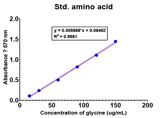
The concentration of free amino groups in the ovomucoid hydrolysates was determined using the Ninhydrin assay, based on the method by (McGrath., 1972). Ovomucoid hydrolysate samples (0.131 mg/mL) were analyzed. For the assay, the samples were diluted with distilled water to a total volume of 5 mL. The absorbance of the resulting solutions was measured using a spectrophotometer at an absorbance of 570 nm. The concentration of free amino groups was calculated using the standard linear regression equation of the amino acid glycine (Y = 0.009888 X + 0.06462) (Figure 1).
DPPH radical scavenging assay
The DPPH (2,2-diphenyl-1-picrylhydrazyl) radical scavenging activity was determined following the procedure outlined by . Briefly, 300 μL of the ovomucoid hydrolysate sample (from the 0, 15, 30, and 60 minute hydrolysis times) was mixed with 300 μL of 0.1 mM DPPH solution in a microtube (1:1 ratio, v/v). The mixture was incubated at room temperature (approx. 25oC) in the dark for 30 minutes. The absorbance of the reaction mixture was measured using a spectrophotometer at 517 nm. The scavenging activity was calculated using the following formula:
Figure 4.
ABTS radical scavenging assay
The ABTS (2,2’-azino-bis(3-ethylbenzothiazoline-6-sulfonic acid)) radical scavenging activity was assessed following the method of . with modifications. The ABTS working solution, prepared and incubated for 24 hours, was used for the assay. (Note: The 1:19 ratio mentioned conflicts with the volumes below, so this sentence is simplified). A 300 μL aliquot of the ovomucoid hydrolysate sample (from the 0, 15, 30, and 60 minute hydrolysis times) was mixed with 950 μL of the ABTS working solution. The mixture was then incubated at room temperature in the dark for 30 minutes. The absorbance of the reaction mixture was measured using a spectrophotometer at 734 nm. The scavenging activity was calculated using the following formula:
Figure 3.
Reducing power assay (rpa)
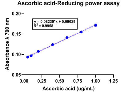
The reducing power of the ovomucoid hydrolysates was determined using the method described by . The ovomucoid hydrolysate samples (from 0, 15, 30, and 60 minutes hydrolysis) were dissolved in 200 μL of 0.2 M Phosphate Buffered Saline (PBS,pH 6.6). Subsequently, 200 μL of 1% Potassium Ferricyanide was added, and the mixture was incubated in a water bath at 50oC for 20 minutes. The reaction was terminated by adding 200 μL of 10% Trichloroacetic Acid (TCA) solution and centrifuged at 10,000 rpm for 10 minutes. Then, 300 μL of the resulting supernatant was collected, mixed with 300 μL of distilled water and 60 μL of 0.1% Ferric Chloride (FeCl3) solution. The absorbance was measured using a spectrophotometer at 700 nm. The reducing power was calculated using the standard linear regression equation derived from ascorbic acid: (Y = 0.08230 X + 0.09029) (Figure 2).
Total antioxydant capacity (phosphomolybdenum assay)
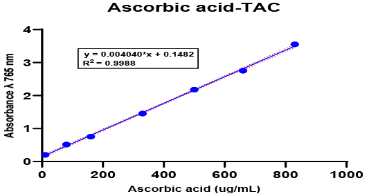
The Total Antioxidant Capacity (TAC) of the ovomucoid hydrolysates was determined using the phosphomolybdenum assay, based on the methods of and (Kpemesi et al., 2023). For the TAC assay, 70 μL of the ovomucoid hydrolysate (from 0, 15, 30, and 60 minute hydrolysis) was mixed with 700 μL of the phosphomolybdate reagent (1:10, v/v). The mixture was then incubated at 95oC for 90 minutes. Following incubation, the reaction mixture was centrifuged at 3,000 rpm for 5 minutes, and the supernatant was collected. The absorbance was measured using a spectrophotometer at 765 nm. The total antioxidant capacity was quantified using the standard linear regression equation derived from ascorbic acid: (Y = 0.08230 X + 0.09029) (Figure 3).
Anti-inflammatory egg albumen denaturation assay
Anti-inflammatory activity was assessed using the Egg Albumen Denaturation Inhibition Assay, based on the method reported by Kpemissi et al. (2023). The reaction mixture consisted of 200 μL of fresh chicken egg albumen, 2,800 μL of Phosphate Buffered Saline (PBS, pH 6.4), and 2,000 μL of the ovomucoid hydrolysate (from 0, 15, 30, and 60 minute hydrolysis). The mixture was initially incubated at 37oC for 15 minutes, then subjected to heating at 70oC for 5 minutes (to induce denaturation of the target protein (albumin) as a model of inflammation), followed by immediate cooling to 37oC. The absorbance of the mixture was measured using a spectrophotometer at 660 nm. The percentage inhibition of albumen denaturation was calculated using the following formula:
Figure 2.
Statistical analysis
The data obtained were analyzed using Analysis of Variance (ANOVA). Significant differences between the treatment means were subsequently analyzed using Duncan’s Multiple Range Test (DMRT). All statistical procedures were performed using the Statistical Product and Service Solution (SPSS) software, Version 26.0.
RESULTS
Yield of ovomucoid
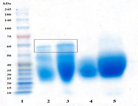
The ovomucoid isolated from goose egg white via ethanol extraction exhibited an average yield (%) of 13.00±0,82 of the total egg white mass (Table 1). The resulting purified protein, obtained through the ethanol extraction and heat treatment steps, is visually represented in Figure 4.
Table 1: Yield of ovomucoid from goose eggs.
Indicator
Goose Eggs
Mean±SD
Egg 1
Egg 2
Egg 3
Egg weight (g)
105,62
135,67
139,6
126,96±18,58
Albumen (g)
55,51
55,54
44,4
51,81±6,42
Yolk (g)
55,58
51,06
77,6
61,41±14,19
Ovomucoid (g)
7,59
7,36
5,367
6,77±1,22
Yield of ovomucoid (%)
13,68
13,25
12,08
13,00±0,82
Purity of ovomucoid by SDS-page
SDS-PAGE analysis of the isolated ovomucoid revealed protein bands primarily in the range of 30 kDa and 40 kDa (Figure 4). This observation confirms the successful purification of ovomucoid, as its molecular weight typically falls within this region.
Total protein hydrolysate of ovomucoid using ninhydrin protein assay
The initial ovomucoid protein concentration, determined using the Bradford assay, was found to be 0.23 mg/mL (corresponding to 1.15 mg of total protein in the 0.2 initial solution). The concentration of free amino acid groups of ovomucoid using ninhydrin protein assay increased over time: (210 μg/mL) at 15 minutes, (340 μg/mL) at 30 minutes, and reached the highest concentration of (510 μg/mL) at 60 minutes (Figure 5). This result suggests rapid and near-complete hydrolysis by Alcalase.
DPPH radical scavenging activity
The ovomucoid protein hydrolysate exhibited the highest DPPH radical scavenging activity at the 60 minute hydrolysis time, reaching 75.83%. This optimal activity was statistically significant (P < 0.05) when compared to other hydrolysis times (Figure 6).
ABTS radical scavenging activity
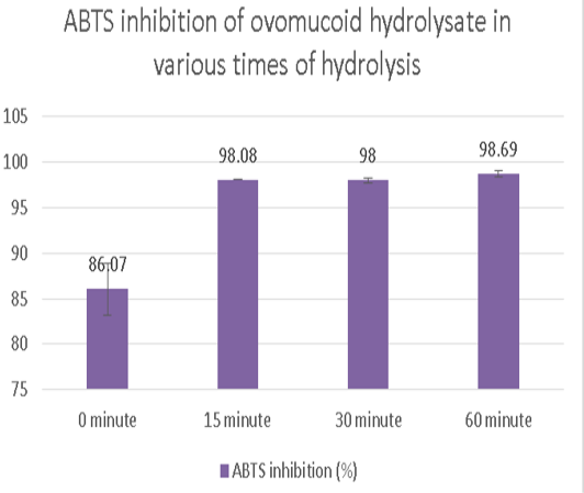
The ovomucoid protein hydrolysate demonstrated the highest ABTS radical scavenging activity at the 60 minute hydrolysis time, achieving an average inhibition percentage of 95.21%. This maximum activity was found to be statistically significant (P < 0.05) (Figure 7).
Reducing power assay (RPA)
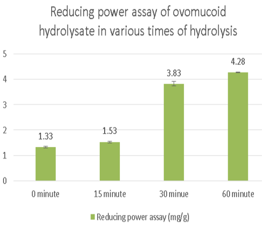
The reducing power of the ovomucoid hydrolysate showed a significant dependence on the hydrolysis time (P < 0.05$, Figure 8). The maximum reducing power was observed at the longest hydrolysis time, reaching an average value of 2.74 mg/g (as Ascorbic Acid Equivalents).
Total antioxidant capacity (Tac)
The Total Antioxidant Capacity (TAC) exhibited a significant dependence on the hydrolysis time (P < 0.05) Figure 9. The maximum TAC value was recorded at the 60 minute hydrolysis time, reaching 58.30 mg/g (as Ascorbic Acid Equivalents).
The anti-inflammatory activity, assessed via the egg albumen denaturation inhibition assay, showed a significant effect (P < 0.05) Figure 10. on inhibiting protein denaturation. The highest inhibition of protein denaturation 17.78% was exhibited by the native ovomucoid (unhydrolyzed protein), indicating the intrinsic anti-inflammatory potential of the whole molecule. Among the hydrolysates, the 15 minute sample displayed the greatest inhibitory effect, reaching 13.32 inhibition.
DISCUSSION
The results reported by Nurliyani et al. (2023). indicate that ovomucoid is a protein with a theoretical molecular weight of 28 kDa. However, the SDS-PAGE bands showed the apparent molecular weight of ovomucoid to be in the range of 30-40 kDa. The high purity is mainly because of the unique chemical properties of ovomucoid. The combination of ethanol extraction and heat treatment effectively denatures and precipitates most egg white proteins, such as ovalbumin, leaving only the more stable ovomucoid and ovotransferrin in solution . Ovomucoid’s exceptional stability to heat (up to 90–100°C) and organic solvents like ethanol is linked to its tertiary structure, which is reinforced by nine disulfide bonds . The initial ovomucoid solution 0.2% was determined to have a protein concentration of 0.23 mg/mL (or 1.15 mg total protein), measured using the Bradford assay. This assay relies on the binding of Coomassie Brilliant Blue G-250 dye to basic amino acid residues, particularly arginine, histidine, phenylalanine, tryptophan, and tyrosine, via hydrophobic interactions, resulting in a color shift to blue . Variations in protein concentration and composition within the egg white, which subsequently affect the final isolated yield, can be influenced by several factors. These factors include genetics, nutrition, the poultry rearing system, the physiological status of the hen, egg storage conditions, heat treatment applied, and inherent variability among domestic poultry species ; .
The hydrolysis process converts the large ovomucoid protein into simpler protein molecules, or peptides . Previous studies suggest that ovomucoid can be hydrolyzed maximally over a prolonged period, typically around 3 hours . The efficacy of the hydrolysis in this study is attributed to the protease used. Alcalase exhibits high specificity toward peptide bonds containing aromatic (e.g., Phe, Trp, and Tyr), acidic (Glu), sulfur-containing (Met), aliphatic (Leu and Ala), hydroxyl (Ser), and basic (Lys) amino acid residues . The SDS-PAGE results confirm the extensive degradation of the ovomucoid; the disappearance or significant fading of the ovomucoid band, combined with the emergence of high soluble protein concentration at 60 minutes, suggests that the protein was nearly or completely hydrolyzed into smaller fragments . This band fading occurs because larger proteins migrate more slowly than smaller protein molecules during electrophoresis, while very small peptides may exit the gel entirely (Roy and Kumar., 2014). The resulting small fragments are likely bioactive peptides, which are defined as biomolecules derived from proteins containing typically 2 to 20 amino acid residues and possessing a molecular weight of less than 6000 Da . The presence of these small, soluble peptides are crucial for the observed increase in antioxidant activity.
The DPPH assay is a common method to evaluate the antioxidant capacity of a substance. The mechanism involves the reduction of the stable DPPH free radical upon receiving electrons or hydrogen radicals from the antioxidant, which is visually quantified by a correlated decrease in absorbance (color change). The high antioxidant activity observed in the ovomucoid hydrolysates is primarily influenced by the molecular weight and structure of the resulting peptides ; . As established, these are typically small fragments containing 2 to 20 amino acid residues . Bioactive peptides from egg hydrolysates work in two main ways to neutralize free radicals that allow them to effectively neutralize free radicals . Firstly, the hydrolysis process is crucial as it reduces the size of the protein, significantly increasing the accessibility of internal functional side chains (R-groups) to reactive oxygen or nitrogen species. Secondly, the fragments function as radical terminators through the Hydrogen Atom Transfer (HAT) mechanism. The hydroxyl group within the aromatic structure of Tyrosine acts as a highly effective hydrogen donor, neutralizing the free radical and effectively breaking the radical chain.
Ovomucoid hydrolysate is what you get when ovomucoid protein is broken down into smaller fragments. These fragments possess bioactive properties and are referred to as bioactive peptides that can prevent oxidative reactions and scavenge free radicals, specifically Reactive Oxygen Species (ROS). Peptides present in egg white protein hydrolysates can inactivate ROS through electron exchange reduction (Jhony et al., 2021; Sureh., 2022).
Egg white hydrolysate exhibits hydrogen/electron donor activity, enabling it to interact with free radicals, thereby halting chain reactions or preventing radical formation . The antioxidant activity of goose egg ovomucoid hydrolysate, with an average value of 2.74 mg/g (as measured by RPA) and a steep increase between the 15th and 30th minutes, demonstrates a superior electron donor capacity compared to duck egg hydrolysates, which averaged 1.105 mg/g (Sureh., 2022). The bioactive peptides in the ovomucoid hydrolysate exert reducing power by facilitating electron transfer, which reduces the Fe3+ species to the Fe2+ form (Jhony et al., 2021; Sureh., 2022).
The bioactive peptides within ovomucoid hydrolysates act as electron donors, enabling them to break the radical chain and convert reactive free radicals into more stable, non-reactive products (Arabshahi-Delouee and Urooj., 2007). Bioactive peptides resulting from ovomucoid hydrolysis demonstrate the ability to scavenge free radicals, partly through the transfer of electrons to species like the phosphomolybdate complex (Hamez et al., 2018). This mechanism, particularly in the TAC assay, is based on the reduction of phosphomolybdate ions in the presence of antioxidants, resulting in the formation of a green-blue phosphate/MoV complex . The resulting antioxidant activity is directly proportional to the concentration of bioactive peptides in the hydrolysate. Specifically, the bioactive peptides interact with the yellow MoVI species, reducing it to the greenish-blue MoV form (MoVI + AOH MoV + AO), which confirms the formation of the phosphomolybdate (V) complex ; .
Stopping protein denaturation is a major sign in evaluating anti-inflammatory potential in in vitro assays ; . Compounds capable of inhibiting protein denaturation are considered potential sources of anti-inflammatory agents . The decrease in inhibition observed in the hydrolysates suggests that the smaller ovomucoid peptides may be less resistant to high temperatures during the incubation process. Hydrolysis breaks the larger protein into small peptides and amino acids, which can alter the overall structure, potentially rendering the fragments more susceptible to thermal denaturation. Thermally- or heat-induced protein denaturation, which can lead to auto-antigen formation, causes structural changes by disrupting disulfide, hydrogen, and electrostatic hydrophobic bonds ; ; . In biological systems, effective anti-inflammatory activity often requires the protection of the lysosomal membrane to mitigate the inflammatory response . Anti-inflammatory substances stabilize liposomal membranes, thereby preventing the release of lysosomal components (such as hydrolytic enzymes) that otherwise increase inflammatory responses and cause tissue damage .
CONCLUSION
Alcalase effectively broke down goose egg ovomucoid, as indicated by the progressive increase in free amino groups from 210 to 510 μg/mL and the disappearance of ovomucoid protein bands during hydrolysis. Extending the hydrolysis time significantly enhanced antioxidant activity, with the 60 min hydrolysate exhibiting the highest DPPH scavenging activity (75.83%), ABTS scavenging activity (95.21%), reducing power (2.74 mg AAE/g), and total antioxidant capacity (58.30 mg AAE/g). These results demonstrate that prolonged hydrolysis promotes the release of antioxidant bioactive peptides from goose egg ovomucoid. Conversely, anti-inflammatory activity decreased following hydrolysis, with native ovomucoid showing the greatest inhibition of protein denaturation (17.78%), while the highest activity among hydrolysates was observed after 15 min of hydrolysis (13.32%). Therefore, a 60 min hydrolysis is optimal for maximizing antioxidant potential, whereas shorter hydrolysis times better preserve anti-inflammatory activity. However, it should be noted that with a larger sample size, the observed trend might become statistically significant. These findings highlight goose egg ovomucoid as a promising source of functional peptides for antioxidant applications. However, these results are from in vitro assays only, and further studies in living systems are needed.
ACKNOWLEDGEMENTS
The authors gratefully acknowledge the Center for Tropical Animal Studies, Faculty of Animal Science, Universitas Gadjah Mada, for providing facilities and research support. This study received no external funding.
NOVELTY STATEMENT
The novelty of this study lies in comparing the effect of hydrolysis time on the antioxidant and anti-inflammatory potential of ovomucoid from goose (Anser anser) eggs, thereby identifying the hydrolysis duration that optimizes each bioactivity.
AUTHOR’S CONTRIBUTION
Ekie Maulana Al Buruuj: Methodology, Formal analysis, Investigation, Resources, Data curation, Visualization, Writing-original draft; Muhammad Fathin Hanif: Writing-review and editing; Winda Fatma Sari: Investigation, Writing-review and editing; Mohammad Zainal Abidin: Methodology, Supervision, Conceptualizing; Writing-review and editing; Nurliyani: Methodology, Supervision, Conceptualizing; Writing-review and editing
Generative AI and AI assisted technology
statement
The authors declare that no generative AI and AI assisted
technology was used in the creation of this manuscript.
Conflict of interest
The authors declare that there is no conflicts of interest.
Figures
Figure 1:.
Graphic of glycine amino acid standard.
Figure 2:.
Graphic of ascorbic acid-reducing power assay standard.
Figure 3:.
Graphic of ascorbic acid-total antioxidant capacity standard.
Figure 4:.
Ovomucoid protein electrophoresis in various concentrations, lane 1: protein marker; lane 2: ovomucoid solution with citric acid extract (10 times dilution), Extraction with the addition of citric acid was carried out in the preliminary study to determine the purity level of ovomucoid; lane 3: ovomucoid solution with citric acid extract (5 times dilution); lane 4: ovomucoid solution with ethanol extract (10 times dilution), Extraction with the addition of citric acid was carried out in the preliminary. study to determine the purity level of ovomucoid; lane 5: ovomucoid solution with ethanol extract (5 times dilution).
Figure 5:.
Graphic of ovomucoid hydrolysate concentration in various incubation times (0 minute (0 minutes, no reaction occurred (0 μg/mL); 15 minutes (210 μg/mL); 30 minutes (340 μg/mL); 60 minutea (510 μg/mL)).
Figure 6:.
Graphic of percentage DPPH inhibition of ovomucoid hydrolysate in various incubation times of hydrolysis (0 minutes (49.51±6.15a); 15 minutes (71.21±1.05b); 30 minutes (71.46±0,46b); 60 minutes (75.83±0,30b)).
Figure 7:.
Graphic of percentage ABTS inhibition of ovomucoid hydrolysate in various incubation times of hydrolysis (0 minutes (86,07±2,86a); 15 minutes (98,08±0,01b); 30 minutes (98,00±0,31b); 60 minutes (98,69±0,35b)).
Figure 8:.
Graphic of total Reducing Power Assay of ovomucoid hydrolysate in various incubation times of hydrolysis (0 minutes (1.33±0,03a); 15 minutes (1.53±0,03b); 30 minutes (3.83±0,09c); 60 minutes (4.28±0,01d)).
Figure 9:.
Graphic of Total antioxidant capacity of ovomucoid hydrolysate in various incubation times of hydrolysis (0 minutes (27.29±0,94a); 15 minutes (45.94±1,92b); 30 minutes (50.82±1,32c); 60 minutes (58.30±2,09d)).
Figure 10:.
Graphic of percentage denaturation inhibition of ovomucoid hydrolysate in various incubation times of hydrolysis (0 minutes (17.78±2,08a); 15 minutes (13.32±4,91ab); 30 minutes (4.76±1,87bc); 60 minutes (6.32±2,99c)).
References
Abeyrathne EDNS, Lee HY, Suh WJ, Anh DU (2014). Separation of ovotransferrin and ovomucoid from chicken egg white. Poultr. Sci., (93): 1010–1017. https://doi.org/10.3382/ps.2013-03649[CrossRef]
Abeyrathne EDNS, Lee HY, Jo C, Suh JW, Ahh DU (2015). Enzymatic hydrolysis of ovomucoid and the functional properties of its hydrolysates. Poulty. Sci., 94: 2280-2287. https://doi.org/10.3382/ps/pev196[CrossRef]
Abeyrathne EDNS, Huang, X, Anh DU (2018). Antioxidant, angiotensin-converting enzyme inhibitory activity and other functional properties of egg white proteins and their derived peptides – A review. Poultr. Sci., (97): 1462–1468. https://doi.org/10.3382/ps/pex399[CrossRef]
Abeyrathne EDNS, Lee HY, Anh DU (2013). Egg white proteins and their potential use in food processing or as nutraceutical and pharmaceutical agents—A review. Poultr. Sci., (92): 3292–3299. https://doi.org/10.3382/ps.2013-03391[CrossRef]
Annadurai P, Annadurai V, Yongkun M, Pugazhendhi A, Dhandayuthapani K (2021). Phytochemical composition, antioxidant and antimicrobial activities of Plecospermum spinosum Trecul. Process Biochem., (100): 107-116. https://doi.org/10.1016/j.procbio.2020.09.031[CrossRef]
Anokwah D, Kwatia EA, Amponsah IK, Jibira Y, Harley BK, Ameyaw EO, Obese E, Biney RP, Mensah AY (2022). Evaluation of the anti-inflammatory and antioxidant potential of the stem bark extract and some constituents of Aidia genipiflora (DC.) dandy (Rubiaceae). Heliyon, 8 (8): e10082. https://doi.org/10.1016/j.heliyon.2022.e10082[CrossRef]
AOAC (2005). Official Methods of Analysis. 18th ed. AOAC International, Gaithersburg, MD.
Arabshahi-Delouee S, Urooj A (2007). Antioxidant properties of various solvent extracts of mulberry (Morus indica L.) leaves. Food Chem., (102): 1233–1240
Chalamaiah M, Keskin S, Hong H, Wu J (2019). Regulatory requirements of bioactive peptides (protein hydrolysates) from food proteins. J. Funct. Foods, (58): 123–129. https://doi.org/10.1016/j.foodchem.2006.07.013[CrossRef]
Chen C, Chi Y, Zao Y, Lv L (2012). Purification and identification of antioxidant peptides from egg white protein hydrolysate. Amino Acids J., (43): 457–466. https://doi.org/10.1007/s00726-011-1102-0[CrossRef]
Chew LY, Toh GT, Ismail A (2019). Chapter 15 - Application of proteases for the production of bioactive peptides. Enzymes Food Biotechnol., 247-261. https://doi.org/10.1016/B978-0-12-813280-7.00015-3[CrossRef]
Cruz-Casas DE, Aguilar CN, Ascacio-Vald´es JA, Rodríguez-Herrera R, Chavez-Gonzalez ML, Flores-Gallegos AC (2021). Enzymatic hydrolysis and microbial fermentation: The most favorable biotechnological methods for the release of bioactive peptides. Food Chemistry: Mol. Sci., (3): 100047. https://doi.org/10.1016/j.fochms.2021.100047[CrossRef]
Cui Q, Sun Y, Zhou Z, Cheng J, Guo M (2021). Effects of enzymatic hydrolysis on physicochemical properties and solubility and bitterness of milk protein hydrolysates. Foods, (10): 2462. https://doi.org/10.3390/foods10102462[CrossRef]
da Silva AC, Queiroz AESDF, Oliveira JTC, Medeiros EV, de Souza-Motta CM, Moreira KA (2019). Antioxidant activities of chicken egg white hydrolysates obtained by new purified protease of aspergillus avenaceus URM 6706. Food/Feed Sci. Technol., (62): e19180062. https://doi.org/10.1590/1678-4324-2019180062[CrossRef]
Das K, Asdaq SMB, Khan MS, Amrutha S, Alamri A, Alhomrani M, Alsanie WF, Bhaskar A, Shree GC, Harshitha P (2022). Phytochemical investigation and evaluation of in vitro anti-inflammatory activity of euphorbia hirta ethanol leaf and root extracts: a comparative study. J. King Saud. Univ.Sci., 34 (7): 102261. https://doi.org/10.1016/j.jksus.2022.102261[CrossRef]
Fernandez-Tome S, Montalban-Arques A, Díaz-Guerra A, Galvan-Roman JM, Marin AC, Mora-Gutierrez I, Ortega Moreno L, Santander C, Sanchez B, Chaparro M, Gisbert JP, Bernardo D (2019). Peptides encrypted in the human intestinal microbial-exoproteome as novel biomarkers and immunomodulatory compounds in the gastrointestinal tract. J. Funct. Foods, (52): 459–468. https://doi.org/10.1016/j.jff.2018.11.036[CrossRef]
Hamzeh A, Rezaei M, Khodabandeh S, Motamedzadegan A, Noruzinia M (2018). Antiproliferative and antioxidative activities of cuttlefish (Sepia pharaonis) protein hydrolysates as affected by degree of hydrolysis. J. Food Meas. Charact., (12): 721–727. https://doi.org/10.1007/s11694-017-9685-0[CrossRef]
Hasan MM, Islam ME, Hossain MS, Akter M, Rahman MAA, Kazi M, Khan S, Parvin MS (2024). Unveiling the therapeutic potential: evaluation of anti-inflammatory and antineoplastic activity of magnolia champaca linn’s stem bark isolate through molecular docking insights. Heliyon, (10): e22972. https://doi.org/10.1016/j.heliyon.2023.e22972[CrossRef]
He Y, Pan X, Chi CF, Sun KL, Wang B (2019). Ten new pentapeptides from protein hydrolysate of miiuy croaker (Miichthys miiuy) muscle: Preparation, identification, antioxidant activity evaluation. LWT - Food Sci. Technol., (105): 1–8. https://doi.org/10.1016/j.lwt.2019.01.054[CrossRef]
Jan S, Khan MR, Rashid U, Bokhari J (2013). Assessment of antioxidant potential, total phenolics and flavonoids of different solvent fractions of monotheca buxifolia fruit. Osong Public Health Respect, 4 (5): 246-254. https://doi.org/10.1016/j.phrp.2013.09.003[CrossRef]
Ji S, Ahn DK, Zhao Y, Lia K, Li S, Huang X (2020). An easy and rapid separation method for five major proteins from egg white: successive extraction and MALDI-TOF-MS identification. Food Chem., (315): 126207. https://doi.org/10.1016/j.foodchem.2020.126207[CrossRef]
Johny LC, Kudre TG, Suresh PV (2021). Production of egg white hydrolysate by digestion with pineapple bromelain: optimization, evaluation and antioxidant activity study. J. Food Sci. Technol., (59): 1769–1780. https://doi.org/10.1007/s13197-021-05188-0[CrossRef]
Karabulut O (2021). Relationship between weight, volume and specific gravity of goose eggs before incubation. J. Adv. Vet.Bio. Sci. Tech., 6 (2), 90-99. https://doi.org/10.31797/vetbio.929031[CrossRef]
Kielkopf CL, Bauer W Urbatsch IL (2020). Bradford assay for determining protein concentration. Cold Spring Harb Protoc., pp. 1-4. https://doi.org/10.1101/pdb.prot102269[CrossRef]
Kokoszyński D (2017). Guinea fowl, goose, turkey, ostrich, and emu eggs. In: Hester PY (ed). Egg Innovations and Strategies for Improvements. Elsevier, pp. 33–43. https://doi.org/10.1016/B978-0-12-800879-9.00004-4[CrossRef]
Kpemissi M, Kantati YT, Veerapur VP, Eklu-Gadegbeku K, Hassan Z (2023). Anti-cholinesterase, anti-inflammatory and antioxidant properties of Combretum micranthum G. Don: Potential implications in neurodegenerative disease. IBRO Neurosci. Rep., (14): 21–27. https://doi.org/10.1016/j.ibneur.2022.12.001[CrossRef]
Liu W, Chen X, Li H, Zhang J, An J, Liu X (2022). Anti-Inflammatory function of plant-derived bioactive peptides: Rev. Foods, (11): 2361. https://doi.org/10.3390/foods11152361[CrossRef]
Liu W, Zhan J, Miyakawa T, Li G, Gu R, Tanokura M (2021). Antioxidant properties and inhibition of angiotensin-converting enzyme by highly active peptides from wheat gluten. Sci. Rep., (11): 5206. https://doi.org/10.1038/s41598-021-84820-7[CrossRef]
McCarthy A.L, O’Callaghan YC, O’Brien NM (2013). Protein hydrolysates from agricultural crops—bioactivity and potential for functional food development. Agriculture, (3): 112-130. https://doi.org/10.3390/agriculture3010112[CrossRef]
McGrath R (1972). Protein Measurement by ninhydrin determination of amino acids released by alkaline hydrolysis. Anal. Biochem., (49): 95-102. https://doi.org/10.1016/0003-2697(72)90245-X[CrossRef]
Nurliyani, Erwanto Y, Rumiyati Sukarno AS (2023). Characteristics of protein and amino acid in various poultry egg white ovomucoid. Food Sci. Tech. Campinas, (43): e101722. https://doi.org/10.1590/fst.101722[CrossRef]
Nutten S (2016). Proteins, peptides and amino acids: Role in infant nutrition. Nestle Nutr. Inst. Workshop Ser., (86): 1–10. https://doi.org/10.1159/000442697[CrossRef]
Palupi NW, Windrati WS, Tamtarini (2010). The effect of enzymatic hydrolysis on the properties of protein hydrolysate from paddy mushroom. Makara J. Technol., (14): 73-76. https://doi.org/10.7454/mst.v14i2.695[CrossRef]
Parameswari P, Devika R, Vijayaraghavan P (2019). In vitro anti-inflammatory and antimicrobial potential of leaf extract from Artemisia nilagirica (Clarke) Pamp. Saudi J. Biol. Sci., (26): 460–463. https://doi.org/10.1016/j.sjbs.2018.09.005[CrossRef]
Prihanto AA, Nurdiani R, Bagus AD (2019). Production and characteristics of fish protein hydrolysate from parrotfish (Chlorurus sordidus) head. PeerJ, 20 (7): e8297. https://doi.org/10.7717/peerj.8297[CrossRef]
Razi SM, Fahim H, Amirabadi S, Rashidinejad A (2023). An overview of the functional properties of egg white proteins and their application in the food industry. Food Hydroco. J., (135): 108183. https://doi.org/10.1016/j.foodhyd.2022.108183[CrossRef]
Roy S, Kumar V (2012). A Practical approach on sds page for separation of protein. Int. J. Sci. Res., pp. 2319-7064.
Sajjad A, Zia M, Xiao X, Olsson RT, Capezza AJ, Rasheed F (2023). Wheat gluten hydrolysates with embedded Ag-nanoparticles; a structure-function assessment for potential applications as wound sorbents with antimicrobial properties. Polym. Test., (118): 107896. https://doi.org/10.1016/j.polymertesting.2022.107896[CrossRef]
Sanlier N, Ustun D (2021). Egg consumption and health effects: A narrative review. J. Food Sci., (86): 4250–4261. https://doi.org/10.1111/1750-3841.15892[CrossRef]
Sarantidi E, Ainatzoglou A, Papadimitriou C, Stamoula E, Maghiorou K, Miflidi A, Trichopoulou A, Mountzouris KC, Anagnostopoulos AK (2023). Egg white and yolk protein atlas: new protein insights of a global landmark food. Foods, (12): 3470. https://doi.org/10.3390/foods12183470[CrossRef]
Sukarno AS, Nurliyani, Erwanto Y, Rakhmatulloh S, Rifqi (2023). Antibacterial and antioxidant activity of protein hydrolysate extracted from different indonesian avian egg white. J. Sain Peternakan Indones., (18): 1. https://doi.org/10.31186/jspi.id.18.1.27-33[CrossRef]
Sureh PV (2022). Protein hydrolysate from duck egg white by Flavourzyme® digestion: Process optimisation by model design approach and evaluation of antioxidant capacity and characteristic properties. LWT Food Sci. Technol., (156): 113018. https://doi.org/10.1016/j.lwt.2021.113018[CrossRef]
Tacias-Pascacio VG, Morellon-Sterling R, Siar E, Tavano O, Berenguer-Murcia A, Fernandez-Lafuente R (2020). Use of Alcalase in the production of bioactive peptides: A review. Int. J. Biol. Macromol., 165 Part B: 2143-2196. https://doi.org/10.1016/j.ijbiomac.2020.10.060[CrossRef]
Vandenplas Y, Ksiazyk J, Luna MS, Migacheva N, Picaud L, Ramenghi LA, Singhal A, Wabitsch M (2022). Partial hydrolyzed protein as a protein source for infant feeding: do or don’t. Nutrients, (14): 1720. https://doi.org/10.3390/nu14091720[CrossRef]
Xia X, Li J, Liang R, Li Y, Ma X, Yang Y, Lozano-Ojalvod D (2024). Effects of unfolding treatment assisted glycation on the IgE/IgG binding capacity and antioxidant activity of ovomucoid. Food Funct., Issue 1. https://doi.org/10.1039/D3FO04035F[CrossRef]
Xue H, Wang J, Xie J, Xiong C, Li J, Tu Y, Zhao Y (2019). Isolation, purification, structure identification of antioxidant peptides from embryonated eggs. Poultr. Sci., 98 (6): 2360-2370. https://doi.org/10.3382/ps/pey606[CrossRef]
Yubolphan R, Phongsiri K, Mongkhammee N, Pidech J, Roytrakul S, Suwattanasophon C, Choowongkomon K, Daduang S, Khunkitti W, Jangpromma N (2025). Anti-inflammatory effects of hen egg white hydrolysate and its specific peptides is8, pa11, pk8 on lps-induced macrophage inflammation. Food Sci. Nutr., 3; 13 (8): e70713. https://doi.org/10.1002/fsn3.70713[CrossRef]