
The present study aimed at measuring the antibody titres against H9N2 influenza viruses and its molecular detections in asymptomatic commercial layers from Faisalabad district of Punjab, Pakistan. Overall 120 blood samples, 24 tissue samples from each organ (trachea, lung and intestine) were collected from the 12 commercial layer flocks selected randomly at the age of 35-50 weeks without any direct clinical manifestation of avian influenza. Serum collected from 120 birds was tested for antibodies against H9N2 by using Haemagglutination Inhibition assay. Calculated geometric mean titers of 5.37 revealed the infectivity of the flock with H9N2 Influenza virus. To investigate the presence of virus in the study population, trachea, lung and intestine tissue samples were processed for RNA isolation and subjected to molecular detection of H9 gene of H9N2 subtypes using one step Reverse Transcriptase Polymerase Chain Reaction (RT-PCR). Out of 24 samples taken from each organ, 22, 21, and 18 samples from trachea, lungs, and intestine showed viral RNA, respectively. Taken together, results showed that the H9N2 is endemic and widely distributed in asymptotic layers. Furthermore results indicated that H9N2 subtype may survive in layers without showing any symptoms.
Short Communication · Volume 49, Issue 1, 21 January 2017 · Open Access
Molecular and Serological Detection of Avian Influenza H9N2 Virus in Asymptomatic Commercial Layers in Faisalabad District, Punjab
· · · · · ·
Affiliations & Notes ⌄
- 1Institute of Microbiology, University of Agriculture, Faisalabad
- 2Department of Microbiology, Government College University, Allama Iqbal Road, Faisalabad
- 3School of Biological Sciences, University of the Punjab, Quaid-I-Azam Campus, Lahore
- 4Department of Anatomy and Histology, Baqai Veterinary College, Karachi
- 5National Institute for Biotechnology and Genetic Engineering, Jhang Road, Faisalabad * Corresponding author: drbilalaslam@gcuf.edu.pk
Article Info ⌄
- Journal:
- Pakistan Journal of Zoology
- Published:
- 21 January 2017

From the article
- The results of the investigation revealed that overall seroprevelance was 60% (72/120) and geometric mean titer of 5.37 (Table I) indicating the natural exposure of the flocks to the H9N2 subtype of avian influenza Virus.
- The table shows seroprevelance and antibody titer against H9N2 AIV in asymptomatic layer flock tested by HI.
- The pproposed study was conducted with the objectives of the estimation of antibodies against H9N2 and confirmation of the virus in asymptomatic commercial layers of various flocks located in district Faisalabad, Punjab by using RT-PCR.
- RT-PCR results showed a surprisingly high prevalence of live virus; 92 % trachea, 88% lungs and 75% intestine samples were found positive for H9N2 RNA.
Selected from the article's own text.
Abstract
Materials and methods
A total of 120 blood samples (10 samples from each flock) and 24 tissue samples (Trachea, lungs, and Intestine) were collected randomly from various layer flocks reared in Faisalabad district of Pakistan. For serum collection, 1 ml blood was taken from the wing vein by using appropriate syringe. Serum was isolated from clots by centrifuging at 4000 rpm/m for 10 min. For tissue collection, birds were slaughtered and then trachea, Intestines and lungs were aseptically removed. Rinsed in tap water and immediately transported to the lab of in cold conditions for further processing.
To test the presence of H9N2 specific antibodies in the serum, Hemagglutination Inhibition (HI) test was performed in V-bottom shaped 96-well microtiter plates. The NOBILIS Influenza H9N2 Vaccine virus (Novus International® USA) was used in the study along with 1% of chicken erythrocytes according to standard protocol . The antibody titer was calculated according to the last well reading which showed the complete inhibition of haemaggutinin activity induced by the H9N2 subtype used in the study.
For RT-PCR analysis trachea, lung, and intestine samples were processed for RNA extraction according to Mannhalter et al. (2000). The RNA pellet was solubilized in 50µl of DEPC treated water. Its quality and quantity was measured by using NanoDrop®.
The RT-PCR was performed to amplify H9 specific 808-bp fragment of avian influenza virus using Forward 5’- AGCAAAAGCAGGGGAACTCC-3’Reverse 3’- CCATACCATGGGGCAATTAG-5’ primers as describe by Dybkaer et al. (2004). The 50µl reaction mixture was prepared by adding 25µl QuickTM Master Mix, 1µl (5u) of AMV Reverse Transcriptase (Sigma, USA), 5µl RNA template, 1µl of 1.0µM upstream and downstream primers. The conditions used in one step RT-PCR were include reverse transcription with the Incubation at 45°C for 45 min, initial denaturation at 95°C for 5 min, and 40 cycle each of 95°C for 40 sec, annealing at 56°C for 1 min, primer extension at 72°C for 30 sec followed by final extension at 72°C for 10 min. The amplified PCR products were visualized on 0.8% agarose gel, stained with ethidium bromide.
Results and discussion
The results of the investigation revealed that overall seroprevelance was 60% (72/120) and geometric mean titer of 5.37 (Table I) indicating the natural exposure of the flocks to the H9N2 subtype of avian influenza Virus. The positive results showed the amplification of 808 bp fragment of H9 from H9N2 subtype (Fig. 1). Out of 24 tissue samples each from trachea, lung, and intestine samples tested, 92% (22/24), 88% (21/24), 75% (18/24) were found positive, respectively. The results reveal the presence of live virus in the asymptomatic birds. Moreover, results show that the respiratory tract is the preferred path of virus multiplication.
Table I.- Seroprevelance and antibody titer against H9N2 AIV in asymptomatic layer flock. The table shows seroprevelance and antibody titer against H9N2 AIV in asymptomatic layer flock tested by HI. The results are presented as geometric mean titer GMT of log 2 titers.
| Flock ID | No. of blood samples (n) | No. of positive samples (%) | HI Titer mean (Log2) |
| 1 | 10 | 56 | 5.1 |
| 2 | 10 | 57 | 4.6 |
| 3 | 10 | 64 | 3.9 |
| 4 | 10 | 48 | 6.05 |
| 5 | 10 | 56 | 5.3 |
| 6 | 10 | 60 | 7.0 |
| 7 | 10 | 68 | 5.5 |
| 8 | 10 | 54 | 5.5 |
| 9 | 10 | 71 | 5.1 |
| 10 | 10 | 57 | 5.0 |
| 11 | 10 | 65 | 7.0 |
| 12 | 10 | 63 | 4.5 |
| Total | 120 | Av: 60% | Av: 5.37 |
In Asia, prevalence of H9N2 has been established since 1990 . This virus belongs to LPAI group and is associated with mild infection in poultry. On the other hand, in the presence of opportunistic pathogens it may cause severe morbidity and sometimes leads to death . The pproposed study was conducted with the objectives of the estimation of antibodies against H9N2 and confirmation of the virus in asymptomatic commercial layers of various flocks located in district Faisalabad, Punjab by using RT-PCR.
In the current study, we collected samples from randomly selected 12 layer flocks with the age of 35-50 weeks. These flocks were apparently healthy and had no signs and symptoms of avian influenza. Results of HI revealed 60% seroprevalence with a GMT of 5.37. It indicates the birds were naturally exposed to H9N2 AIV. Natural exposure to the virus may be due to the risk factors associated with the environment which include the rearing of the domestic poultry in the vicinity of layer farms, because the prevalence of AI in domestic poultry and its transmission through aerosol has been established .
Presence of H9N2 was further verified by RT-PCR based molecular detection of viral RNA in various organs of birds. RT-PCR results showed a surprisingly high prevalence of live virus; 92 % trachea, 88% lungs and 75% intestine samples were found positive for H9N2 RNA. Kwon et al. (2008) reported the detection of AIV A/Chicken/HS/K5/01(H9N2) in various organs including bursa, spleen, kidneys, lungs and trachea. It has also been observed that H9N2 has the ability to stay in various tissues like spleen and kidneys of the commercial layers ; . Results of the present study are also in accordance with a study conducted by Shamseddini et al. where the researchers detected H9N2 of Iranian origin in numerous tissues include lungs, trachea and kidneys .
In conclusion, results of the present study show that the healthy layer flocks can be naturally exposed to AIV H9N2. Furthermore, our findings affirm the fact that AIV is disseminated to various body organs. In continuation of this study prospective studies should be planned with the objective of screening of all poultry population and live poultry markets (LPMs) as well with special reference to AIV at national level.
Acknowledgments
We appreciate the National Agriculture Research Council for the permission to use their laboratory facilities.
Statement of conflict of interest
Authors have declared no conflict of interest.
Figures
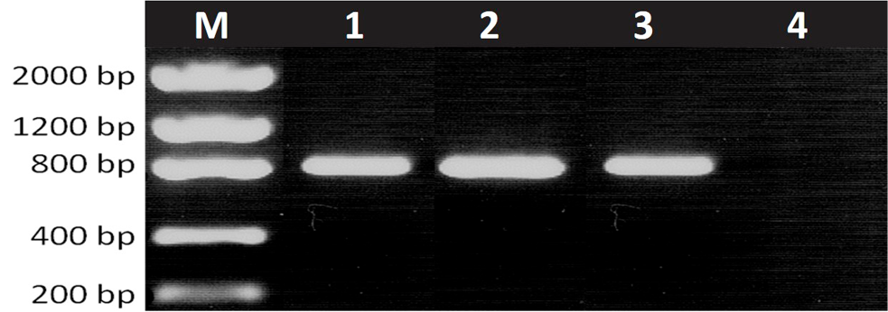
RT-PCR Amplification of AIV H9 gene. Representative results of H9 RT-PCR amplification (808 bp) from different tissue (Trachea, Lung, Intestine). Lane M, Marker; Lane 1, Trachea; Lane 2, Lungs; Lane 3, Intestine; Lane 4, Negative control.
References
- Alexander, D.J., 2007. Vaccine, 25: 5637–5644. http://dx.doi.org/10.1016/j.vaccine.2006.10.051[CrossRef]
- Bonfante, F., Patrono, L.V., Aiello, R., Beato, M.S., Terregino, C. and Capua, I., 2013. Vet. Microbiol., 165: 177–183. http://dx.doi.org/10.1016/j.vetmic.2013.03.014[CrossRef]
- Dybkaer, K., Munch, M., Handberg, K.J. and Jorgensen, P.H., 2004. J. Virol. Methods, 119: 151-158. http://dx.doi.org/10.1016/j.jviromet.2004.03.014[CrossRef]
- Guan, Y., Shortridge, K.F., Krauss, S. and Webster, R.G., 1999. Proc. natl. Acad. Sci., 96: 9363–9367. http://dx.doi.org/10.1073/pnas.96.16.9363[CrossRef]
- Huang, Y., Zhang, H., Li, X., Hu, S., Cai, L., Sun, Q., Li, W., Deng, Z., Xiang, X., Zhang, H., LI, F. and Gao, L., 2015. PLoS One, 10: 1-12. http://dx.doi.org/10.1371/journal.pone.0140812[CrossRef]
- Khawaja, J.Z., Naeem, K., Ahmed Z. and Ahmed, S., 2005. Int. J. Poult. Sci., 4: 39-43. http://dx.doi.org/10.3923/ijps.2005.39.43[CrossRef]
- Kwon, J.S., Lee, H.J., Lee, D.H., Lee, Y.J., Mo, I.P., Nahm, S.S., Kim, M.J., Lee, J.B., Park, S.Y., Choi, I.S. and Song, C.H.S., 2008. Virus Res., 133: 187-194. http://dx.doi.org/10.1016/j.virusres.2007.12.019[CrossRef]
- Lee, Y.J., Shin., J.Y., Song, M.S., Lee, Y.M., Choi, J.G., Lee, E.K., Jeong, O.M., Sung, H.W., Kim, J.H., Kwon, Y.K., Kwon, J.H., Kim, C.J., Webby, R.J., Webster, R.G. and Choi, Y.K., 2007. Virology, 359: 313-323. http://dx.doi.org/10.1016/j.virol.2006.09.025[CrossRef]
- Lee, D.H. and Song, C.S., 2013. Clin. exp. Vaccine Res., 2: 26–33. http://dx.doi.org/10.7774/cevr.2013.2.1.26[CrossRef]
- Mannhalter, C., Koizar, D. and Mitterbauer, G., 2000. Clin. Chem. Lab. Med., 38: 171-177. http://dx.doi.org/10.1515/CCLM.2000.026[CrossRef]
- Mosleh, N., Dadras, H. and Mohammadi, A., 2009. Iran. J. Vet. Res., 10: 12-20.
- Naeem, K., Siddique, N., Ayaz, M. and Jalalee, M.A., 2007. Avian Dis., 51: 189-193. http://dx.doi.org/10.1637/7617-042506R.1[CrossRef]
- Nili, H. and Asasi, K., 2003. Avian Dis., 47: 828-831. http://dx.doi.org/10.1637/0005-2086-47.s3.828[CrossRef]
- Shamseddini, M.V., Marandi, M., Pourbakhsh, S.A., Gharagozlo, M., Bahmani-nejad, M. and Khazraee-NIA, P., 2002. Arch, Razi Inst., 53: 11-21.
- Swayne, D.E., 1997. Vet. Pathol., 7: 557-567. http://dx.doi.org/10.1177/030098589703400603[CrossRef]
- Richard, M., Herfst, S., van den Brand, J. M., Lexmond, P., Bestebroer, T.M., Rimmelzwaan, G.F., Koopmans, M., Kuiken, T. and Fouchier, R.A., 2015. PLoS One, 10: e0129827. http://dx.doi.org/10.1371/journal.pone.0119034[CrossRef]
- Sohaib, M., Siddique, M., Muhammad, K., Rabbani, M., Altaf, I. and Hanif A., 2010. Prevalence of avian influenza virus (H5) in poultry layer flocks in and around Faisalabad, Punjab, Pakistan. Pakistan J. Zool., 42: 325-329.
- Sun, Y. and Liu, J., 2015. Protein Cell, 6: 18–25. http://dx.doi.org/10.1007/s13238-015-0145-5[CrossRef]
- Swayne, D.E. and Halvorson, D.A., 2008. In: Disease of poultry (eds. Y.M. Saif, H.J. Barnes, A.M. Fadl, J.R. Glisson, L.R McDougald, and D.E. Swayne). 12th Edn. Iowa State University Press, Ames, Iowa, pp. 153-184.
- Tanner, W.D., Toth, D. J.A. and Gundlapalli, A.V., 2015. Epidemiol. Infect., 143: 3359–3374. http://dx.doi.org/10.1017/S0950268815001570[CrossRef]
- Vasfi, M.M. and Bozorgmehrifard, M.H., 1999. An outbreak of non-highly pathogenic avian influenza in chickens in Iran. Proceedings of 61st meeting of world veterinary association. France.
Services
Article usage statistics
Loading…