Influenza virus type A is the only member of the family Orthomyxoviridae that have been reported to cause a serious threat to the poultry lead to causing respiratory or systemic diseases that vary in severity. Avian influenza viruses are divided into two groups, LPAI and HPAI (highly pathogenic AI) viruses. Both HPAI and LPAI virus infections are present all over the world and associated with high morbidity and low mortality (Aamir et al., 2007; Suarez, 2010).
Virus samples with accession number (MH368755.1) that used in the current study was propagated well in allantoic fluid of embryonated eggs, and produced high titers in both HA and EID50, the method of propagation, detection and titration of propagated virus are the same that be mentioned by may reports (Pansota et al., 2013; Ahmad et al., 2014). The propagated stock virus was confirmed as H9N2 by the use of specific hyper immune serum in HI test. This test was used widely in identification and differentiation AIV depending on the antiserum against AIV, in the same time used to determine the level of immunity for any tested flock. (Dormitorio et al., 2009; EL Zowalaty et al., 2011).
The results of current study revealed that there were obvious clinical signs in group A that was challenge with LPAI H9N2 and not vaccinated against the AIV characterized by severe nasal discharge, prominent depression, mild edema of the head and ruffling feather, also there were mortality of 2 birds with ratio of 3.3% comparing to other group. These findings are consistent with those of previous works (Soliman, 2014; Dabbour, 2015) who found that AIV serotype H9N2 affected many commercial flocks in the Middle East and most of the flocks of broiler chickens that experimentally infected with the LPAI H9N2 showed typical manifestation that characterized by respiratory signs which included rals, sneezing with nasal and ocular discharge, whitish to greenish diarrhea, depression and anorexia with 20% mortality. The low mortality and minimal clinical signs observed in experimentally infected broilers of present study consistent with other published studies of (El Zowalaty et al., 2011; Franca et al., 2014) who reported that LPAI H9N2 induces minimal or no clinical signs in controlled specific pathogen-free (SPF) challenge experiments. Zhang et al. (2008) reported that to control the spread of H9N2 avian influenza, a vaccination program for the H9N2 influenza virus has been widely implemented over the past two decades throughout the world by using inactivated vaccine for H9N2. Vaccination against H9N2 influenza virus will not prevent the shedding of this strain (H9N2) and may play a role in driving the evolution and spread of H9N2 influenza virus. The second scenario under which H9N2 avian influenza virus is transmitted relies on the ability of the H9N2 virus to re-assort with other influenza virus subtypes to generate new influenza viruses. In this manner, an H9N2 virus supplied internal genes to an H5N1 virus that emerge and also to other strain like H7N9 and H10N8 which is considered as highly pathogenic AIV (Ye et al., 2016). The results of the present study reinforce the previous study by (Mosleh et al., 2009) who reported that the predominant infection due to experimental challenge of broiler chickens with intranasally (IN) of 100µl allantoic fluid LPAI H9N2, the clinical signs appeared in the respiratory tract at 3-4 days post inoculation and all the sings represented in the respiratory and gastrointestinal tract.
On the other hand the results of the present study were unparalleled with the other studies (Guo et al., 2000; Khamas, 2008) who reported that AIV H9N2 was more pathogenic to broiler chickens, and it produced more severe gross and histologic changes in the respiratory system compared with SPF chickens on the affected farms. Mixed infections of AIV with other viral diseases were more pathogenic in older birds compared with younger birds, and they had caused more than 70% to 80% mortality in broiler chickens.
The current study exhibited that, in group B from vaccinated group with (available commercial H9N2 inactivated vaccine) the clinical signs were appeared after 12 days post inoculation. The clinical signs characterized by mild nasal discharge, mild depression and no mortality rate. These results were agreed with the findings of (Bano et al., 2003; Banet-Noach et al., 2007) who reported that the pathogenicity of AIV H9N2 is variable and depends on the presence of other pathogens in the field. Bublot et al. (2007) and Capua and Alexander, 2008 found that inactivated AI vaccines (Influenza H9N2) are capable of inducing antibody response, which aids in the protection of the infected birds from mortality, and egg production decline that was compatible with the current study for group B which vaccinated (with influenza H9N2) and challenge with H9N2.
ELISA is the most common serologic assay allows potentially a rapid and specific assay for processing of large numbers of serum samples and to increase the diagnostic sensitivity lead to provide useful information on environmental exposure to pathogens and response to vaccination and also to measure the maternal antibody of new hatching chickens (Sharma, 2003; Zhang et al., 2006).
Maternal antibodies are immunoglobulins that are transferred from vaccinated or naturally infected breeder hens to the progeny through the egg, which provide passive immunity to progeny and protect them against infectious agents that cannot be cleared by the immature immune system (Alizadeh et al., 2016).
Maternal IgG level against AIV in broilers of present study was determined at age of 3 days, and found to be high in all groups. This comes in agreement with many studies as the peak levels of maternal IgG in the circulation of the newly hatched chick reach around 2–3 days of age. Maternally derived antibodies decline after 2–5 weeks (Alizadeh et al., 2016). The same findings were also reported by Sharma, (2003) and Hamal et al. (2006) who found that the high level of IgG in the serum of broiler chickens at 3 days old might be attributed to vertical transmission of maternal antibodies from parent to the progeny against certain pathogens lead to provide critical protection. Such level of IgG ant AIV was reduced on following weeks significantly (P≤0.05) when compared to maternal antibody levels This finding was in agreement with results of Gharaibeh and Mahmoud (2013) and Ming et al. (2019) who displayed that the maternal derived antibodies (MDA) that present in the hen’s plasma declined, subsequently after 2-3 weeks.
In experimentally infected broiler chicks in the current study, group A and B were vaccinated with NDV (La-Sota) and AIV H9N2 (inactivated bivalent vaccine) showed high level of antibodies with the mean titers of 1173± 177.57 and 2503.77±388.56 respectively comparing with group C (control group) which was not vaccinated (373.94 ±183.73) in 14 days old. Group B was significantly (P≤ 0.05) higher than those of group A and C, whereas in 35 days group B showed little drop in mean titer (2303.22±398.30) compared to group A and C (337.77±155.88 and 174.27±117.66) respectively. It seemed that the vaccinated group with ND-AIV H9N2 (inactivated bivalent vaccine) led to increase the antibodies titer against AIV in group B broilers. These results come in consistent with finding of many workers (Song et al., 2010; Lee et al., 2011) who demonstrated that H9N2 inactivated oil-emulsion vaccine have been used successfully against AIV outbreaks and elicited high levels of antibody responses to the virus as shown by hemagglutination inhibition activity.
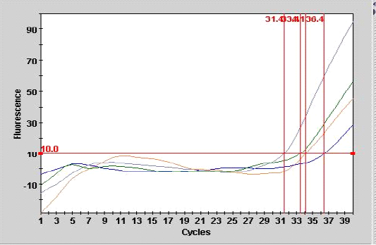
The current study showed that the one-step RRT-PCR was highly sensitive and reliable technique for detection the matrix gene (M gene) by using forward primer (M25F) and reverse primer (M124R) and specific probe (FAM M64), this finding was in agreement with the with many previous works (Jackwood and Sommer, 2002; Mosleh et al., 2009) who found that because of the high speed and relatively low cost and sensitive methods, there is considerable interest in using RT-PCR-based methods directly targeted to the different subtypes of AIV as primary diagnostic tests. In contrast Mosleh et al., (2009) found that by using real time PCR for detection LPAI H9N2 in chickens experimentally infected with AIV, the presence of the virus is restricted to the trachea and lung with ratio of 40% and 20% respectively. Inconsistent to above mentioned result, Swayne and Halvorson, (2003) stated that the LPAI viruses produce infections that are localized to the respiratory and gastrointestinal (GI) tracts of chickens indicating that H9N2 AI virus is pneumotropic and nephrotropic following IN inoculation.
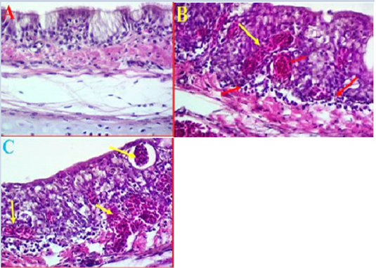
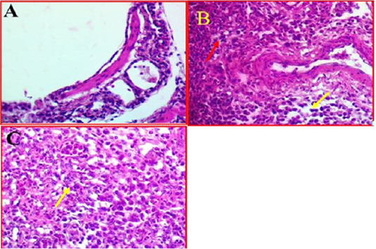
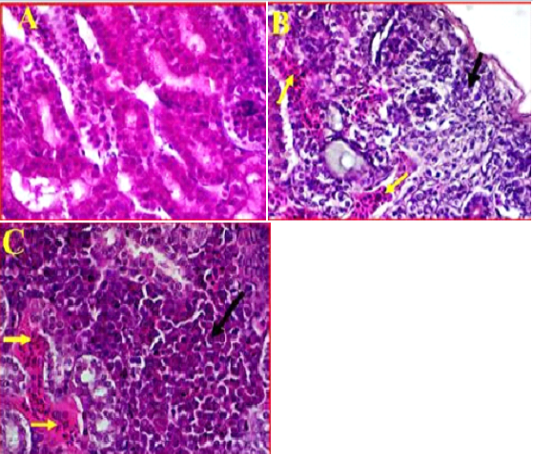
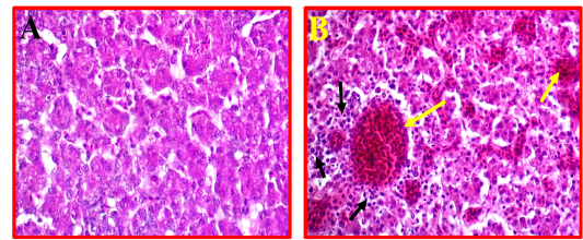
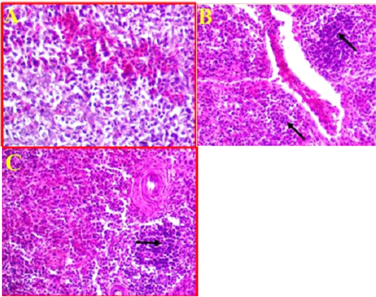
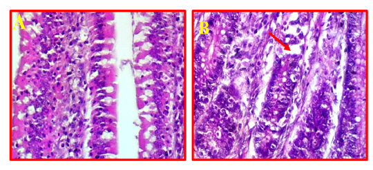
Avian influenza viruses can induce two different types of pathological changes based on their virulence and pathogenicity (Gharaibeh, 2008; Subtain et al., 2011; Eladl et al., 2019). The present study investigated on the pathophysiological impacts of H9N2 virus on certain tissues. In this study, the common obvious finding to emerge from the histological examinations was that a clear inflammation and necrosis accompanied with infiltration and congestion of trachea, lung and kidney (interstitial tubular nephritis), and to a lesser extent observed in liver and spleen. In accordance with the present results, previous studies worked on H9N2 found that tracheitis, pneumonia and pancreatitis were the most frequent and prominent histological changes (Gharaibeh, 2008; Aslam et al., 2015). In the current findings indicated that infected chickens with H9N2 virus caused sever tubular atrophy and nephritis which broadly supports the work of other studies in this area linking the majority of death of poultry farms to renal damages (Hadipour et al., 2011). It can thus be suggested that these results provide further support for the hypothesis that there is a straight pathogenic and pathophysiologic mechanism in the respiratory system and/or urinary system. In this study, experimental infected chickens with H9N2 were found to cause multifocal necrosis with mild to moderate congestion and edema, in addition to infiltration with mononuclear inflammatory cells in trachea, lung, kidney, spleen and liver simultaneous with histological lesions reveal direct pathologic impacts of H9N2 virus (Hablolvarid et al., 2004; Hassan et al., 2017).
It was concluded from above mentioned findings that H9N2 local isolate of AIV produced mild infections in both vaccinated and unvaccinated broilers with available commercial vaccine, but the vaccine reduced the severity of the infection and the mortality. It also led to high levels of anti H9N2 antibodies that might have the key role of protection. Circulation of other influenza A viruses in the same area, and especially the high pathogenic strain may led to emerging of new strains of AIV due to reassortment or genetic shift and drift. Accordingly, applying of epidemiological molecular and serological studies to screen different Iraqi provinces for the presence of HPAIV and LPAIV is too important.