ResearchersLinks

Short Communication · Volume 14, Issue 4, 16 September 2026 · Open Access

Molecular Detection and Histopathological Investigations of Sarcocystis Species in Domestic Chicken (Gallus gallus domesticus) Meat

·

Affiliations & Notes ⌄
  1. 1Department of Biology, Faculty of Education, University of Al-Qadisiyah, Iraq. *Correspondence | Ahmed Mohammed Kadhim, Department of Biology, Faculty of Education, University of Al-Qadisiyah, Iraq
  2. 2Email: edu.bio.posta53@qu.edu.iq
Article Info ⌄
Journal:
Journal of Animal Health and Production
Published:
16 September 2026

From the article

  • In the current study, a total of 59 samples, previously identified infected with Sarcocystis by both macroscopic and microscopic methods were collected for molecular diagnosis using nested-PCR.
  • The results of the current study showed 11 gross infections in the pectoral muscles (27.5%), 5 gross infections in the esophagus muscles (12.5%), and only one infection in the thigh muscles (2.5%) with total infection 14.1%.
  • The results of the current study indicated that there were no significant differences between the sex variable and the percentage of microscopic infection in poultry.
  • Sarcocystis wenzeli is prevalent in local chickens in Babylon Governorate, and nested-PCR proved to be efficient and accurate compared to traditional methods.

Selected from the article's own text.

Abstract

Sarcocystis, a genus of protozoan parasites, causes sarcocystosis, which is a zoonotic disease and is widespread around the globe. Sarcocystis species are obligate parasites requiring two hosts including a carnivorous definitive host and an herbivorous intermediate host such as a bird. It poses economic losses and public health risks through food and water contamination and may cause sudden death in the poultry. In order to assess the presence of sarcocystis, a total of 59 tissue samples including pectoral, esophagus and thigh muscles) from local chickens were collected. A total DNA was extracted from cysts and the presence of Sarcocystis was performed using nested-PCR targeting the 18S rRNA gene. The molecular detection and analysis indicated that nested-PCR detected infection in 37 of 59 (62.71%) samples. Specifically, we identified one species, Sarcocystis wenzeli, in higher abundance with a 750 bp gene band. The breast samples showed the highest infection rate (27.5%), followed by the esophagus (12.5%) and thigh (2.5%), with no significant differences by sex or location. Histological examination revealed lymphocytic aggregation, muscle fiber shrinkage, and vascular congestion with cysts containing bradyzoites. Taken together, Sarcocystis is prevalent in local chickens in Babylon Governorate, and nested-PCR proved to be efficient and accurate compared to traditional methods.
 
Keywords | Sarcocystis, Local chickens, Babylon Governorate, Nested-PCR, 18S rRNA gene
Introduction

Sarcocystis is a genus of protozoan parasites that causes sarcocystosis, also known as sarcosporidiosis . It is a zoonotic disease and is widespread throughout the world ; . Sarcocystis species are obligate parasites, requiring two hosts to complete their life cycle. The definitive host is a carnivorous animal, such as cats, dogs, or even humans. The intermediate host is an herbivore, such as birds, cattle, sheep, and even reptiles .

Infection of definitive hosts occurs through the ingestion of raw or undercooked meat containing mature cysts that are mechanically digested in the stomach and intestines. Intermediate hosts acquire infection through the consumption of food and water contaminated with sporocysts which are shed in the feces of the definitive host .

Sarcocystis is an opportunistic infection, and the parasite exists in various forms within the muscle tissues of its host, such as birds . Gross cysts are found within skeletal and cardiac muscles and exhibit several shapes depending on the species, some are thread-like in S. muris, rice-grain-like in S. rileyi, spindle-shaped in S. fusiformis, or spherical in S. gigantea . Large cysts visible to the naked eye are called macroscopic cysts, while small cysts visible only under a microscope are called microscopic cysts . Macroscopic cysts are milky white and can be seen within the muscle fibers of infected animals . Microscopic cysts are characterized by internal septa that form small compartments containing numerous slow-reproducing bradyzoites, they have a vesicular nucleus and a crescent shape . The parasite does not have a specific infection period; it can occur throughout the year. The cysts remain active outside the host for a more extended period in warm, humid environments compared to cold, dry ones.

Various types of animals are affected and in birds including chickens, it causes acute myositis, in addition to encephalitis, fever, loss of appetite and may sometimes lead to sudden death . The economic importance of the parasite lies in the fact that it causes significant annual financial losses, which are spent on control and treatment programs using anticoccidials and sulfa compounds. Despite this, the disease remains widespread and poses a threat to public health through water and food contamination .

This study is conducted to understand the presence of sarcocystis in domestic chickens, to identify associated pathologies and to ascertain the feasibility of molecular detection in screening and prevalence of Sarcocystis in the poultry.

Materials and Methods

The current study was executed between July and December 2024. Samples were collected from chickens in the city center and the districts of Babylon Governorate. Molecular analysis of positive samples from macroscopic cysts and microscopic samples was performed using nested PCR to detect the Sarcocystis spp.

Total DNA was extracted using a specialized extraction kit previously manufactured by Add Bio, Korea. DNA was extracted according to the company’s instructions. After DNA extraction, DNA purity was measured using a nanodrop spectrophotometer. The nested PCR master mix was prepared using the GoTaq Green PCR Master Kit according to the instructions provided by Promega, USA, and then placed in a PCR thermocycler. Subsequently, the master mix for the second round of nested PCR was prepared and placed in a PCR thermocycler. The primers used to detect the parasite are provided in the Table 1. The products of the cross-linked polymerase chain reaction were analyzed by electrophoresis on an agarose gel.

Results

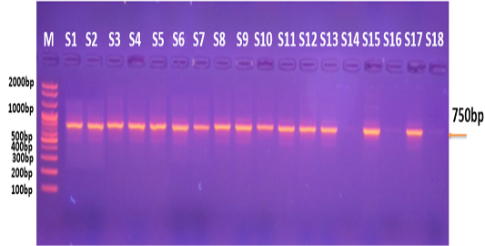
In the current study, a total of 59 samples, previously identified infected with Sarcocystis by both macroscopic and microscopic methods were collected for molecular diagnosis using nested-PCR. These 59 samples were amplified using primers specific to the 18SrRNA gene of Sarcocystis species, which infects local chickens. Following electrophoresis on a 2% agarose gel, as shown in Figure 1, the reaction results were obtained, revealing 37 positive samples (62.71%).

The results of the current study showed 11 gross infections in the pectoral muscles (27.5%), 5 gross infections in the esophagus muscles (12.5%), and only one infection in the thigh muscles (2.5%) with total infection 14.1%. The results also showed no significant differences (P < 0.05) in gross infection rates between the organs infected with the parasite, as shown in Table 2.

Table 1: Primers used in Sarcocystis molecular identification.

Primers

Sequence 5’-3’

Product size

PCR gene Primer

F= AAGTATAAGCTTTTATACGGCGAAA

688bp

R= TCGCAGTAGTTCGTCTTTAACAAA

Nested PCR gene Primer

F= TGGATAACCGTGGTAATTCTATG

456bp

R= TCCCTATTAATCATTACTTCAGTCCTA

Table 2: The gross infection rates and numbers of Sarcocystis in different organs of chickens.

P. value

X value

Percentage ( %)

Gross infections No.

Examined No.

Organs

0.0138

8.5688

27.5

١١

40

Pectoral muscles

12.5

٥

40

Esophagus muscles

2.5

١

40

Thigh muscles

14.1

17

120

Total

*Significant variations (P<0.05) between organs.

Table 3: Numbers and percentage of Sarcocystis infection according to sex.

P. value

X value

Percentage %

Infected No.

Examined No.

Sex

N.S 0.4669

0.5294

20

10

٥٠

Male

10

7

٧٠

Female

14.1

17

١٢٠

Total

*No significant variations (P<0.005) between gross infected sex.

Table 4: The percentages and numbers of gross infections distributed by geographical location in Babylon Governorate.

P. value

X value

Percentage ٪

Infected No,

Examined No.

Region

N.S 0.8856

0.6471

16.6%

5

٣٠

Al-Hashimiya

16.6%

5

٣٠

Al-Mahawil

10%

3

٣٠

Hillah

13.3%

4

٣٠

Al-Musayyib

14.1

17

١٢٠

Total

*No significant variations ( P<0.005) between regions.

The study results also showed no statistically significant differences (P < 0.05) in the percentage of gross infected of chickens (males and females) with the Sarcocystis. Ten gross infective cases were recorded in males (20%), while seven gross infective cases were recorded in females (10%), as shown in Table 3.

The current study, which compared the sample collection areas in Babylon Governorate for gross infections, showed no statistically significant differences at a significance level (P<0.05). The number of infections was recorded as follows: in Al-Hashimiya District, there were 5 gross infections (16.6%); in Al-Mahawil District, there were also 5 infections (16.6%); in the governorate’s capital (Hillah), there were 3 infections (10%); and in Al-Musayyib District, there were 3 infections (13.33%), as shown in Table 4.

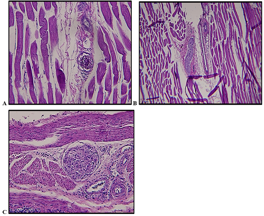
Histological study of the breast muscles in chickens revealed infection with the Sarcocyst is. This study revealed a high concentration of lymphocytes surrounding the muscle fibers, accompanied by contraction of the transverse muscle fibers and congestion of blood vessels. It also indicated programmed cell death of myofibroblasts, (Figure 2). In the thigh muscles, cysts containing numerous bradyzoites appeared. These oval cysts were located near the blood vessels surrounding the muscle fibers, causing fibrosis and damage, as well as an accumulation of lymphocytes near the infected tissue.

The pathological changes observed in the esophageal muscles of chickens infected with the Sarcocystis included the presence of oval-shaped cysts within and between the tissue layers. Congestion and infiltration of inflammatory cells were observed in the blood vessels, with inflammatory cells accumulating near the parasite cyst (Figure 2).

Discussion

The results of the current study demonstrated the molecular detection of 59 positive samples (macroscopic and microscopic) using nested-PCR technology based on the small subunit gene of ribosomal RNA. This method is characterized by its high sensitivity compared to conventional PCR.

The current molecular study results indicated that 37 out of the 59 samples tested using nest-PCR were positive, representing 62.7% of the positive samples, while 37.3% were negative. The presence of some negative samples is attributed to several factors, including storage conditions, inaccurate microscopic identification of some samples, errors in the DNA extraction process, mistakes in one of the stages of the DNA amplification program, the presence of inhibitors that bind to the polymerase and thus hinder its reading by PCR, or sample damage due to temperature fluctuations that may impede the amplification process.

These results are not significantly different from some previous studies that recorded high rates of molecular detection, such as the study by Sazmand et al. (2025), which recorded a rate of 94.78% in local chickens in Iran, and Vahedi et al. (2019) in his study of the Sarcocystis in local poultry in Iran, where the infection rate reached 96.5%.

The difference in infection rates across age groups may be attributed to the weakened immune systems of older adults, making them more susceptible to infection. Significant differences in infection rates were recorded between different age groups at a significance level (P>0.005). The results presented in the current study are consistent with those of Vahedi et al. (2019) study on detecting the Sarcocystis muscariformis in local poultry in Mazandaran, Iran. The study targeted age groups and recorded infection rates of 80% in the under-6-months age group, 97.91% in the over-6-months age group, and 100% in the over-1-year age group. The results of the current study indicated that there were no significant differences between the sex variable and the percentage of microscopic infection in poultry. This suggests that the reason may be that the parasite is not inclined towards a particular sex, and the spread of pathogens is equal to both sexes. This study differed from, Mac et al. (2018), where the percentage of infection with the Sarcocystis in female poultry was higher than in males (67.5% in females and 32.5% in males).

The concentration of macroscopic infection in the pectoral muscles (27.5%) and esophagus (12.5%) is attributed to the physiological activity of these muscles. Sarcocystis preferentially in tissues with high blood supply and continuous motor activity, such as the respiratory and swallowing muscles. This explains the lower infection rate in the thigh muscles (2.5%) compared to the pectoral muscles . The parasite is also an obligate parasite of the intermediate host (chicken) to form cysts, which may be macroscopic or microscopic depending on the species .

Histological examination revealed a dense accumulation of lymphocytes around the muscle fibers, which is attributed to the host’s inflammatory immune response to the presence of the parasitic cysts . Muscle fiber contraction and vascular congestion result from the mechanical pressure exerted by cyst growth within the muscle fiber, in addition to the parasite’s metabolic byproducts that induce programmed cell death . The presence of slow-reproducing stages (bradyzoites) within oval cysts near blood vessels causes fibrosis and damage to surround tissues due to infiltration of inflammatory cells .

Conclusion

Sarcocystis wenzeli is prevalent in local chickens in Babylon Governorate, and nested-PCR proved to be efficient and accurate compared to traditional methods.

Acknowledgment

The authors express their gratitude and appreciation to the Department of biology at the College of Education, University of Al-Qadisiyah, Iraq, for providing all the necessary laboratory facilities and technical support to complete this manuscript.

Novelty Statement

This study represents the first molecular and histopathological identification of Sarcocystis wenzeli in domestic chickens in Babylon governorate, Iraq. Using nested-PCR, we report a high prevalence (62.71%) and provide critical evidence linking specific muscle tissue damage to bradyzoite cysts, highlighting previously undocumented public health and economic risks in the region.

AUTHOR’S CONTRIBUTIOn

Conceptualization: AMK and KTM. Methodology and laboratory analysis: AMK. Data curation and histopathological examination: KTM. Writing original draft preparation: AMK. Writing review and editing: KTM. All authors have read and agreed to the published version of the manuscript.

Generative AI and AI assisted technology statement

The authors declare that no generative AI and AI assisted technology was used in the creation of this manuscript.

Conflict of interest

The authors have declared no conflict of interest.

Figures

Figure 1:
Figure 1:.

Electrophoresis of the Nested-PCR product on agarose gel for the 18SrRNA gene of Sarcocystis extracted from local chicken tissue samples. The DNA sequence is M-track, with a 100-base-pair run. The S1-S18 trajectory represents the PCR results for 18 chicken tissue samples. A 750-base-pair (BPG) specificity band was successfully amplified. 

Figure 2:
Figure 2:.

Cross-sections of chicken muscle tissues infected with Sarcocystis spp. (A) Thigh muscle: An oval-shaped cystic stage is observed near the blood vessels surrounding the muscle fibers (40x). (B) Pectoral muscle: Congestion of blood vessels with accumulated inflammatory cells and destruction of muscle fibers are observed (40x). (C) Esophageal muscle: Congestion of blood vessels and inflammatory cell infiltration are observed (40x).

References

  1. Bahari P, Salehi M, Seyedabadi M, Mohammadi A (2014). Molecular identification of macroscopic and microscopic cysts of sarcocystis in sheep in north khorasan province, iran. Int. J. Mol. Cell. Med., 3(1): 51–56. https://pubmed.ncbi.nlm.nih.gov/24551821/
  2. Dubey J, Moré G, Van Wilpe E, Calero-Bernal R, Verma S, Schares G (2016). Sarcocystis rommeli n. sp. (Apicomplexa: Sarcocystidae) from cattle (Bos taurus) and its differentiation from Sarcocystis hominis. J. of Eukaryot. Microbiol., 63: 62–68. https://doi.org/10.1111/jeu.12248[CrossRef]
  3. Dubey J, Speer C, Charleston W (1989). Ultrastructural differentiation between sarcocystis of Sarcocystis hirsuta and Sarcocystis hominis. Vet. Parasitol., 34(1-2): 153–157. https://doi.org/10.1016/0304-4017(89)90177-5[CrossRef]
  4. Farhang-Pajuh F, Yakhchali M, Mardani K (2014). Molecular determination of abundance of infection with Sarcocystis species in slaughtered sheep of Urmia, Iran. Vet. Res. Forum Int. Quart. J., 5: 181–186. https://pmc.ncbi.nlm.nih.gov/articles/PMC4279651/
  5. Fayer R (2004). Sarcocystis spp. in human infections. Clin. Microbiol. Rev., 17: 894–902. https://doi.org/10.1128/CMR.17.4.894-902.2004[CrossRef]
  6. Fayer R, Esposito D, Dubey J (2015). Human infections with Sarcocystis species. Clin. Microbiol. Rev., 28: 295–311. https://doi.org/10.1128/CMR.00113-14[CrossRef]
  7. Gareh A, Soliman M, Saleh A, El-Gohary F, El-Sherbiny H, Mohamed R, Elmahallawy E (2020). Epidemiological and histopathological investigation of Sarcocystis spp. in slaughtered dromedary camels (Camelus dromedarius) in Egypt. Vet. Sci., 7: 162. https://doi.org/10.3390/vetsci7040162[CrossRef]
  8. Kirillova V, Prakas P, Calero-Bernal R, Gavarāne I, Fernández-García J, Martínez-González M, Rudaitytė-Lukošienė E, Martínez-Estéllez M, Butkauskas D, Kirjušina M (2018).Identification and genetic characterization of Sarcocystis arctica and Sarcocystis lutrae in red foxes (Vulpes vulpes) from Baltic States and Spain. Parasit. Vectors, 11: 1–9. https://doi.org/10.1186/s13071-018-2694-y[CrossRef]
  9. Mac PA, Ibrahim TM, Buba DM, Airiohuodion PE, Ambu S (2018) Prevalence of Sarcocystis Species in Meat of Cattle, Pigs and Birds slaughtered in North Central Nigeria. J Vet Sci Med Diagn 7:5.
  10. Mutalib A, Keirs R, Maslin W, Topper M, Dubey J (1995). Sarcocystis-associated encephalitis in chickens. Avian Dis., 39: 436-440. https://doi.org/10.2307/1591891[CrossRef]
  11. Sazmand A, Moradi L, Almasi P, Nabavi R, Prakas P (2025). First report of Sarcocystis halieti in Asia: The genetic confirmation in muscles of the Eurasian sparrowhawk (Accipiter nisus) from Iran. Sci. Rep., 15: 6989. https://doi.org/10.1038/s41598-025-90666-0[CrossRef]
  12. Swar S, Shnawa B (2021). Recent advances in molecular characterization of Sarcocystis species in some meat-producing animals: An updated review. Asian J. Agric. Biol., 1.
  13. Vahedi NN, Salehi A, Razavi M, Masoumi M (2019). Poultry carcasses investigation of parasitic Sarcocystis infection in native in Mazandaran (North part of Iran). Anim. Husb. Dairy Vet. Sci., 3: 1–3. https://doi.org/10.15761/AHDVS.1000165[CrossRef]

Services

Article usage statistics

Loading…