
The present study was conducted to detect, isolate and characterize the Newcastle disease virus (NDV) in ducks. For this purpose, a total of 100 samples of cloacal and 100 oropharyngeal swabs of ducks (Anas Platyrhynchos domesticus) were collected from different villages of four tahsils of Sheikhupura during period (May to July 2017). NDV was isolated from field samples by inoculating the suspected samples into embryonated eggs of 9-11 days. Isolated virus was confirmed through haemagglutination and haemagglutination inhibition test. Virus isolation rate of NDV from cloacal swab was 12% (6/50) and from oropharygeal swab was 8% (4/50). The 238-bp region of F gene of NDV was amplified by using specific primers of reverse transcriptase-polymerase chain reaction (RT-PCR). Out of 50 field samples 11 cloacal swabs (34%), were positive and out of 50, nine oropharyngeal swabs (18%) were positive for NDV through RT-PCR. DNA sequencing was performed by amplifying the RT-PCR amplicon targeted the F gene. Phylogenetic tree was constructed. It was concluded that virulent strains were existed in ducks and more than one genotype of NDV was prevalent in ducks.
Short Communication · Volume 52, Issue 1, 13 November 2019 · Open Access
Detection and Molecular Characterization of Virulent Newcastle Disease Virus in Ducks (Anas platyrhynchos domesticus)
· · · · · · · · ·
Affiliations & Notes ⌄
- 1Department of Pathobiology, KBCMA, CVAS, Sub Campus University of Veterinary And Animal Sciences, Narowal 2Department of Zoology, University of the Punjab, Lahore 3Department of Pathology, University of Veterinary and Animal Sciences, Lahore 4Department of Biotechnology, Baluchistan University of Information Technology, Engineering Management Science, Quetta 5Department of Zoology, University of Education, Lahore 6ADL Office,16 Cooper Road, Lahore 7IMBB Department, University of Lahore , * Corre
Article Info ⌄
- Journal:
- Pakistan Journal of Zoology
- Published:
- 13 November 2019

From the article
- Virus isolation rate of NDV from cloacal swab was 12% (6/50) and oropharnygeal swab was 8% (4/50).
- Out of 50 field samples 11 cloacal swabs (34%), were positive and out of 50, nine oropharyngeal swabs (18%) were positive for NDV through RT-PCR (Supplementary Fig. 1).
- It is concluded virulent strain of Newcastle disease virus existed in domestic ducks of district Shiekhupura.
- More than one genotype of NDV prevalent in ducks and carrier ducks are the one of the source of transmission of virus in backyard chicken.
Selected from the article's own text.
Abstract
Materials and methods
A total of 100 samples of cloacal swabs and oropharyngeal swabs were collected from ducks (Anas Platyrhynchos domesticus), from different villages in four different Tahsil (Sheikhupura, Muridke, Sharqpor, Ferozwala) of district Sheikhupura. The four villages were named as Kotnoor shah, ila star shah, Khanpur, BurjAttari and Chak-23. Samples were collected from May to July 2017. The collected samples were preserved under refrigeration and were processed for molecular diagnosis and virus isolation.
The fifty collected samples of cloacal swab and oropharyngeal swabs were processed for virus isolation. Swabs were dipped in tube contained PBS solution to the 1ml as well as antibiotics (10,000 1U/mL penicillin, 1 mg/ml streptomycin sulphate, 1 mg/ml gentamicin sulphate). For isolation of NDV virus, 9 day old embryonated chicken eggs were inoculated through allantoic cavity at dose rate of 0.2 ml of NDV inoculum having titer of (ELD50 107.32 /100µl) (0.1ml virus suspension + 0.1ml antibiotic mixture) ; . Allantoic fluid was examined by using egg Candler (bright light in dark room), and mortality of embryonated inoculated eggs observed after 24hrs to 48hrs of incubation . Allantoic fluid was also confirmed through RT-PCR.
Haemagglutination inhabitation test was performed to each row of microtiter plate. 25µl of phosphate buffered saline and 25µl of the chicken serum was also dispensed into the first well only. Serial dilutions of the sera were made. A volume of 25µl of NDV containing 4 haemagglutination units was dispensed into all the wells. A positive control reference serum was included on the last row of the micro plate. At room temperature plates were incubated for 30 minutes and 25µl of 1 % chicken RBC was dispensed into each well. The plates were shaken gently and incubated at room temperature. The test was read after 45 minutes. End point was indicated by a red button of RBC at the bottom of the well while a negative test was indicated by a matt at the bottom of the well .
The fifty clocal and oropharyngealswabs were directly processed for molecular detection. The viral RNA of the ND viruswas extracted from FavorPrep™Viral Nucleic Acid Extraction Kit (Favorgen, FisherBiotech, Australia). The RNA was extracted in 50 μl of elution buffer and used as a template directly for RT-PCR assay and stored at -80ºC until further use.
A commercial cDNA synthesis kit (Fermantas,USA) was used to make cDNA. The procedure was adopted from instruction manual of manufacturer. To amplify a 238 bp fragment of F gene hypervariable region, Forward primer5’GCCCAGAGTCTACACCAT -3’ and Reverse primer 5’- CCCGGATTATGTCTTTGA -3’ were used. The amplification products were detectedby gel electrophoresis in 1.5% agarose gel in TAE buffer. Gels were run for 1.5 h at 80 Volts, stained with ethidium bromide (0.5μg/ml), exposed to ultraviolet light and photographed (Visi-Doc-It system, UVP, UK). A commercial 100-bp DNA ladder (Fermentas) was used as molecular-weight marker in each gel .
DNA sequencing was performed by amplifying the RT-PCR amplicon targeted the F gene. Five positive amplicon (ND1, ND2, ND3, ND4 and ND5) were sequenced. Phylogenetic tree was constructed. The data was analyzed by using NCBI BLAST and Bioedit software 7.2 V. Electropherograms of NDV samples obtained were compared with reference sequence present at NCBI. Any mutations present in the sequences were noted. All the 5 NDV sequences were aligned with each other to find the homology by use of online Clustal W software and the phylogenetic trees were constructed by use of Phylogenetic software MEGA 7. The reported sequences of different countries including Pakistan were retrieved from the NCBI and were compared with the sequences of NDV obtained in this research work. Phylogeny was formed to conclude the origin of NDV obtained from Pakistani ducks included in this study.
Results and discussion
A total of 100 cloacal and oropharyngeal field samples from ducks were collected. Virus isolation rate of NDV from cloacal swab was 12% (6/50) and oropharnygeal swab was 8% (4/50). For NDV isolation samples were inoculated in embryonated eggs through Allantoic cavity route. Embryonated eggs that showed mortality after 48 hours of incubation were selected for collection. In second and third passages 100% mortality observed in inoculated embryonated eggs after this time period due to virulent NDV . NDV was confirmed after each passage from triturated embryo and allantoic fluid (AF) through plate haemagglutination inhabitation test ; . Egg infectivity dose (ELD50) was estimated through the described procedure described by Reed and Muench (1938). Infectivity titer was 10 /100µl (107.32) which also reported that NDV could be propagated in embryonated chicken eggs ; . Newcastle disease virus was confirmed from field oropharyngeal and cloacal swab samples and AF after each passage through the reverse transcriptase polymerase chain reaction (RT-PCR), primers of 238 bp were used. Out of 50 field samples 11 cloacal swabs (34%), were positive and out of 50, nine oropharyngeal swabs (18%) were positive for NDV through RT-PCR (Supplementary Fig. 1). BK specific primers confirmed the NDV in F gene tissue samples . Molecular identification of NDV was done by using RT-PCR and it is more sensitive method. To identify the strain of NDV existed in field, DNA sequencing was done using five amplicons (ND1, ND2, ND3, ND4 and ND5) targeted F genes. The phylogenetic tree represent that sequence No5 has closeness with already reported sequences of virulent strain of NDV Shandong, India and Egypt. This sequence has some closeness with the virulent NDV (Supplementary Fig. 2). Sequences of F region of NDV of other countries like China, Russia, Spain. Israel, Chimalhualan, Finland, Muktesar, Mexico, Egypt, Belize, USA, Mexico, Shanxi, Liaoning. Beijing, Shandong, Guangxi, Luxemboung, India, Egypt, Hellongjian, Malko Tarnrovo retrieved from NCBI. Virulent NDV has very good similarity with the NDV isolates obtained from NARC Pakistan. Similarly, NDV 3 is also surrounded by China, India and Egypt isolates and virulent NDV and Pakistani NARC sequences are very close neighbors with each other but are far neighbors of sequences 3 and 5. The virulent NDV strain was isolated from healthy ducks ; . On the other hand, NDV isolates No 2&4 are close neighbors without any gap and these isolates are surrounded by China in phylogenetic tree while the sequence 1 is the most recent one and surrounded by already reported Pakistani sequences and new vaccines should be prepared for these isolates. Evolutionary analysis was conducted in MEGA7 (Fig. 1). The analysis involved 131 nucleotide sequences. Codon positions included were 1st+2nd+3rd+Noncoding. All positions containing gaps and missing data were eliminated. There were a total of 157 positions in the final dataset. The similarity matrix of all the five NDV isolated from ducks clearly shows that each sample is identical (ID) with its own sequence only, while it has different similarity matrix with each other. The sample No 1 has 0.828 value of similarity index with NDV No2 which shows 82.8%. The each similarity matrix value should be multiplied with 100 to get the %age similarity index and all five samples have variations of similarity ranging from 75%-97% (Fig. 2). The Clustal W alignment of all 5 NDV isolates also showing that all six NDV have a wide difference in similarity among each other, representing the fact the NDV F gene is undergoing in the process of mutations very rapidly (Supplementary Fig. 3) .
Conclusion
It is concluded virulent strain of Newcastle disease virus existed in domestic ducks of district Shiekhupura. More than one genotype of NDV prevalent in ducks and carrier ducks are the one of the source of transmission of virus in backyard chicken. The current research indicated more work should be done in NDV endemic areas. Due to rapid mutations in NDV, even vaccinated birds get infections with NDV and shows mortality. So there is need to make new vaccines.
Supplementary material
There is supplementary material associated with this article. Access the material online at: http://dx.doi.org/10.17582/journal.pjz/2020.52.1.sc1
Statement of conflict of interest
The authors declare no conflict of interest.
Figures
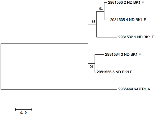
Evolutionary relationships of taxa. The evolutionary history was inferred using the Neighbor-Joining method. The optimal tree with the sum of branch length = 1.61196123 is shown. The percentage of replicate trees in which the associated taxa clustered together in the bootstrap test (1000 replicates) are shown next to the branches. The tree is drawn to scale, with branch lengths in the same units as those of the evolutionary distances used to infer the phylogenetic tree. The evolutionary distances were computed using the Maximum Composite Likelihood method and are in the units of the number of base substitutions per site. The analysis involved 6 nucleotide sequences. All positions containing gaps and missing data were eliminated. There were a total of 212 positions in the final dataset. Evolutionary analyses were conducted in MEGA7.
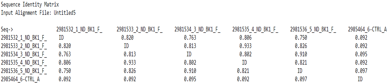
The similarity matrix of all the five NDV isolated from ducks.
References
- Aldous, E.W. and Alexander, D.J., 2001. Avian Pathol., 30: 117-128. https://doi.org/10.1080/03079450120044515[CrossRef]
- Alexander, D.J., 2001. Br. Poult. Sci., 42: 5–22. https://doi.org/10.1080/713655022[CrossRef]
- Alexander, D.J. and Gough, R.E., 2003. In: (eds. Saif, Y.M., Barnes, H.J., Glisson, J.R., Fadly, A.M., McDougal, L.R. and Swayne, D.E.) Disease of poultry. 11th Edition, Iowa State University Press, Ames, pp. 63-87.
- Alexander, D.J. and Senne, D.A., 2008. In: (ed. Saif, Y.N.) Disease of poultry. 12th ed. IOWA, USA, Blackwell Publishing. p75-94.
- Bouzari, M., 2014. Vet. Res. Forum Int. Quart. J., 5: 145.
- Chen, H.T., Zhang, J., Sun, D.H., Zhang, J.L., Cai, X.P., Liu, X.T., Ding, Y.Z., Ma, L.N., Yang, S.H., Jin, L. and Liu, Y., 2008. Vet. Res. Commun., 32: 491-498. https://doi.org/10.1007/s11259-008-9052-z[CrossRef]
- Dai, Y., Cheng, X., Liu, M., Shen, X., Li, J., Yu, S., and Ding, C., 2014. BMC Vet. Res., 10: 164. https://doi.org/10.1186/1746-6148-10-164[CrossRef]
- Dawid, N., Edyta, W., Krzysztof, S., Boguslaw, S. and Zenon, M., 2013. PCR, 60:475-480.
- Goldhaf, T.M., 1980. Avian Dis., 24: 297-301. https://doi.org/10.2307/1589696[CrossRef]
- Kattenbelt, J.A., Meers, J., Gould, A.R., 2006. Vet. Microbiol., 114: 134-141.
- Khan, T.A., Rehmani, S.F., Ahmed, A., Lone, N.A. and Khan, M.N., 2012. Pakistan. J. Zool., 44: 443-448.
- Lanre, S.K., Omolade, O.A., Smaila, I., 2011. Afri. J. Biotech., 10: 4256-8.
- Mabiki, F.P., Mdegela, R.H., Mosha, R.D. and Magadula, J.J., 2013. J. Med. Pl. Res., 7:863-870.
- OIE, Office International des Epizootie, 2012. Newcastle disease. OIE Terrestrial Manual. Office International des Epizootie, New Brunswick
- Otim, O.M., Christensen, H. and Mukiibi, G.M., 2006. Trop. Anim. Hlth. Prod., 38: 285-289. https://doi.org/10.1007/s11250-006-4309-4[CrossRef]
- Panda, A., Hung, Z., Elankumaran, S., Rockemann, D.D. and Samal, S.K., 2004. Microbial. Pathog., 36: 1-10. https://doi.org/10.1016/j.micpath.2003.07.003[CrossRef]
- Rasool, M.H., Afzal, A.M. and Siddique, A.B., 2015. Pakistan J. agric. Res., 28: 200-8.
- Rena, B. and Peter, C.,., 2015. J. Vis. Exp., 97: 52421.
- Reed, L.J. and Muench, H., 1938. Am. J. Hyg., 27: 493-497. https://doi.org/10.1093/oxfordjournals.aje.a118408[CrossRef]
- Sadiq, M., 2004. World Poult., 20: 10-11.
- Seal, B.S., King, D.J,, Sellers, H.S., 2000. Dev. Comp. Immunol., 24: 257–268 https://doi.org/10.1016/S0145-305X(99)00077-4[CrossRef]
- Shabbir, M.Z., Abbas, M., Yaqub, T., Mukhtar, N., Subhani, A., Habib, H. and Munir, M., 2013. Virus Genes, 46: 309-315. https://doi.org/10.1007/s11262-012-0862-2[CrossRef]
- Wajid, A., Shafqat, F.R., Muhammad, W., Asma, B.A., Tasra, B., Saima, A., Kiril, M. and Dimitrov, C.L., 2016. Am. Soc. Microbil., 4: 1-2.
- Zhang, S., Wang, X. Zhao, C., Liu, D., Hu, Y.J. and Zhang, G., 2011. PLoS ONE, 6: e25000. https://doi.org/10.1371/journal.pone.0025000[CrossRef]
Services
Article usage statistics
Loading…