Research Article · Volume 14, Issue 11, 29 September 2026 · Open Access
Molecular and Pathological Characterization of Chicken Infectious Anemia Virus in Egyptian Broiler Flocks with Runting–Stunting Syndrome
· · · ·
Affiliations & Notes ⌄
1Department of Poultry Diseases, Faculty of Veterinary Medicine, Cairo University, Giza, 12211 Egypt
2Genome Research Unit, Animal Health Research Institute (AHRI), Agriculture Research Center (ARC), Dokki, Giza, 12618 Egypt. *Correspondence | Omnia Mohamed Khattab, Genome Research Unit, Animal Health Research Institute (AHRI), Agriculture Research Center (ARC), Dokki, Giza
CIAV was detected in four of the 100 investigated commercial broiler farms showing clinical signs consistent with runting–stunting syndrome.
Two CIAV isolates were successfully propagated in one-day-old SPF chicks and were associated with anemia, lymphoid depletion, bone-marrow hypocellularity, and characteristic gross and histopathological lesions.
Whole-genome analysis showed that the two isolates were identical to each other and closely related to previously reported Egyptian CIAV strains, while also showing substantial nucleotide similarity to the Del-Ros vaccine strain.
These findings confirm the continued circulation of CIAV among the investigated Egyptian broiler flocks and support its potential contribution to immunosuppressive disease complexes in young chickens.
Selected from the article's own text.
Abstract
Chicken infectious anemia virus (CIAV) is an important immunosuppressive pathogen of poultry worldwide. This study investigated CIAV in Egyptian broiler flocks showing clinical signs consistent with runting–stunting syndrome (RSS). Between January 2023 and March 2024, samples were collected from 100 commercial broiler farms in ten Egyptian provinces and screened for CIAV DNA by conventional PCR. Four farm-level pooled samples, originating from Giza and Beheira provinces, were PCR-positive. Tissue homogenates from the four positive cases were inoculated intramuscularly into one-day-old specific-pathogen-free (SPF) chicks, and CIAV was successfully propagated from two samples. Experimental infection was associated with anemia, lymphoid depletion, bone-marrow hypocellularity, and characteristic gross and histopathological lesions. CIAV-specific antibodies were detected by ELISA following experimental infection. Whole-genome sequences of two isolates, deposited in GenBank under accession numbers PV686782 and PV686783, were identical to each other and clustered within genotype III according to the classification used in this study. They showed 99.8–100% nucleotide identity with selected previously reported Egyptian strains, 98.4% identity with the Del-Ros vaccine strain, and lower identity with selected Chinese and Australian strains. The findings confirm the circulation of CIAV among the investigated Egyptian broiler flocks and highlight the importance of continued molecular surveillance and investigation of its contribution to immunosuppressive disease complexes in broilers.
Chicken infectious anemia (CIA) is an economically important immunosuppressive disease of poultry caused by chicken infectious anemia virus (CIAV). Infection can result in growth retardation, increased mortality, higher susceptibility to secondary infections, poor vaccination responses, increased antimicrobial use, and carcass condemnation, particularly when young or immunologically susceptible birds are affected , 2017; . CIAV infection in broiler chickens can therefore contribute to considerable health and production problems. Both horizontal and vertical transmission play important roles in maintaining the virus within poultry populations .
CIAV was first reported in Japan in 1979 . In Egypt, the virus was first identified in 1990 , and subsequent studies have documented its continued detection in commercial chicken flocks ; , 2014; . More recent studies have focused on the molecular detection and genetic characterization of CIAV circulating in Egyptian poultry, providing further information on its epidemiology and genetic diversity ; , 2024; ; .
Chicken infectious anemia virus belongs to the genus Gyrovirus within the family Anelloviridae . It is a non-enveloped virus with a circular, single-stranded DNA genome of approximately 2,298–2,319 nucleotides . The CIAV genome contains three major partially overlapping open reading frames (ORFs) encoding the viral proteins VP1, VP2, and VP3, with reported coding lengths of approximately 1,350, 651, and 366 nucleotides, respectively . VP1 is the major capsid protein and is an important target of neutralizing antibodies. It also contains variable regions associated with viral genetic diversity and has therefore been widely used for molecular characterization of CIAV strains ; Schat and van Santen, 2008). Considerable amino acid variation has been reported within VP1, particularly around residues 139–151.
Several phylogenetic classification systems have been proposed for CIAV. Earlier VP1-based analyses identified three major genotypes, designated I, II, and III . Genotypes II and III have been reported widely, whereas genotype I was historically associated predominantly with Australian isolates . Subsequently, an additional genotype, designated genotype IV, was proposed and was suggested to have arisen through recombination between viruses assigned to genotypes II and III . Because CIAV genotype nomenclature has varied among studies, genotype assignments should be interpreted in relation to the reference sequences and classification approach used in each phylogenetic analysis.
VP2 is a multifunctional non-structural protein with dual-specificity phosphatase activity and contributes to the proper folding and assembly of the VP1 capsid protein . VP3, commonly known as apoptin, induces apoptosis in susceptible cells, including hematopoietic cells and thymocytes, and is closely associated with CIAV pathogenesis and immunosuppressive effects .
Chickens are considered the principal natural host of CIAV, although serological and molecular evidence of gyrovirus exposure or detection has also been reported in other avian species . Infection may occur in either clinical or subclinical forms . Young susceptible chickens, particularly during the first few weeks of life, may develop anemia, growth retardation, weakness, and increased mortality. Typical gross lesions include thymic atrophy, bone-marrow depletion or pallor, and subcutaneous or muscular hemorrhages . Subclinical infection is also important because immunosuppression may impair vaccine responses and increase susceptibility to concurrent infections .
Accurate diagnosis of CIAV is important for surveillance and control in poultry populations. Polymerase chain reaction (PCR) has been widely used for molecular detection of CIAV in Egyptian poultry and provides a rapid and sensitive method for identifying viral DNA in clinical samples ; . For primary propagation of CIAV, bioassay in susceptible one-day-old specific-pathogen-free (SPF) chicks following intramuscular inoculation has been described as a sensitive approach (Schat and van Santen, 2020). Serological assays such as ELISA are also useful for monitoring CIAV antibody responses within poultry flocks. In addition, nucleotide sequencing and phylogenetic analysis allow investigation of the genetic relationships among circulating field strains, previously reported isolates, and vaccine strains, and can provide useful information on CIAV molecular epidemiology .
Pathologically, CIAV infection is characterised by lesions involving hematopoietic and lymphoid tissues. Reported findings include yellowish or gelatinous bone marrow, generalized lymphoid depletion, particularly in the thymus, liver enlargement or mottling, congestion, and hemorrhages affecting internal organs and skeletal muscles. Hemorrhagic lesions may also occur in the breast and thigh muscles, proventricular mucosa, heart, and subcutaneous tissues . Control of CIAV in commercial poultry relies largely on appropriate breeder immunization to ensure transfer of protective maternal antibodies to progeny. Breeder vaccination programs in Egypt have included CIAV vaccine strains such as Cux-1, Del-Ros, and 26P4 ; .
Runting–stunting syndrome (RSS) is a multifactorial condition affecting broiler chickens, particularly during early life, and is characterized by poor growth, uneven flock performance, ruffled feathers, and diarrhea . CIAV has previously been detected in broiler flocks affected by RSS in Egypt and has been proposed as one of several infectious agents that may contribute to the development or severity of this syndrome . However, detection of CIAV in RSS-affected birds does not by itself establish the virus as the sole cause of the syndrome. Accordingly, the present study aimed to detect and identify CIAV in Egyptian broiler flocks showing clinical signs consistent with RSS, propagate selected PCR-positive viruses in one-day-old SPF chicks, evaluate their hematological, serological, and pathological effects, and genetically characterize the recovered CIAV isolates in comparison with previously reported Egyptian field strains, international reference strains, and vaccine strains. This approach was intended to improve understanding of the molecular and pathological characteristics of CIAV circulating in Egyptian broiler flocks and to support further investigation of its role in immunosuppressive disease complexes and RSS.
Materials and Methods
Field sample collection and preparation
Field samples were collected from 100 commercial broiler farms comprising non-vaccinated flocks aged 1–4 weeks and located in ten Egyptian provinces: Giza, Menoufia, Beheira, Fayoum, Beni Suef, Qalyubia, Sharqia, Dakahlia, Assiut, and Minya. Sampling was conducted between January 2023 and March 2024 (Table 4). The investigated birds showed clinical signs including depression, pallor, growth retardation, diarrhea, and mortality. Postmortem examination revealed lesions including thymic atrophy, muscular hemorrhages, and pallor of the liver.
Table 1: Hematological changes in SPF chicks experimentally infected with CIAV at 14 and 21 days post-inoculation.
Parameter
Control
(n = 10)
14 dpi
(n = 10)
21 dpi
(n = 10)
P value
RBC (×10⁶/µL)
2.80±0.07
1.40±0.05
1.85±0.03
<0.001
Hb (g/dL)
12.94±0.03
11.74±0.04
10.73±0.02
<0.001
PCV (%)
26.15±0.47
15.30±0.45
16.22±0.47
<0.001
Values are presented as mean ± SD. Differences were considered statistically significant at P < 0.05.
Under aseptic conditions, tissue samples were collected from the liver, thymus, spleen, bone marrow, and bursa of Fabricius. The tissues were homogenized using a sterile mortar and pestle in phosphate-buffered saline (PBS; pH 7.4) containing Antibiotic-Antimycotic Solution (100×; Thermo Fisher Scientific, Cat. No. 15240062) to prepare a 20% tissue homogenate. Samples collected from each farm were pooled for PCR investigation. The homogenates were subjected to three freeze–thaw cycles and centrifuged at 3,000 rpm for 20 min. The resulting supernatants were transferred aseptically into sterile tubes and stored at −80°C until DNA extraction and conventional PCR testing.
DNA extraction and PCR detection of CIAV
Viral DNA was extracted from the prepared tissue homogenate supernatants using the GeneDireX Viral Nucleic Acid Isolation Kit (GeneDireX®, Inc., Taiwan; Cat. No. NA016-0100) according to the manufacturer’s instructions. The extracted DNA was used as template for amplification of a 418-bp CIAV genomic fragment using the forward and reverse oligonucleotide primers listed in Table 5. The primer binding region was reported at nucleotide positions 633–1052.
Table 2: Histopathological lesion scores in SPF chicks experimentally infected with CIAV strains 1 and 2 at 14 days post-inoculation.
Organ
Histopathological lesion
Strain 1 (Mean ± SD)
Strain 2 (Mean ± SD)
P value
Liver
Congestion
1.60 ± 0.55
1.60 ± 0.55
1.000
Hepatocellular degeneration
1.60 ± 0.55
1.40 ± 0.55
0.580
Necrosis
0.80 ± 0.45
0.60 ± 0.55
0.550
Lymphocytic infiltration
1.40 ± 0.85
1.40 ± 0.85
1.000
Spleen
Lymphocytic depletion
1.60 ± 0.55
1.80 ± 0.45
0.550
Necrosis
1.20 ± 0.45
1.40 ± 0.55
0.550
Thymus
Congestion
1.80 ± 0.45
1.60 ± 0.55
0.550
Lymphocytic depletion
2.00 ± 0.71
2.20 ± 0.84
0.690
Bone marrow
Cellular depletion
2.20 ± 0.45
2.40 ± 0.55
0.550
Bursa of Fabricius
Lymphocytic depletion
1.80 ± 0.45
2.20 ± 0.45
0.195
Necrosis
1.40 ± 0.55
1.80 ± 0.84
0.397
Hyperplasia of the lining epithelium
0.60 ± 0.55
0.80 ± 0.45
0.550
Histopathological lesions were scored on an ordinal scale from 0 to 4: 0 = normal, 1= mild, 2= moderate, 3= severe, and 4= extremely severe. Data are presented as mean ± SD. Differences were considered statistically significant at P < 0.05. No statistically significant differences were detected between strains 1 and 2 for the evaluated lesions at 14 dpi.
Table 3: Histopathological lesion scores in SPF chicks experimentally infected with CIAV strains 1 and 2 at 21 days post-inoculation.
Organ
Histopathological lesion
Strain 1, Mean ± SD
Strain 2, Mean ± SD
P-value
Liver
Congestion
1.80 ± 0.45
1.80 ± 0.45
1.000
Hepatocellular degeneration
2.60 ± 0.55
2.20 ± 0.84
0.397
Necrosis
1.80 ± 0.84
1.60 ± 0.89
0.720
Lymphocytic infiltration
2.40 ± 0.89
2.20 ± 0.45
0.670
Spleen
Lymphocytic depletion
2.20 ± 0.45
2.20 ± 0.45
1.000
Necrosis
2.00 ± 0.71
2.20 ± 0.45
0.610
Thymus
Congestion
2.20 ± 0.45
1.60 ± 0.59
0.095
Lymphocytic depletion
2.40 ± 0.55
2.60 ± 0.55
0.850
Bone marrow
Cellular depletion
2.60 ± 0.55
2.80 ± 0.45
0.550
Bursa of Fabricius
Lymphocytic depletion
2.40 ± 0.55
2.40 ± 0.55
1.000
Necrosis
2.40 ± 0.55
2.40 ± 0.55
1.000
Hyperplasia of the lining epithelium
2.00 ± 0.71
2.40 ± 0.55
0.350
Histopathological lesions were scored on an ordinal scale from 0 to 4: 0 = normal, 1= mild, 2= moderate, 3= severe, and 4= extremely severe. Data are presented as mean ± SD. Differences were considered statistically significant at P < 0.05. No statistically significant differences were detected between strains 1 and 2 for the evaluated lesions at 21 dpi.
Table 4: Summary of field samples collected from commercial broiler farms across ten Egyptian provinces between January 2023 and March 2024.
Province
Number of farms investigated
Mortality range (%)
Body-weight
range (g)
Birds sampled per farm
CIAV PCR results
Giza
54
2–10
200–700
10
2 positive farms
Beheira
8
3–6
300–500
10
2 positive farms
Fayoum
8
2–4
250–500
10
Negative
Menoufia
6
2–3
350–400
10
Negative
Minya
6
2–5
200–600
10
Negative
Sharqia
4
2–4
150–300
10
Negative
Dakahlia
4
3–4
250–400
10
Negative
Qalyubia
4
3–4
400–600
10
Negative
Beni Suef
3
1–3
350–500
10
Negative
Assiut
3
1–3
150–500
10
Negative
Total
100
—
—
10 per farm
4 positive farms
Body weight (B.W.) is expressed in grams. PCR results refer to farm-level pooled tissue samples.
Table 5: Oligonucleotide primer sequences used for CIAV detection and genome sequencing.
Primer
Oligonucleotide sequence (5′→3′)
Product size
Reference / Purpose
F
5’-CTA AGA TCT GCA ACT GCG GA-3’
418 bp
Hussein et al. (2016)- CIAV detection
R
5’-CCT TGG AAG CGG ATA GTC AT-3’
F1-1
5’ CCGAGTGGTTACTATTCCATCACC 3’
668 bp
Abdel-Mawgod et al. (2018)- CIAV genome sequencing
R1-668
5’ CAGCGATAGAGTGATTGTAATTCC 3’
F2-502
5’ TTCAGGCCACCAACAAGTTCACGG 3’
698 bp
R2-1200
5’ CCGCAATCAACTCACCGGCGATGG 3’
F3-1350
5’ ATGCAGCCCACGGACTCTTGCCGG 3’
900 bp
R3-2250
5’ CTGGGGGGGAATCCCCCCCAGGGG 3’
Primer sequences are presented in the 5′→3′ orientation. The F/R primer pair was used for conventional PCR detection of CIAV, while the remaining primer sets were used for sequencing of the CIAV genome.
PCR amplification was performed using EmeraldAmp® GT PCR Master Mix (Takara Bio Inc., Japan; Cat. No. RR310A) in a final reaction volume of 25 μL containing 12.5 μL of master mix, 5.5 μL of PCR-grade water, 1 μL of each primer, and 5 μL of template DNA. The thermal cycling conditions consisted of initial denaturation at 94°C for 5 min, followed by 35 cycles of denaturation at 94°C for 30 s, annealing at 50°C for 40 s, and extension at 72°C for 40 s, followed by a final extension at 72°C for 10 min.
PCR products were separated by electrophoresis on a 1.5% agarose gel, stained with ethidium bromide, and visualized using a transilluminator. Amplicon sizes were estimated using a GeneRuler 100 bp DNA Ladder (Thermo Scientific™, Thermo Fisher Scientific Baltics UAB, Lithuania; Cat. No. SM0243).
Experimental propagation of CIAV in one-day-old SPF chicks
For experimental propagation of CIAV, fifty one-day-old specific-pathogen-free (SPF) chicks obtained from Kom Oshim, Fayoum, were divided into five groups designated C1, C2, C3, C4, and negative control, with ten chicks per group. Each of the four experimental groups was inoculated intramuscularly with 0.1 mL of a 20% tissue homogenate prepared from one of the four PCR-positive field samples, according to the experimental approach described by Bahmaninejad et al. (2012). The negative-control group was inoculated with PBS only. The experiment was conducted in chicken isolators at the Experimental Research Center, BSL-3 facility, Animal Health Research Institute (AHRI), Agricultural Research Center (ARC), Dokki, Giza, Egypt. Chicks were maintained for 21 days under hygienic conditions with continuous lighting and ad libitum access to feed and water. All groups were monitored daily following inoculation for clinical signs, changes in body weight, and mortality.
Sample collection
Blood samples were collected at 14 and 21 days post-inoculation (dpi). One sample was collected into EDTA-containing tubes for hematological examination, while a second sample was collected for serum separation and stored at −20 °C until serological testing.
At 14 and 21 dpi, five chicks from each group were euthanized following sedation with diazepam at 0.5 mg/kg body weight administered intramuscularly. Postmortem examination was performed, and samples of bone marrow, thymus, liver, spleen, and bursa of Fabricius were collected for PCR, genetic characterization, and histopathological examination. Histopathological lesions were scored according to Gibson-Corley et al. (2013) using an ordinal scale of 0–4, where 0 represented normal tissue, 1 mild lesions, 2 moderate lesions, 3 severe lesions, and 4 extremely severe lesions.
Hematological examination
Blood smears were stained with Wright’s stain for differential leukocyte evaluation. Packed cell volume (PCV) was determined using the microhematocrit method described by Coles (1967).
Serological examination
Serum samples collected at 14 and 21 dpi were tested for CIAV-specific antibodies using a commercial CAV antibody ELISA kit (BioChek B.V., The Netherlands; Cat. No. CK126 CAV) according to the manufacturer’s instructions. The assay was considered valid when the mean negative-control absorbance was <0.3 and the difference between the mean positive- and negative-control absorbance values was >0.15. Samples with an S/P ratio ≥0.35, corresponding to an antibody titre ≥725, were considered positive .
Histopathological examination
Tissue samples were fixed in 10% neutral-buffered formalin, embedded in paraffin, sectioned at 5 μm, and stained with hematoxylin and eosin (H & E) according to standard procedures described by Suvarna et al. (2013).
Confirmation of CIAV propagation by PCR
Tissue samples collected after experimental inoculation were examined by conventional PCR to confirm the presence of CIAV DNA using the PCR protocol described above.
The original manuscript does not provide further detail under this heading, so I would not add specific tissues or time points unless the authors confirm them.
Whole-genome sequencing and phylogenetic analysis
PCR products selected for sequencing were purified from agarose gels using the QIAquick Gel Extraction Kit (QIAGEN GmbH, Germany). Sanger sequencing was performed using the primers listed in Table 5 and the BigDye® Terminator v3.1 Cycle Sequencing Kit (Applied Biosystems™, Thermo Fisher Scientific, USA) on an ABI 3500 Genetic Analyzer (Life Technologies, USA). Sequences were assembled and edited using Geneious software. Similarity searches were performed using NCBI BLAST/MegaBLAST. Representative CIAV field and vaccine strains were retrieved from GenBank, and multiple-sequence alignment and nucleotide identity analyses were performed using BioEdit . Phylogenetic analysis was conducted using the neighbor-joining method in MEGA6 with 1,000 bootstrap replicates .
Statistical analysis
Quantitative data were analysed using one-way analysis of variance (ANOVA), followed by Fisher’s least significant difference (LSD) post hoc test for multiple comparisons. Differences were considered statistically significant at P < 0.05.
Table 6: Reference and field CIAV strains retrieved from GenBank and used for sequence alignment and phylogenetic analysis.
Accession number
Strain
Country
Year
Sample source
Genome length (bp)
DQ991394
01-4201
USA
2007
Broiler chickens
2298
AF311892
98D02152
USA
2016
Broiler chickens
2298
MK089243
17SY0902
China
2019
Domestic cats
2298
AY040632
3-1P60
Malaysia
2003
—
2298
AY846844
TJBD40
China
2004
—
2298
JX260426
GD-1-12
China
2012
Human
2298
KU645512
HN1504
China
2016
—
2298
OQ116656
5-IM201910
China
2023
—
2298
JF507715
CIAV89-69
South Korea
2011
—
2298
OP038322
SD201103
China
2023
—
2298
PP354991
JS2007Yao
China
2024
—
2298
PP354992
JS2009Shi
China
2024
—
2298
OP038331
FJ21425
China
2023
—
2298
MT268633
El Beheira2019
Egypt
2021
—
2298
MH001553
CAV-EG-2
Egypt
2018
Broiler chickens
2298
MH001563
CAV-EG-27
Egypt
2018
Broiler chickens
2298
MH001562
CAV-EG-23
Egypt
2018
Broiler chickens
2298
M55918
Cux-1
Germany
1991
Chickens
2298
D10068
26P4
The Netherlands
—
Chickens
2319
PV686782
TO1-2024
Egypt
2024*
Broiler chickens
2298
PV686783
TO2-2024
Egypt
2024*
Broiler chickens
2298
*Strains PV686782 and PV686783 were isolated and sequenced in the present study. “—” indicates information not provided in the original table.
Results
Clinical signs and postmortem findings in field broilers
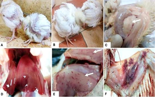
Broiler chickens aged 1–4 weeks from the investigated farms showed pallor of the combs, wattles, shanks, eyelids, and beaks, together with ruffled feathers, lethargy, general weakness, and growth retardation. Gross lesions included marked thymic atrophy, pallor of the liver and bone marrow, and hemorrhages in the breast and thigh muscles. Hemorrhages were also observed on the ventral surface of the wings in some affected birds (Figure 1).
The 100 investigated farms were distributed across ten Egyptian provinces. Giza accounted for 54 farms, followed by Beheira and Fayoum with eight farms each, Menoufia and Minya with six farms each, Sharqia, Dakahlia, and Qalyubia with four farms each, and Beni Suef and Assiut with three farms each. Reported flock mortality ranged from 1% to 10%, while the recorded body-weight ranges varied from 150 to 700 g according to province (Table 4).
Detection of CIAV in field samples by PCR
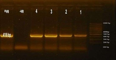
Conventional PCR analysis of pooled tissue samples from the 100 investigated farms detected CIAV DNA in four farm-level samples, giving an overall detection rate of 4%. Two positive samples originated from Giza and two from Beheira, whereas samples collected from Fayoum, Menoufia, Minya, Sharqia, Dakahlia, Qalyubia, Beni Suef, and Assiut were negative. Agarose gel electrophoresis demonstrated the expected 418-bp PCR products in all four positive samples. In Figure 2, lanes 1 and 2 correspond to positive samples from Giza and lanes 3 and 4 to positive samples from Beheira, together with the positive and negative controls and a 100-bp DNA ladder.
Experimental propagation of CIAV in one-day-old SPF chicks
The four PCR-positive field tissue homogenates were inoculated separately into SPF chick groups C1–C4. Successful CIAV propagation was demonstrated in two groups, C1 and C2, by detection of the expected 418-bp PCR product. Successful propagation was not reported for groups C3 and C4.
Clinical signs and gross lesions in experimentally infected chicks
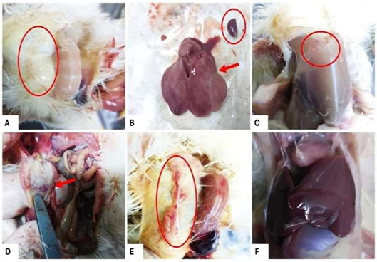
Chicks in the CIAV-positive groups developed general weakness, lethargy, anorexia, growth retardation, and pallor of the combs and wattles. Postmortem examination revealed severe thymic atrophy, an enlarged pale liver and spleen, yellowish fatty bone marrow, muscular hemorrhages, and hemorrhage at the junction between the proventriculus and gizzard. Figure 3 illustrates severe thymic atrophy, enlargement and pallor of the liver and spleen, petechial hemorrhages in the breast muscle, and hemorrhage at the proventriculus–gizzard junction. In contrast, the negative-control birds showed a normal thymus and liver.
Hematological findings
Hematological examination showed marked reductions in red blood cell counts and packed cell volume in the CIAV-infected chicks compared with the control group. The RBC count was 2.80 ± 0.07 × 10⁶/µL in controls, compared with 1.40 ± 0.05 × 10⁶/µL at 14 dpi and 1.85 ± 0.03 × 10⁶/µL at 21 dpi. Hemoglobin concentrations were 12.94 ± 0.03 g/dL in controls, 11.74 ± 0.04 g/dL at 14 dpi, and 10.73 ± 0.02 g/dL at 21 dpi. Similarly, PCV decreased from 26.15 ± 0.47% in the control group to 15.30 ± 0.45% at 14 dpi and 16.22 ± 0.47% at 21 dpi. Differences among the groups were reported as highly significant for RBC, Hb, and PCV (P < 0.001; Table 1).
Serological findings
CIAV-specific antibodies were detected by ELISA in serum samples collected from the two CIAV-positive experimental groups. At 14 dpi, the reported antibody titres were 877 and 853, while at 21 dpi they increased to 973 and 943, respectively. These values were above the stated ELISA positivity threshold of 725, confirming seroconversion following experimental infection.
Histopathological findings
Histopathological examination showed normal tissue architecture in the negative-control birds. In contrast, chicks infected with strains 1 and 2 developed lesions in the liver, spleen, thymus, bone marrow, and bursa of Fabricius. No statistically significant differences were detected between the two strains for the individual lesions evaluated at either 14 or 21 dpi (Tables 2 and 3).
At 14 dpi, the liver showed vascular congestion, vacuolar hepatocellular degeneration, focal necrosis, and lymphocytic infiltration. Congestion scores were 1.60 ± 0.55 for both strains, while hepatocellular degeneration scores were 1.60 ± 0.55 and 1.40 ± 0.55 for strains 1 and 2, respectively. Necrosis scores were 0.80 ± 0.45 and 0.60 ± 0.55, and lymphocytic infiltration was 1.40 ± 0.85 for both strains. None of these differences was statistically significant.
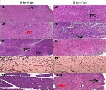
At 21 dpi, hepatic lesions were generally more pronounced. Congestion scored 1.80 ± 0.45 for both strains, hepatocellular degeneration scored 2.60 ± 0.55 and 2.20 ± 0.84, necrosis scored 1.80 ± 0.84 and 1.60 ± 0.89, and lymphocytic infiltration scored 2.40 ± 0.89 and 2.20 ± 0.45 for strains 1 and 2, respectively. Figure 4 illustrates vacuolar degeneration and focal lymphocytic infiltration in the liver, together with focal necrotic areas and marked hepatocellular degeneration.
The spleen showed lymphoid depletion and inflammatory cell infiltration at 14 dpi. Lymphoid-depletion scores were 1.60 ± 0.55 and 1.80 ± 0.45, while necrosis scores were 1.20 ± 0.45 and 1.40 ± 0.55 for strains 1 and 2, respectively. At 21 dpi, lymphoid-depletion scores increased to 2.20 ± 0.45 for both strains, while necrosis scores were 2.00 ± 0.71 and 2.20 ± 0.45. Histologically, marked capsular thickening and lymphocytic necrosis were also evident at the later time point (Figure 4).
The thymus showed cortical lymphoid depletion, congestion of medullary blood vessels, and a mild increase in Hassall’s corpuscles at 14 dpi. Congestion scores were 1.80 ± 0.45 and 1.60 ± 0.55, while lymphoid-depletion scores were 2.00 ± 0.71 and 2.20 ± 0.84 for strains 1 and 2, respectively. At 21 dpi, marked lymphoid depletion and loss of corticomedullary demarcation were evident. Congestion scores were 2.20 ± 0.45 and 1.60 ± 0.59, while lymphoid-depletion scores were 2.40 ± 0.55 and 2.60 ± 0.55. The difference in thymic congestion at 21 dpi approached, but did not reach, statistical significance (P = 0.095).
Bone marrow lesions were characterized by marked depletion of hematopoietic cells, increasing hypocellularity, focal aggregation of reticular cells, and replacement of hematopoietic tissue by adipose tissue. At 14 dpi, cellular-depletion scores were 2.20 ± 0.45 and 2.40 ± 0.55 for strains 1 and 2, respectively. At 21 dpi, these scores increased to 2.60 ± 0.55 and 2.80 ± 0.45. Figure 4 demonstrates moderate and severe bone-marrow hypocellularity, with prominent adipose replacement in the more severe lesion.
The bursa of Fabricius showed mild to moderate lymphoid depletion at 14 dpi, with scores of 1.80 ± 0.45 and 2.20 ± 0.45 for strains 1 and 2, respectively. Necrosis scores were 1.40 ± 0.55 and 1.80 ± 0.84, while epithelial hyperplasia scores were 0.60 ± 0.55 and 0.80 ± 0.45. At 21 dpi, lymphoid-depletion scores were 2.40 ± 0.55 for both strains, necrosis scores were also 2.40 ± 0.55 for both strains, and epithelial hyperplasia scores increased to 2.00 ± 0.71 and 2.40 ± 0.55. Histological examination also demonstrated severe follicular lymphoid depletion, follicular necrosis, epithelial hyperplasia, and cyst formation (Figure 4).
Overall, the lesion scores showed numerically greater severity at 21 dpi than at 14 dpi for several pathological parameters. However, Tables 2 and 3 provide statistical comparisons between the two strains at each individual time point rather than direct statistical comparisons between 14 and 21 dpi; therefore, the temporal increase should be described as a numerical or descriptive trend rather than a statistically significant increase.
Whole-genome sequence and nucleotide identity analysis
Complete genome sequences were obtained for two CIAV isolates, designated TO1-2024 and TO2-2024, and deposited in GenBank under accession numbers PV686782 and PV686783, respectively. Both genomes were 2,298 bp in length (Table 6).
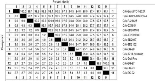
Pairwise nucleotide identity analysis showed that PV686782 and PV686783 were 100% identical to each other. The isolates showed 99.8–100% identity with the previously reported Egyptian strains CAV-EG-23, CAV-EG-22, and CAV-EG-27, 98.4% identity with the Del-Ros vaccine strain, and 95.7–96.4% identity with the Chinese strains SD2017 and SD2102. The lowest nucleotide identities reported were 94.5–94.8%, observed in comparison with the Australian strain CAV-3711 and the Egyptian strain CAV-EG-28 (Figure 5). The identity matrix further demonstrates the very close genetic relationship between the two isolates recovered in the present study.
Phylogenetic analysis
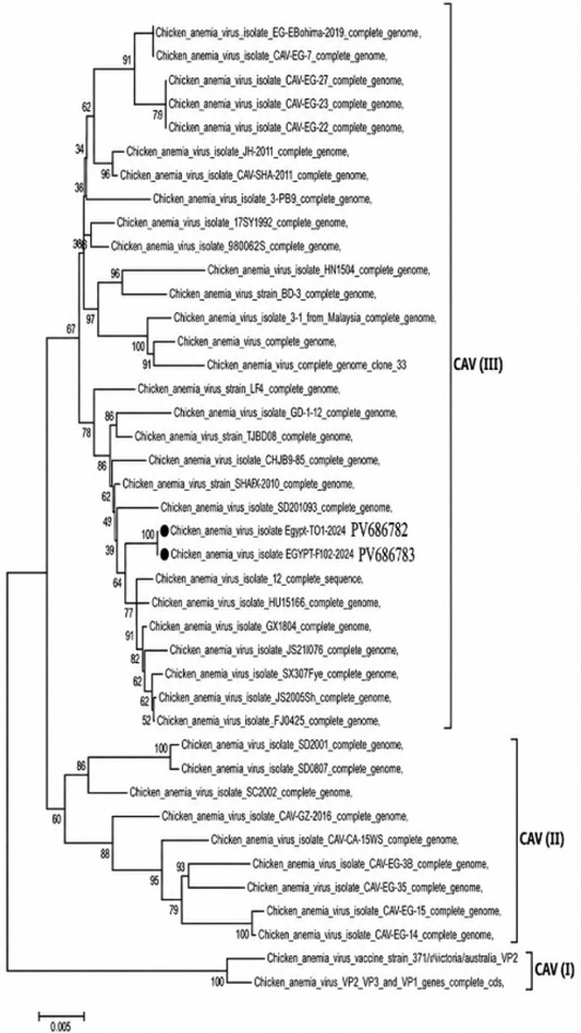
Whole-genome phylogenetic analysis grouped the two isolates obtained in the present study, PV686782 and PV686783, within genotype III according to the classification scheme applied in this analysis. The two isolates clustered closely together and within a broader cluster containing CIAV sequences from Egypt and other countries, including China, the USA, Malaysia, and South Korea, together with the Del-Ros vaccine strain. The sequences generated in the present study are indicated by black circles in the phylogenetic tree (Figure 6).
Discussion
Chicken infectious anemia virus (CIAV) remains an important immunosuppressive pathogen of poultry and can cause considerable production losses, particularly in young susceptible birds. Reported mortality is commonly in the range of 10–20%, although substantially higher mortality has been described in severe outbreaks ; . The virus mainly targets hematopoietic and lymphoid tissues, resulting in anemia, lymphoid depletion, and impaired immune competence ; . Consequently, CIAV-infected flocks may become more susceptible to secondary bacterial and viral infections and may show reduced responses to vaccination. In older birds, infection is often subclinical, but both horizontal and vertical transmission contribute to persistence of the virus within poultry populations ; .
In Egypt, CIAV has been associated with immunosuppression, vaccination failure, and recurrent disease problems in commercial poultry despite the use of breeder vaccines such as Del-Ros and 26P4. Maternal antibodies derived from vaccinated breeders are important for protecting young chicks during the period of greatest susceptibility. PCR remains one of the most sensitive and practical methods for detecting CIAV DNA in tissues from suspected cases. In the present study, conventional PCR detected the expected 418-bp amplicon in pooled tissue samples from four broiler farms in Giza and Beheira, in agreement with previous reports demonstrating the usefulness of PCR for CIAV detection in Egyptian poultry , 2016).
Bioassay in one-day-old SPF chicks has been described as a sensitive approach for primary propagation of CIAV (Schat and van Santen, 2003). In the present study, two of the four PCR-positive field samples were successfully propagated in SPF chicks and subsequently confirmed by PCR. The infected chicks developed clinical signs consistent with CIAV infection, including weakness, anemia, growth retardation, feather ruffling, and pallor of the shanks, beaks, combs, wattles, and eyelids. Similar clinical manifestations have been reported in experimentally and naturally infected chickens ; .
The gross lesions observed in experimentally infected chicks were also compatible with previously reported CIAV pathology. Thymic atrophy was a prominent finding and is consistent with the marked susceptibility of thymic lymphoid cells to CIAV infection ; . Pallor of the liver and bone marrow was also observed, together with replacement of hematopoietic tissue by adipose tissue, as previously described in CIAV-infected chickens . Hemorrhages involving the breast and thigh muscles and the ventral surface of the wing were consistent with earlier pathological observations ; . Damage to hematopoietic precursor cells, including those involved in thrombocyte production, may contribute to the hemorrhagic tendency observed during CIAV infection .
Hematological findings in the present study showed reductions in red blood cell counts, hemoglobin concentration, and packed cell volume in infected chicks compared with controls. These findings support the known suppressive effect of CIAV on hematopoiesis and are consistent with recent observations describing impaired hematopoietic development following CIAV infection . Previous work has suggested that markedly reduced hematocrit values during the second and third weeks after infection are characteristic of clinically significant CIAV infection . However, interpretation of hematocrit values should take into account the age of the birds and the corresponding values in the control group.
Histopathological examination demonstrated lymphoid depletion in the thymus, spleen, and bursa of Fabricius, together with marked depletion of hematopoietic cells in the bone marrow. These lesions agree with previous reports describing the principal pathological effects of CIAV in lymphoid and hematopoietic organs ; . The pronounced depletion of thymic lymphocytes and bone-marrow precursor cells is consistent with the immunosuppressive nature of CIAV infection. VP3, also known as apoptin, has been implicated in apoptosis of susceptible lymphoid and hematopoietic cells and is considered an important contributor to CIAV-associated tissue damage ; . The liver also showed vacuolar degeneration, vascular and sinusoidal congestion, focal lymphocytic infiltration, and areas of hepatocellular necrosis. Similar hepatic changes have been described previously in CIAV-infected chickens ; . The absence of significant differences in lesion scores between the two experimental isolates suggests that both produced broadly comparable pathological changes under the conditions used in this study. Nevertheless, the apparent increase in lesion severity between 14 and 21 dpi should be interpreted cautiously unless a direct statistical comparison between the two time points is performed Table 2 and 3.
Serological examination demonstrated CIAV-specific antibodies following experimental infection, with higher titres observed at 21 dpi than at 14 dpi. This pattern is consistent with the development of a progressive humoral response after infection and agrees with previous work reporting increasing CIAV antibody titres during the weeks following inoculation, with higher titres detected at later sampling times .
Phylogenetic analysis showed that the two complete genome sequences obtained in this study clustered together within genotype III according to the classification approach applied here. The two isolates were 100% identical to each other and showed 99.8–100% nucleotide identity with previously reported Egyptian strains CAV-EG-23, CAV-EG-22, and CAV-EG-27. They also showed 98.4% identity with the Del-Ros vaccine strain and 95.7–96.4% identity with the Chinese strains SD2017 and SD2102. The lowest reported identities, 94.5–94.8%, were observed with the Australian strain CAV-3711 and CAV-EG-28 . These findings indicate a close genetic relationship between the isolates identified in the present study and CIAV strains previously circulating in Egypt.
Several studies have demonstrated long-term circulation and considerable genetic diversity of CIAV in Egyptian poultry since its initial detection in the country ; , 2016; Abo-Elkhair et al., 2014). Previous molecular studies based on partial VP1 sequences showed that Egyptian strains could cluster with viruses reported from several geographic regions, suggesting multiple introductions and continued evolution of CIAV within Egyptian poultry populations (Abo-Elkhair et al., 2014; . More recent studies have also identified Egyptian isolates closely related to both previously reported field strains and vaccine-associated sequences .
Whole-genome studies have further demonstrated substantial genetic heterogeneity among Egyptian CIAV strains and have provided evidence of recombination involving field and vaccine-related viruses . Earlier work also reported high genetic similarity between some Egyptian CIAV isolates and the Del-Ros vaccine strain ; . However, the 98.4% similarity observed between the present isolates and Del-Ros should not, by itself, be interpreted as evidence that the current viruses are vaccine-derived. The even higher identity of the present isolates with previously reported Egyptian field strains suggests closer genetic continuity with locally circulating viruses. Demonstration of vaccine origin or recombination would require dedicated recombination and ancestry analyses beyond simple pairwise sequence identity. Conversely, CIAV strains detected in Egypt during 2020 were reported to cluster separately from commonly used vaccine strains and to contain distinct sequence variations, further illustrating the genetic heterogeneity of CIAV circulating in the country . Together, these studies emphasize the need for continued molecular surveillance and careful comparison of contemporary field viruses with historical Egyptian strains and vaccine strains.
The association between CIAV and runting–stunting syndrome should also be interpreted cautiously. CIAV has previously been reported in broiler flocks affected by RSS; however, RSS is a multifactorial condition and may involve several infectious, nutritional, and management-related factors. Therefore, detection of CIAV in the investigated RSS-affected broiler flocks does not establish the virus as the sole cause of the syndrome. Rather, its immunosuppressive and hematopoietic effects may contribute to disease severity, impaired growth, and increased susceptibility to concurrent infections.
Although live attenuated CIAV vaccines can induce effective immunity in breeder flocks, control depends on appropriate vaccination of breeders, adequate transfer of maternal antibodies, biosecurity, and continued surveillance of circulating viruses. Further molecular characterization of contemporary Egyptian CIAV strains, together with evaluation of their pathogenicity and genetic relationship to vaccine strains, will be important for improving control strategies in the poultry industry ; .
Study limitations
This study had several limitations. Only four of the 100 investigated farms were CIAV-positive, and successful experimental propagation was achieved from only two samples, limiting the number of isolates available for detailed comparison. The field sampling was also unevenly distributed among provinces, and the study did not systematically investigate other infectious or non-infectious factors associated with runting–stunting syndrome. In addition, the relatively small number of experimentally infected chicks and the absence of dedicated recombination or antigenic analyses limit broader conclusions regarding pathogenic diversity, vaccine origin, or the epidemiological significance of the detected strains.
Conclusion
CIAV was detected in four of the 100 investigated commercial broiler farms showing clinical signs consistent with runting–stunting syndrome. Two CIAV isolates were successfully propagated in one-day-old SPF chicks and were associated with anemia, lymphoid depletion, bone-marrow hypocellularity, and characteristic gross and histopathological lesions. Whole-genome analysis showed that the two isolates were identical to each other and closely related to previously reported Egyptian CIAV strains, while also showing substantial nucleotide similarity to the Del-Ros vaccine strain. These findings confirm the continued circulation of CIAV among the investigated Egyptian broiler flocks and support its potential contribution to immunosuppressive disease complexes in young chickens. However, because RSS is a multifactorial condition, the detection of CIAV in affected flocks should not be interpreted as evidence that CIAV is the sole cause of the syndrome. Further molecular surveillance, together with broader investigation of concurrent infections, breeder vaccination history, maternal antibody status, and the genetic relationship between circulating field strains and vaccine strains, is needed to better define the epidemiological significance of CIAV and improve prevention and control strategies in Egyptian poultry.
Acknowledgements
The authors gratefully acknowledge Prof. Dr. Mostafa Bastami, Professor of Poultry Diseases, Faculty of Veterinary Medicine, Cairo University; Prof. Dr. Magdy El Kady, Professor of Poultry Diseases, Faculty of Veterinary Medicine, Beni Suef University; Prof. Dr. Wafaa Abdel-Ghany, Professor of Poultry Diseases, Faculty of Veterinary Medicine, Cairo University; and Prof. Dr. Heba Mohamed Salem, Professor of Poultry Diseases, Faculty of Veterinary Medicine, Cairo University, for providing the facilities and support required to complete this research.
Novelty Statement
This study reports recent whole-genome characterization of CIAV isolates from Egyptian broiler flocks showing signs consistent with runting–stunting syndrome, together with their hematological, serological, and pathological effects in SPF chicks. The findings add updated evidence on the genetic characteristics and pathogenic significance of CIAV currently circulating in Egypt.
Authors Contribution
All authors contributed equally to the study design, methodology, investigation, data analysis, interpretation of the results, and manuscript preparation. All authors reviewed and approved the final manuscript.
Ethical approvals
The experimental procedures were approved by the Scientific Research Ethics Committee of the Faculty of Veterinary Medicine, Cairo University, Egypt, under approval number Vet CU 290720251224.
Generative AI and AI-assisted technology statement
The authors declare that generative AI and AI-assisted tools were used solely for language editing and grammatical refinement. All scientific content, data analysis, interpretation, and conclusions were developed and verified independently by the authors.
Conflict of interest
The authors have declared that they have no conflicts of interest related to the publication of this work. All data were generated and analysed independently to maintain objectivity and research integrity.
Figures
Figure 1:.
Clinical signs and gross lesions observed in 1–4-week-old broiler chickens from field cases investigated for CIAV. (A, B) Lethargy, growth retardation, general weakness, and ruffled feathers. (C) Marked thymic atrophy. (D) Enlarged pale liver (white arrow). (E) Hemorrhages in the breast and thigh muscles (white arrows). (F) Hemorrhages on the ventral surface of the wing.
Figure 2:.
Agarose gel electrophoresis of CIAV PCR products. Lanes 1 and 2 represent CIAV-positive samples from Giza Governorate, and lanes 3 and 4 represent CIAV-positive samples from Beheira Governorate. The positive and negative controls are indicated by (+) and (−), respectively. M indicates the 100-bp DNA ladder. The expected amplicon size was 418 bp.
Figure 3:.
Gross lesions in one-day-old SPF chicks experimentally infected with CIAV compared with negative-control birds. (A) Severe thymic atrophy. (B) Enlarged pale liver and spleen. (C) Petechial hemorrhages in the breast muscle (red circle). (D) Hemorrhage at the proventriculus–gizzard junction (red arrow). (E) Normal thymus from a negative-control chick. (F) Normal liver from a negative-control chick.
Figure 4:.
Histopathological lesions in SPF chicks experimentally infected with CIAV. (1) Liver showing vacuolar degeneration and focal lymphocytic infiltration. (2) Liver showing focal necrosis and marked hepatocellular degeneration. (3) Spleen showing lymphoid depletion and inflammatory cell infiltration. (4) Spleen showing capsular thickening and lymphoid necrosis. (5) Thymus showing medullary vascular congestion and lymphoid depletion. (6) Thymus showing marked lymphoid depletion and loss of corticomedullary demarcation. (7) Bone marrow showing moderate hypocellularity. (8) Bone marrow showing severe depletion of hematopoietic cells with adipose replacement. (9) Bursa of Fabricius showing lymphoid depletion. (10) Bursa of Fabricius showing severe lymphoid depletion, follicular necrosis, epithelial hyperplasia, and cyst formation.
Figure 5:.
Pairwise whole-genome nucleotide identity (%) among CIAV isolates PV686782 and PV686783 and selected reference and vaccine strains. The two isolates obtained in the present study were 100% identical to each other and showed 98.4% nucleotide identity with the DelRos vaccine strain.
Figure 6:.
Phylogenetic analysis of complete CIAV genome sequences, including Egyptian isolates PV686782 and PV686783 obtained in the present study and selected reference and vaccine strains. The tree was constructed using the neighbor-joining method in MEGA6 with 1,000 bootstrap replicates. The two sequences generated in this study are indicated by black circles and clustered within genotype III according to the classification scheme used in this analysis.
References
AbdAlla HA, Tantawy LAEG, Hussein HA, Elsamadony HA, Hassan NRA (2022). Pathogenesis of two chicken infectious anemia virus (CIAV) vaccines in lymphoid and non-lymphoid organs. Vet. Med. J. Giza, 68(1): 1-24.
Abdelhalim A, Samir A, Yehia N (2021). Molecular characterization of chicken anaemia virus circulating in commercial poultry flocks in Egypt during 2020. World Vet. J., 11(2): 235-241. https://doi.org/10.54203/scil.2021.wvj30[CrossRef]
Abdel-Mawgod S, Adel A, Arafa AS, Hussein HA (2018). Full genome sequences of chicken anemia virus demonstrate mutations associated with pathogenicity in two different field isolates in Egypt. Virus Dis., 29(3): 333-341. https://doi.org/10.1007/s13337-018-0467-z[CrossRef]
Abdel-Mawgod S, Zanaty A, Elhusseiny M, Said D, Samir A, Elsayed MM, Mahana O, Said M, Hussein AM, Hassan HM, Selim A, Shahien MA, Selim K (2024). Genetic heterogeneity of chicken anemia virus isolated in selected Egyptian provinces as a preliminary investigation. Front. Vet. Sci., 11: 1362219. https://doi.org/10.3389/fvets.2024.1362219[CrossRef]
Abdien HM, Hamed DM, Elfeil WK, Selim AA, Elhalous DS, Abdallah MS (2022). Current genomic characterization of circulating chicken infectious anemia virus in backyard and commercial chicken flocks in Ismailia and Sharkia Provinces, Egypt. J. Adv. Vet. Res., 12(5): 513-519.
AboElkhair M, Abd El-Razak AG, Metwally AEY (2014). Molecular characterization of chicken anemia virus circulating in chicken flocks in Egypt. Adv. Virol., 2014: 797151. https://doi.org/10.1155/2014/797151[CrossRef]
Aly MM, Hassan MK, Lüschow D, Hafez HM (2004). Stunting syndrome associated with avian leukosis virus subgroup J and chicken infectious anemia in broiler flocks in Egypt. Europ. Poult. Sci., 68(2): 57-61. https://doi.org/10.1016/S0003-9098(25)00023-2[CrossRef]
Aziz ARA, Elbayoumi KM, Awad SS, Mosad SM, El-Azm KIA (2025). Molecular characterization of chicken anemia virus and evaluation of maternally derived antibodies defense against commercially available vaccines. Beni-Suef Univ. J. Basic Appl. Sci., 14(1): 30. https://doi.org/10.1186/s43088-025-00620-2[CrossRef]
Bahmaninejad MA, Tavasoly A, Toroghi R, Marjanmehr H, Shoushtari A, Ezzi A (2012). Experimental studies of pathogenicity of chicken infectious anemia virus (3 isolates) in Iran. Arch. Razi Inst., 67(1): 13-19.
Castaño P, Benavides J, Lee MS, Fernández M, Fuertes M, Royo M, Fernández, J.M., Pérez, V. and Ferreras, M.C., (2019). Tissue tropism of chicken anaemia virus in naturally infected broiler chickens. J. Comp. Pathol., 167: 32-40. https://doi.org/10.1016/j.jcpa.2018.11.008[CrossRef]
Cavanagh D (2001). Innovation and discovery: The application of nucleic acid-based technology to avian virus detection and characterization. Avian Pathol., 30(6): 581-598. https://doi.org/10.1080/03079450120092071[CrossRef]
Chandrashekaraiah GB, Karamala S, Doddappaiah NH, Karumuri NK, Kumar V (2020). Pathology of chicken infectious anemia (CIA) with concurrent infections. J. Entomol. Zool. Stud., 8(2): 519-524.
Chettle NJ, Eddy RK, Wyeth PJ, Lister SA (1989). An outbreak of disease due to chicken anaemia agent in broiler chickens in England. Vet. Rec., 124(9): 211-215. https://doi.org/10.1136/vr.124.9.211[CrossRef]
Coles EH (1967). Veterinary Clinical Pathology. W.B. Saunders Company, Philadelphia, USA.
Ducatez MF, Owoade AA, Abiola JO, Muller CP (2006). Molecular epidemiology of chicken anemia virus in Nigeria. Arch. Virol., 151(1): 97-111. https://doi.org/10.1007/s00705-005-0609-7[CrossRef]
El-Lethi AM (1990). Viral contamination of dressed poultry. MVSc Thesis, Cairo University, Egypt.
El-Samadony H, Mekky HM, Fedawy H, Sedeek D, El-Shemy A, Saad A (2025). Genetic differences and pathogenicity of chicken anemia virus strains in broiler chicks. Egypt. J. Vet. Sci., 56(4): 851-861. https://doi.org/10.21608/ejvs.2024.256791.1736[CrossRef]
El-Samadony HA, Mekky HM, Hafez AS, Saad ASA (2020). Detection of gamma interferon and some pro-inflammatory cytokines in SPF chicks experimentally infected with chicken anemia virus. Adv. Anim. Vet. Sci., 8(4): 339-346. https://doi.org/10.17582/journal.aavs/2020/8.4.339.346[Full text][CrossRef]
Erfan AM, Selim AA, Naguib MM (2018). Characterization of full genome sequences of chicken anemia viruses circulating in Egypt reveals distinct genetic diversity and evidence of recombination. Virus Res., 251: 78-85. https://doi.org/10.1016/j.virusres.2018.05.008[CrossRef]
Fang L, Jia H, Hu Y, Wang Y, Cui Z, Qi L, Zhao P (2023). Molecular characterization and pathogenicity study of a highly pathogenic strain of chicken anemia virus that emerged in China. Front. Cell. Infect. Microbiol., 13: 1171622. https://doi.org/10.3389/fcimb.2023.1171622[CrossRef]
Fatoba AJ, Adeleke MA (2019). Chicken anemia virus: A deadly pathogen of poultry. Acta Virol., 63(1): 19-25. https://doi.org/10.4149/av_2019_110[CrossRef]
Franzo G, Legnardi M, Poletto F, Baston R, Faustini G, Cecchinato M, Tucciarone CM (2025). Phylodynamic reconstruction of major chicken infectious anemia virus clades: Epidemiology, dispersal, and evolution. Front. Microbiol., 16: 1527335. https://doi.org/10.3389/fmicb.2025.1527335[CrossRef]
Gelderblom H, Kling S, Lurz R, Tischer I, von Bülow V (1989). Morphological characterization of chicken anemia agent (CAA). Arch. Virol., 109(1-2): 115-120. https://doi.org/10.1007/BF01310522[CrossRef]
Gibson-Corley KN, Olivier AK, Meyerholz DK (2013). Principles for valid histopathologic scoring in research. Vet. Pathol., 50(6): 1007-1015. https://doi.org/10.1177/0300985813485099[CrossRef]
Giotis ES, Rothwell L, Scott A, Hu T, Talbot R, Todd D, Burt DW, Glass EJ, Kaiser P (2015). Transcriptomic profiling of virus-host cell interactions following chicken anaemia virus (CAV) infection in an in vivo model. PLoS One, 10(8): e0134866. https://doi.org/10.1371/journal.pone.0134866[CrossRef]
Goryo M, Suwa T, Umemura T, Itakura C, Yamashiro S (1989). Histopathology of chicks inoculated with chicken anaemia agent (MSB1-TK5803 strain). Avian Pathol., 18(1): 73-89. https://doi.org/10.1080/03079458908418581[CrossRef]
Hall TA (1999). BioEdit: A user-friendly biological sequence alignment editor and analysis program for Windows 95/98/NT. Nucleic Acids Symp. Ser., 41: 95-98.
Haridy M, Goryo M, Sasaki J, Okada K (2009). Pathological and immunohistochemical study of chickens with co-infection of Marek’s disease virus and chicken anaemia virus. Avian Pathol., 38(6): 469-483. https://doi.org/10.1080/03079450903349162[CrossRef]
Hegazy AM, Abdallah FM, Abd-El Samie LK, Nazim AA (2010). Chicken infectious anemia virus (CIAV) in broilers and laying hens in Sharkia Province, Egypt. J. Am. Sci., 6(9): 752-761.
Hegazy AM, Abdallah FM, Abd-El Samie LK, Nazim AA (2014). Incidence of chicken anemia virus in Sharkia Governorate chicken flocks. Assiut Vet. Med. J., 60(142): 75-82. https://doi.org/10.21608/avmj.2014.170966[CrossRef]
Hussein E, Arafa AE, Anwar N, Khafaga A (2016). Molecular and pathological analysis of chicken anemia virus isolated from field infection in three Egyptian provinces. Adv. Anim. Vet. Sci., 4(5): 218-229. https://doi.org/10.14737/journal.aavs/2016/4.5.218.229[Full text][CrossRef]
Hussein HA, Sabry MZ, El-Ibiary EA, El-Safty M, Abd El-Hady AI (2002). Chicken infectious anemia virus in Egypt: molecular diagnosis by PCR and isolation of the virus from infected flocks. Arab J. Biotechnol., 5(2): 263-274.
Hussein HA, Sabry MZ, El-Sanousi AA, El-Sabagh IM (2003). Chicken anemia virus in broiler chickens in Egypt: molecular and pathological studies. Journal of the Egyptian Veterinary Medical Association, 63: 95-106.
Kang KI, El-Gazzar M, Sellers HS, Dorea F, Williams SM, Kim T, Collett S, Mundt E (2012). Investigation into the aetiology of runting and stunting syndrome in chickens. Avian Pathol., 41(1): 41-50. https://doi.org/10.1080/03079457.2011.632402[CrossRef]
Kim HR, Kwon YK, Bae YC, Oem JK, Lee OS (2010). Molecular characterization of chicken infectious anemia viruses detected from breeder and broiler chickens in South Korea. Poult. Sci., 89(11): 2426-2431. https://doi.org/10.3382/ps.2010-00911[CrossRef]
Kraberger S, Opriessnig T, Celer V, Maggi F, Okamoto H, Blomström AL, Cadar D, Harrach B, Biagini P, Varsani A (2021). Taxonomic updates for the genus Gyrovirus (family Anelloviridae): recognition of several new members and establishment of species demarcation criteria. Arch. Virol., 166(10): 2937-2942. https://doi.org/10.1007/s00705-021-05194-9[CrossRef]
Li C, Wang L, Zheng S (2023). Editorial: Immunosuppressive disease in poultry. Front. Immunol. 14: 1215513. https://doi.org/10.1111/trf.17452[CrossRef]
Lucio B, Schat KA, Shivaprasad HL (1990). Identification of the chicken anemia agent, reproduction of the disease, and serological survey in the United States. Avian Diseases, 34(1): 146-153.
Mohamed MA (2010). Chicken infectious anemia status in commercial broiler chicken flocks in Assiut-Upper Egypt: occurrence, molecular analysis using PCR-RFLP and apoptosis effect on affected tissues. Int. J. Poult. Sci., 9(6): 591-598. https://doi.org/10.3923/ijps.2010.591.598[CrossRef]
Mostafa DIA, Hamed RI, Salem SM, Abdallah F, Tolba HMN (2021). Pathological and immunopathological studies on broiler chicks infected with chicken anemia virus. Adv. Anim. Vet. Sci., 9(4): 508-518. https://doi.org/10.17582/journal.aavs/2021/9.4.508.518[Full text][CrossRef]
Najm-ul-Islam NUI, Saleemi MK, Khan MZ, Butt SL, Khan A, Javed I, Awan FS, Rafique S (2013). Molecular diagnosis and pathology of chicken infectious anemia in commercial White Leghorn layer flocks in Pakistan. Pak. Vet. J., 33(3): 378-381.
Ou SC, Lin HL, Liu PC, Huang HJ, Lee MS, Lien YY, Tsai YL (2018). Epidemiology and molecular characterization of chicken anaemia virus from commercial and native chickens in Taiwan. Transbound. Emerg. Dis., 65(6): 1493-1501. https://doi.org/10.1111/tbed.12886[CrossRef]
Rosario K, Breitbart M, Harrach B, Segalés J, Delwart E, Biagini P, Varsani A (2017). Revisiting the taxonomy of the family Circoviridae: Establishment of the genus Cyclovirus and removal of the genus Gyrovirus. Arch. Virol., 162(5): 1447-1463. https://doi.org/10.1007/s00705-017-3247-y[CrossRef]
Saini NS, Dandapat A (2009). Diagnosis and molecular characterization of chicken anemia virus. Vet. World, 2(4): 156.
Schat KA (2003). Marek’s disease, chicken infectious anemia and immunosuppression: A nasty combination. In: Proceedings of the 15th Annual Australian Poultry Science Symposium, Sydney, Australia, pp. 51-57.
Schat KA, van Santen VL (2003). Chicken infectious anemia. In: Saif YM (ed). Diseases of Poultry. 11th ed. Iowa State Press, Ames, Iowa, USA, pp. 182-202.
Schat KA, van Santen VL (2008). Chicken infectious anemia virus and other circovirus infections. In: Saif YM, Fadly AM, Glisson JR, McDougald LR, Nolan LK, Swayne DE (eds). Diseases of Poultry. 12th ed. Blackwell Publishing, Ames, Iowa, USA, pp. 209-249.
Schat KA, van Santen VL (2020). Chicken infectious anemia and circovirus infections in commercial flocks. In: Swayne DE, Boulianne M, Logue CM, McDougald LR, Nair V, Suarez DL (eds). Diseases of Poultry. 14th ed. Wiley-Blackwell, Ames, Iowa, USA, pp. 284-320. https://doi.org/10.1002/9781119371199.ch8[CrossRef]
Sharma RN, Tiwari KP, Chikweto A, Thomas D, DeAllie C, Stratton G, Bhaiyat MI (2014). Seroprevalence of five important viral diseases in commercial chickens in Grenada, West Indies. International Journal of Poultry Science, 13(5): 299-303. https://doi.org/10.3923/ijps.2014.299.303.[CrossRef]
Smyth JA, Moffett DA, McNulty MS, Todd D, Mackie DP (1993). A sequential histopathologic and immunocytochemical study of chicken anemia virus infection at one day of age. Avian Dis., 37(2): 324-338. https://doi.org/10.2307/1591656[CrossRef]
Snoeck CJ, Komoyo GF, Mbee BP, Nakouné E, Le Faou A, Okwen MP, Muller CP (2012). Epidemiology of chicken anemia virus in Central African Republic and Cameroon. Virol. J., 9: 189. https://doi.org/10.1186/1743-422X-9-189[CrossRef]
Suvarna SK, Layton C, Bancroft JD (2013). Theory and Practice of Histological Techniques. 7th ed. Churchill Livingstone Elsevier, Oxford, UK.
Suvarna SK, Layton C, Bancroft JD (2013). Theory and Practice of Histological Techniques. 7th ed. Churchill Livingstone Elsevier, Oxford, UK.
Tamura K, Stecher G, Peterson D, Filipski A, Kumar S (2013). MEGA6: molecular evolutionary genetics analysis version 6.0. Mol. Biol. Evol. 30(12): 2725-2729. https://doi.org/10.1093/molbev/mst197[CrossRef]
Todd D (2000). Circoviruses: immunosuppressive threats to avian species: A review. Avian Pathol., 29(5): 373-394. https://doi.org/10.1080/030794500750047126[CrossRef]
Umar S, Munir MT, Ahsan U, Raza I, Chowdhury MR, Ahmed Z, Shah MAA (2017). Immunosuppressive interactions of viral diseases in poultry. World’s Poult. Sci. J., 73(1): 121-135. https://doi.org/10.1017/S0043933916000829[CrossRef]
Umar S, Ullah S, Yaqoob M, Shah MAA, Ducatez M (2014). Chicken infectious anemia, an immunosuppressive disease of poultry birds. World’s Poult. Sci. J., 70(4): 759-766. https://doi.org/10.1017/S0043933914000828[CrossRef]
van Santen VL, Li L, Hoerr FJ, Lauerman LH (2001). Genetic characterization of chicken anemia virus from commercial broiler chickens in Alabama. Avian Dis., 45(2): 373-388. https://doi.org/10.2307/1592977[CrossRef]
Yan T, Song Y, Zhang D, Wang Z, Li R, Zhang X, Xiang B (2025). Chicken anemia virus inhibits hematopoiesis and development of chicken embryo. Poult. Sci., 104(4): 104857. https://doi.org/10.1016/j.psj.2025.104857[CrossRef]
Yuasa N, Taniguchi T, Yoshida I (1979). Isolation and some characteristics of an agent inducing anemia in chicks. Avian Dis., 23(2): 366-385. https://doi.org/10.2307/1589567[CrossRef]
Zaki M, El-Sanousi A (1994). Chicken anaemia agent in Egypt: I. A serological survey of antibody against chicken anaemia agent in some commercial chicken flocks using indirect immunofluorescent technique. Vet. Med. J. Giza, 42(3): 53-58. https://doi.org/10.21608/vmjg.1994.371636[CrossRef]
Molecular and Pathological Characterization of Chicken Infectious Anemia Virus in Egyptian Broiler Flocks with Runting–Stunting Syndrome · ResearchersLinks