
Breast cancer is the second most common cancer that is diagnosed worldwide as it affects one in eight women approximately during their lifetime and also accounts for 23% of cases among all cancer cases. Deregulation of miRNAs in cell proliferative and cell cycle regulatory pathways is also attributed to resistance towards breast cancer treatments aimed at suppressing cell growth and proliferation. The present study aims to identify relative expression of miRNA221, 222, 146b, 181b in the breast cancer tissue and parallel change in the gene expression of MAPK1, MAP3K9, AKT, ER-α, p53 and EGFR. Breast cancer tissue samples of the patients undergoing mastectomy and lumpectomy were collected along with the patients’ demographic and clinical data. Relative gene expression profiling of miRNA221, 222, 146b, 181b, MAPK1, MAP3K9, AKT, ER-α, p53 and EGFR was carried by RT-PCR and the fold change in gene expression was determined by delta-delta Ct method. Disease associated clinical parameters revealed the complications associated with the patients. Majority of the patients (62.50%) were in pre-menopause age. All the patients belong to stage I and stage II of the breast cancer and seventy percent of the cases reported metastasis. It was observed that the increase in the expression of miR-221 causes the increase in expression of MAPK1 (30%), MAP3K9 (80%) and AKT (70%). While the increase in expression of miR-222 causes the increase in expression of MAPK1 (0%), MAP3K9 (80%) and AKT (60%). Whereas the expression of ER-α increases among 40% cases with increase in the expression of miR-221 and the expression of ER-α changes irrespective of the expression of miR-222. Furthermore, the results revealed that 70% of the disease occurrence was reported on the left side among the patients as compared to the right side (30%). As far as the location of the disease is concerned, higher incidence of disease was reported in outer and upper quadrant of the gland. The current study concludes that the miR-221 and miR-222 regulate the expression of oncogenes MAPK1, MAP3K9, and AKT among the breast cancer patients.
Short Communication · Volume 58, Issue 6, 12 October 2026 · Open Access
miRNAs Authoritarians in Calibrating the MAP Kinases and Serine Threonine Protein Kinases in the Breast Cancer Microenvironment
· · · ·
Affiliations & Notes ⌄
- 1Department of Zoology, Wildlife & Fisheries, PMAS-Arid Agriculture University, Rawalpindi, Pakistan 2Institute of Biomedical and Genetic Engineering, Islamabad, Pakistan. * Corresponding author: dr.ruqia@uaar.edu.pk, samabbasi33@gmail.com
Article Info ⌄
- Journal:
- Pakistan Journal of Zoology
- Published:
- 12 October 2026

From the article
- The gene expression profile revealed that an increase in the expression of miR-221 causes the increase in expression of MAPK1 (30%), MAP3K9 (80%) and AKT (70%).
- Whereas the expression of ER-α increases among 40% cases with increase in the expression of miR-221 and the expression of ER-α changes irrespective of the expression of miR-222 (Fig. 2).
- Overall, our findings suggest that miR-221/miR-222 are oncogenic miRNAs having central role in breast carcinogenesis.
- Future studies should be focused on targeted inhibition of miR-221/miR-222 to mechanistically analyze metastatic dissemination in breast cancer models.
Selected from the article's own text.
Abstract
Materials and methods
Clinically diagnosed breast cancer patients were selected for the study. Only female patients with invasive ductal carcinoma were selected in this study. Stages I and II of breast cancer, along with, metastasis and non-metastasis cases were included. Breast cancer cases having any other disease were not included in the study also the patients not willing to participate or sign the consent form were not targeted in the study. Executing ethical consideration for the study, pregnant females and cases with any psychological illness were not included.
Demographic data including age and age of menarche, marital status, pre and postmenopausal status were recorded. The clinical hallmark i.e., metastasis, type of carcinoma, grade, location, nipple discharge, and type of surgery were also recorded.
Fifty fresh tissue samples of breast cancer, along with adjacent healthy tissue as a control were included in the study. These patients were clinically confirmed for breast cancer at tertiary care hospitals. Samples were collected in 5ml RNA later at the time of surgery and stored at -18°C for further processing.
Total RNA extraction was carried out by using the Trizol method . RNA quantification was done and first-strand cDNA was synthesized from the total RNA extracted following the kit instructions (Revert Aid First Strand cDNA Synthesis Kit, K1622, Thermo Fisher Scientific, USA).
Expression profiling was carried out for miRNAs; miR-221, miR-222, miR-146b and miR-181b along with the target genes i.e., MAPK1, MAP3K9, AKT, ER-α, p53, EGFR through RT-PCR. Expression of GAPDH gene was performed for all samples for relative quantification of all the genes under investigation. The RT-PCR was performed in a final volume of 25µl using gene specific primers 0.5µl of 100pmol/µl of each forward and reverse primer, 12.5µl of Maxima SYBR Green/ROX qPCR Master Mix (Thermo Scientific, Lithuania), 2µl cDNA and 9.5µl PCR water in standard cycling program for SYBR Green on SLAN96 RT-PCR Machine (Sansure, China). A fold change in the expression of the genes was calculated by delta delta Ct method .
Results
Table I shows the demographic data of the breast cancer patients with higher incidence of disease at mean age of 49.55±15 years and 15.33+2 years age of menarche. A high prevalence of the disease (62.50%) occurred in women at pre-menopause stage. More than 70% cases were metastatic and all the cases were equally distributed in grade I and II of breast cancer. The left sided breast was pertinently affected in 70% of the cases. Nipple discharge was observed among 60% of the patients. While, 60% of
Table I. Demographic data of the patients.
|
Description |
Patients (n=50) |
|
Age |
|
|
Mean age |
49.55 ± 15years |
|
Age at menarche |
15.33 ± 2years |
|
Grade |
|
|
I |
50% |
|
II |
50% |
|
Metastasis |
|
|
Yes |
70% |
|
No |
30% |
|
Location |
|
|
Right breast |
30% |
|
Left breast |
70% |
|
Menopausal status |
|
|
Premenopausal |
62.50% |
|
Postmenopausal |
37.50% |
|
Type |
|
|
Invasive ductal carcinoma |
100% |
|
Lactation history |
|
|
Positive |
100% |
|
Oral contraceptive pills |
|
|
Positive |
50% |
|
Negative |
50% |
|
Nipple discharge/deformities |
|
|
Yes |
60% |
|
No |
40% |
|
Type of surgery |
|
|
Mastectomy |
60% |
|
Lumpectomy |
10% |
|
No history for surgery |
30% |
the cases has undergone mastectomy and 10% undergone lumpectomy at some stage of their life. All the patients were married and suffering from invasive ductal carcinoma.
Figure 1 shows relative gene expression in comparison with the housekeeping gene GAPDH. The fold change in the expression for mir-146b, EGFR, MAPK1, and mir-181b showed a two to four fold (2.07, 2.14, 2.36, and 3.92) increase. While an increase of fifteen and twenty-one fold (15.00, 21.21) was observed for p53 and ER-α, respectively. A much higher increase in the expression of mir-221; 27.96 folds and AKT expression up to 29.34 folds was seen. Moreover, the highest increase in expression was observed for MAP3K9 and mir-222 i.e., forty-one to forty-eight fold (41.77-48.38).
Discussion
The breast cancer patients presented in the study are in the borderline age of menopause. More than two third (70%) of the disease occurrence was reported on the left side among the patients as compared to the right-side breast (30%). As far as the location of the disease is concerned, higher incidence of disease was reported in outer and upper quadrant of the gland. The gene expression profile revealed that an increase in the expression of miR-221 causes the increase in expression of MAPK1 (30%), MAP3K9 (80%) and AKT (70%). While the increase in expression of miR-222 causes the increase in expression of MAPK1 (0%), MAP3K9 (80%) and AKT (60%). Whereas the expression of ER-α increases among 40% cases with increase in the expression of miR-221 and the expression of ER-α changes irrespective of the expression of miR-222 (Fig. 2). Existing evidence highlights the activation of AKT/mTOR pathway in miR-221-expressing cancer cells. As miR-221 negatively regulates PTEN thus pro-survival signaling is activated in breast cancer cells . Overexpression of miRNA-221 potentiated the activation of PI3K/AKT pathway which promotes cell transformation. Moreover, an increase in expression of miRNA-221/222 has been associated with the altered response to chemotherapy . miR-221 and miR-222 are downstream effectors of EGFR-RAS-RAF-MEK pathway and fuel the progression of metastatic transformation of breast cancer tumors .
Conclusion
Overall, our findings suggest that miR-221/miR-222 are oncogenic miRNAs having central role in breast carcinogenesis. Inactivation of PTEN mainly because of post-transcriptional processing causes the progression of breast cancer. Future studies should be focused on targeted inhibition of miR-221/miR-222 to mechanistically analyze metastatic dissemination in breast cancer models. Likewise, patients derived xenografts will enable us to develop a better understanding of the complex interplay among miR-221/miR-222, PTEN and AKT/mTOR pathways.
Declarations
Acknowledgment
The authors would like to acknowledge the patients who contributed to this research work. The authors would also like to acknowledge the Holy Family Hospital Rawalpindi Pakistan and Pakistan Institute of Medical Sciences (PIMS) hospital Islamabad, for their cooperation in sampling. The authors would like to acknowledge Department of Zoology, PMAS-Arid Agriculture University, Rawalpindi, Pakistan, and Institute of Biomedical and Genetic Engineering (IBGE), Islamabad, Pakistan for experimental help.
Funding
The study received no external funding.
IRB approval
The study was approved by the Ethical Review Committee, PMAS-Arid AAU, Rawalpindi, Pakistan.
Ethical statement
The study was approved by the ethical committee of PMAS-Arid Agriculture University Rawalpindi, Pakistan, as well as by the ethical committees of hospitals. All the protocols, methods, and experiments used in the study were as per guidelines and approved by IBGE, Islamabad, Pakistan. The patients were requested to participate in this study after informed consent.
Generative AI and AI-assisted technology statement
The authors declare that no generative AI and AI assisted technology was used in the creation of this manuscript.
Statement of conflict of interest
The authors have declared no conflict of interest.
Figures
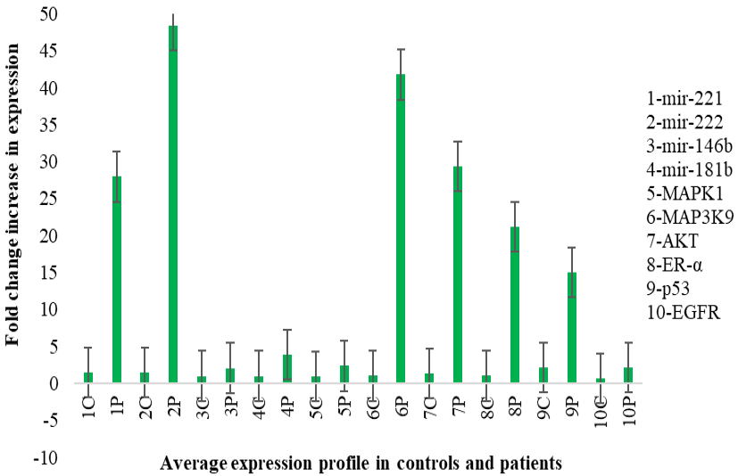
Average expression profile in cases and controls.
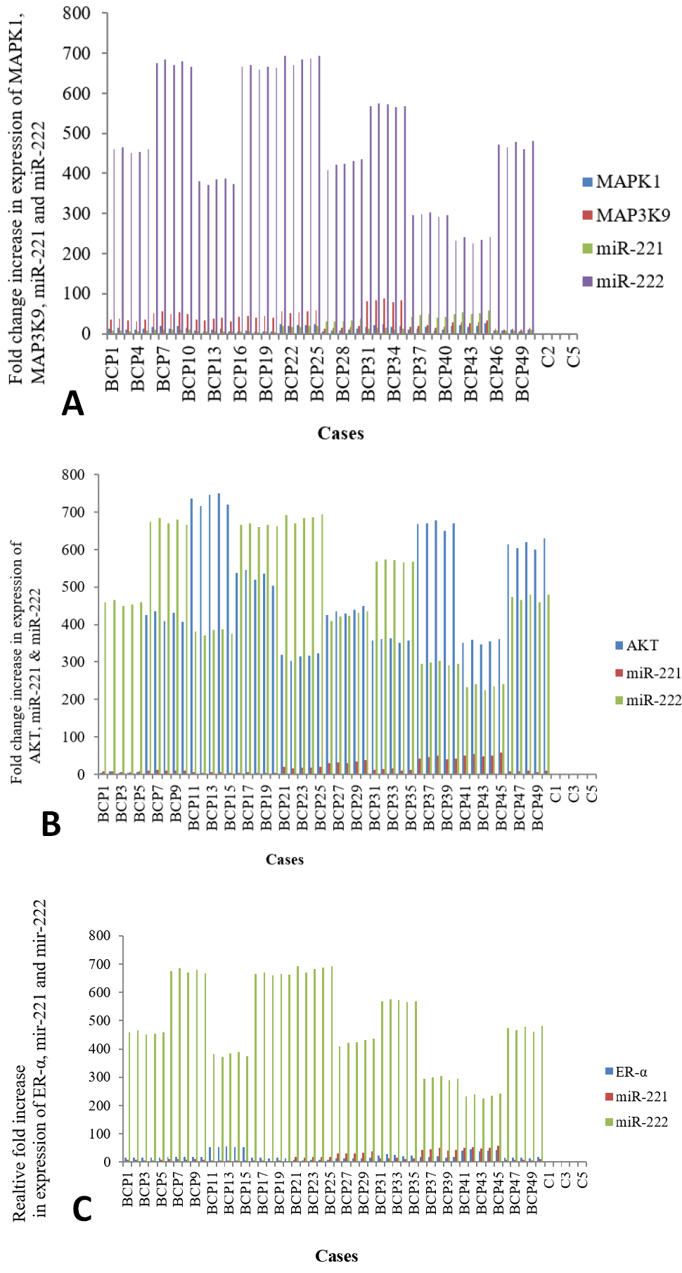
Fold increase in the expression of MAPK1, MAP3K9, miR-221 and miR-222 (A), AKT, miR-221, miR-222 (B), and ER-α, miR-221 and miR-222 (C) among cases and controls.
References
- Apostolou, P., Iliopoulos, A.C., Parsonidis, P. and Papasotiriou, I., 2019. Oncotarget, 10: 3328–3338. https://doi.org/10.18632/oncotarget.26913[CrossRef]
- Di Martino, M.T., Arbitrio, M., Caracciolo, D., Cordua, A., Cuomo, O., Grillone, K., Riillo, C., Caridà, G., Scionti, F., Labanca, C., Romeo, C., Siciliano, M. A., D’Apolito, M., Napoli, C., Montesano, M., Farenza, V., Uppolo, V., Tafuni, M., Falcone, F., D’Aquino, G., Calandruccio, N. D., Luciano, F., Pensabene, L., Tagliaferri, P. and Tassone, P., 2022. Mol. Ther. Nucl. Acids, 27: 1191-1224. https://doi.org/10.1016/j.omtn.2022.02.005[CrossRef]
- Lei, S., Zheng, R., Zhang, S., Wang, S., Chen, R., Sun, K., Zeng, H., Zhou, J. and Wei, W., 2021. Cancer Commun., 41: 1183–1194. https://doi.org/10.1002/cac2.12207[CrossRef]
- Masood, N., Malik, F.A. and Kayani, M.A., 2011. Asain Pac. J. Cancer Prev., 12: 377–382.
- Momenimovahed and Salehiniya, 2019. Breast Cancer Targets Ther., 11: 151–164. https://doi.org/10.2147/BCTT.S176070[CrossRef]
- O’Day, E. and Lal, A., 2010. Breast Cancer Res., 12: 1-10. https://doi.org/10.1186/bcr2484[CrossRef]
- Safa, A., Abak, A., Shoorei, H., Taheri, M. and Ghafouri-Fard, S., 2020. Biomed. Pharmacother., 132: 1-15. https://doi.org/10.1016/j.biopha.2020.110903[CrossRef]
- Shah, M.Y. and Calin, G.A. 2011. Genome Med., 3: 1-4. https://doi.org/10.1186/gm272[CrossRef]
- Sidorova, E.A., Zhernov, Y.V., Antsupova, M.A., Khadzhieva, K.R., Izmailova, A.A., Kraskevich, D.A., Belova, E.V., Simanovsky, A.A., Shcherbakov, D.V., Zabroda, N.N. and Mitrokhin, O.V., 2023. Int. J. mol. Sci., 24: 1-17. https://doi.org/10.3390/ijms24031980[CrossRef]
- Yin, Y., Wang, X., Li, T., Ren, Q., Li, L., Sun, X., Zhang, B., Wang, X., Han, H., He, Y., Cao, Z., Sun, X. and Zhou, Z., 2020. Cancer Med., 9: 1544-1552. https://doi.org/10.1002/cam4.2817[CrossRef]
- Zaheer, S., Shah, N., Maqbool, S.A. and Soomro, N.M., 2019. BMC Publ. Hlth., 19: 1-9. https://doi.org/10.1186/s12889-019-7330-z[CrossRef]
Services
Article usage statistics
Loading…