
Bats are a potential reservoir of zoonotic pathogens. Some strains of bacteria in the gastrointestinal tract of bats are pathogenic to humans. Hypsugo alaschanicus feeds on insects and has a wide geographic distribution in China, and people are in frequent contact with these bats. However, assessing the gut microbiota, especially the potential pathogens, is needed for public health. Thus, this study aimed to explore the microbial diversity of the gastrointestinal tract of H. alaschanicus and to estimate the risk to humans caused by the hosted pathogenic bacteria. The 16S rRNA gene V1−V2 regions were sequenced using MiSeq high-throughput sequencing platform to study the bacterial community of both the stomach and the intestine of H. alaschanicus. A total of 21, 336 and 34, 188 high quality reads of microbiota were obtained from the stomach and intestinal tract of H. alaschanicus, respectively. The phylogenetic analyses showed that the gastrointestinal bacteria were mainly classified into five phyla, dominated by Proteobacteria (27.8% in stomach and 39.7% in intestine) and Firmicutes (59.5% in stomach and 12.7% in intestine). Enterococcus and Bacillus were the two dominant bacterial genera in the stomach, accounting for 46.1% and 7.4% of total bacteria, respectively. Sphingomonas and Mycobacterium were the two dominant genera in the intestine, accounting for 10.5% and 7.3% of total bacteria, respectively. Furthermore, the results revealed that H. alaschanicus carried a large number of human pathogens and thus should be the subject of greater study to prevent transmission of diseases from bats to humans.
Research Article · Volume 51, Issue 5, 27 June 2019 · Open Access
Microbial Diversity in the Gastrointestinal Tract of a Bat, Hypsugo alaschanicus
· · · ·
Affiliations & Notes ⌄
- 1College of Life Science, Henan Normal University, Xinxiang 453007, China 2College of Animal Science, Henan Institute of Science and Technology, Xinxiang 453007, China , * Corresponding authors: hongxingniu@htu.cn
- 2buyanzhen@htu.cn
Article Info ⌄
- Journal:
- Pakistan Journal of Zoology
- Published:
- 27 June 2019

From the article
- These indices suggested that the bacteria abundance and diversity in the intestine were higher than those in the stomach.
- Thus, the bacteria in different bat species included different phyla and genera numbers, and the dominant phyla and genera were also different.
- Some bacteria in the gastrointestinal tract of H. alaschanicus may be pathogenic bacteria or opportunistic pathogens.
- Some species of the predominant genus Sphingomonas and second dominant genus Mycobacterium in the stomach of H. alaschanicus could be pathogenic bacteria.
Selected from the article's own text.
Abstract
Introduction
Bats are a vital part of natural ecosystems as they play essential roles in pollination, seed dispersal and controlling forest and agricultural insect pests, among other roles . Although bats play positive roles in ecosystems, they are considered to be reservoirs of viruses and pathogens. As the number of emerging infectious diseases increases in a manner that is detrimental to human health, a growing number of studies have found that bats act as natural reservoirs for many emerging viruses and pathogens ; ; . A mass of pathogenic viruses colonize bats, such as henipaviruses, SARS (severe acute respiratory syndrome), MERS (Middle East respiratory syndrome), Ebola and Marburg virus or lyssaviruses ; . However, bat bacterial flora and its zoonotic threat remain ill defined . Studies have shown that the best studied microorganisms in mammals are in the gastrointestinal tract . To date, there are only a few studies on the microbiota of bats originating from the gastrointestinal tract ; ; ; ; ; . Most previous approaches have been limited to culturable microbial communities, while systematically analyzing the bat bacterial community using a next-generation sequencing approach has only been reported in recent years. Veikkolainen et al. (2014) found Bartonella spp. in peripheral blood, fecal droppings, and ectoparasites of Myotis daubentonii. de Mandal et al. (2015) first reported on a bacterial community from bat guano using Illumina next-generation sequencing. Banskar et al. (2016) performed a microbiome analysis of the abundance of bacterial pathogens in Rousettusle schenaultii guano. In addition, Hatta et al. (2016) detected Campylobacter jejuni in rectal swab samples from Rousettus amplexicaudatus in Philippines. However, systematic analysis of the bacterial community in Hypsugo alaschanicus using a next-generation sequencing approach has not been reported.
The majority of microbial species could not be cultured in the laboratory using traditional technology, while high-throughput sequencing technologies can overcome the limit and identify the microbial community efficiently even in complex systems, such as the gastrointestinal tract (Human Microbiome Project Consortium, 2012). MiSeq high-throughput sequencing technology combines the advantages of Illumina HiSeq 2500 and Roche 454.
In China, H. alaschanicus is distributed in the central and northeast regions. H. alaschanicus lives mainly on insects and has various habitats. For instance, this species is relatively common in urban or human-associated areas, and can be found roosting in abandoned houses and buildings as well as in natural caves. For the past decades, some bat caves or houses have been explored for tourism, which not only threaten the habitats of bats but also could increase the risk of the transmission of bat pathogenic bacteria to humans. This transmission may cause zoonosis ; ; . With the possibility of human to wildlife transmission, the study of the gastrointestinal microbiota composition in H. alaschanicus and especially the diversity of pathogens are extremely crucial.
To comprehensively analyze the microbiota of the gastrointestinal tract of H. alaschanicus, MiSeq high-throughput sequencing technology was used to sequence the V1−V2 variable regions of the 16s rRNA gene of gastrointestinal tract bacteria.
Materials and methods
Bat collection
In September 2015, bats were captured using mist nets placed along the Weihe River in Xinxiang City, Henan Province, China (N35°35′, E113°92′). In this study, five H. alaschanicus specimens (three males and two females, all were adults) were captured in the field. The bat species were first identified based on the morphology according to “A Guide to the Mammals of China” (Smith and Xie, 2009), and then were verified using sequence analysis of the cytochrome B (cytb) gene amplified from total extracted DNA of the five H. alaschanicus. All fieldwork was conducted in accordance with the Law of the People’s Republic of China on the Protection of Wildlife.
Separation of gastrointestinal contents
An overdose of chloroform was used to euthanize the bats. The bats were then soaked with 70% ethanol for surface sterilization and rinsed with sterile water. Then the stomach and intestinal contents (including the ileum, jejunum, duodenum, and hind gut) were removed using sterile surgical forceps and a scalpel under aseptic conditions. The organs were placed in sterile tubes and immediately stored in liquid nitrogen until DNA extraction.
DNA extraction and sequencing
The genomic DNA of the stomach and intestinal fluid of five H. alaschanicus was extracted using a Stool DNA Kit (Feiyang, Guangzhou, China). The 16S rRNA gene V1−V2 regions were amplified with the primers 8F (5′-GCTTTGATGGACAT GGAAGAAGACAT-3′) and 338R (5′-GAGCCATCCCTCT CAATAATTTCAGG-3′). Polymerase chain reactions (PCRs) were performed in volume of 50 μl. The cycling parameters for the V1−V2 hypervariable regions of the 16S rRNA gene were as follows: 98 °C for 30 s; 98 °C for 10 s, 50 °C for 30 s, and 72 °C for 30 s for 20 cycles; and a final extension at 72 °C for 7 min. Amplicons were extracted from 2% agarose gels and purified using the QIA quick Gel Extraction kit (Comwei, Beijing, China) according to the manufacturer’s instructions. Purified amplicons were paired-end sequenced on an Illumina MiSeq platform (Zhongyijinda, Jiangsu, China).
Data analysis
With the use of the rRNA Detection pipeline in MG - RAST V3.3.6 for data analysis, the original data were subjected to filter processing and sequence optimization, and the optimized sequence was classified according to operational taxonomical unit (OTU) via clustering analysis and classification analysis. Based on the results of OTU clustering analysis, the diversity indices and depth of the sequences were detected. Using this taxonomic information, statistical analyses of the community structure at the level of the phylum and genus were conducted. Calculating the OTU number of each sample at a 97% similarity level, the analyses included optimized sequence statistics and calculated diversity indices and allowed for the generation of a high-throughput sequencing heat map.
Results
Gastrointestinal tract microbiome composition and diversity
After data analysis of gastrointestinal tract bacteria in H. alaschanicus, a total of 21, 336 effective sequences were obtained from the stomach fluid, and the effective sequences were clustered into 2, 468 OTUs with 97% identity. The sequencing coverage rate was 99.001%, indicating that the majority of the bacterial species in the stomach were included. A total of 34, 188 effective sequences were obtained from the intestinal fluid, and the effective sequences were clustered into 3, 511 OTUs with 97% identity. The sequencing coverage rate was 98.890%, indicating that the majority of the bacterial species in the intestine were included. Thus, there were species diversity in the stomach and intestine of H. alaschanicus (Table І).
The richness indices (Chao, 7803.176; ACE, 12051.440) in the stomach were significantly lower than those of the intestinal tract (Chao, 10749.844; ACE, 23799.700). The Shannon diversity index (3.536) in the stomach was lower than that of the intestinal tract (5.472). The Simpson diversity index (0.199) in the stomach was higher than that of intestinal tract (0.027). These indices show that the bacterial abundance and diversity in the intestine are higher than those in the stomach (Table І).
Table I.- Summary of sequencing data from the stomach and intestine.
| Parameters | Intestine | Stomach |
| After trim | 34,188 | 21,336 |
| Sequence depth | 21,336 | 21,336 |
| OTUs | 3511 | 2468 |
| Chao index | 10749.844 | 7803.176 |
| ACE index | 23799.700 | 12051.440 |
| Shannon index | 5.472 | 3.536 |
| Simpson index | 0.027 | 0.199 |
| Goods coverage (%) | 99.001 | 98.890 |
The phylum level structure of gastrointestinal tract flora
The bacteria from the samples (intestine and stomach) of H. alaschanicus belonged to 24 different phyla and were mainly distributed in four phyla: Bacteroidetes, Firmicutes, Proteobacteria and Actinobacteria (Fig. 1). The four phyla were the dominant taxa in the gastrointestinal tract, but the proportion for each group varied. The two dominant phyla were Proteobacteria (accounting for 27.8% of the total bacteria in the stomach and 39.7% of the total bacteria in the intestine) and Firmicutes (accounting for 12.7% of the total bacteria in the stomach and 59.5% of the total bacteria in the intestine) (Table II). More than 80% of the bacteria in the gastrointestinal tract belong to Proteobacteria, Firmicutes, Bacteroidetes and Actinobacteria. The other bacteria phyla only accounted for a small number of the bacteria in the gastrointestinal tract.
Table II.- The composition of bacteria from the stomach and intestine at the phylum level.
| Phylum | Intestine (%) | Stomach (%) |
| Bacteroidetes | 15.6 | 3.9 |
| Firmicutes | 12.7 | 59.5 |
| Proteobacteria | 39.7 | 27.8 |
| Actinobacteria | 15.7 | 2.4 |
| Unclassified | 6.7 | 4.0 |
| Others* | 9.6 | 2.3 |
Only the top 5 enriched class categories are shown in the table. *Others, represents the rare genera.
The genus level structure of gastrointestinal tract flora
The gastrointestinal tract flora diversity at the level of the genus was analyzed by drawing a heat map, and the bacterial genera that were found at concentrations greater than 0.1% in at least one of the samples are shown (the percentage is based on log10 and shown on a heat map) (Fig. 2). The deeper the red color, the higher the percentage of the bacteria. There were 20 bacteria genera present at more than 0.1% in the intestine and stomach (Fig. 2), and the changes in color demonstrates that the genera percentages in the intestine and stomach flora were different. In the stomach, the predominant bacteria genus was Enterococcus, accounting for 46.1% of the flora, and the second largest group was Bacillus, accounting for 7.4% of the flora. In the intestine, the predominant bacteria genus was Sphingomonas, accounting for 10.5% of the flora, and the subdominant group was Mycobacterium, accounting for 7.3% of the flora (Table III).
Table III.- The composition of bacteria from the stomach and intestine at genus level.
| Genus | Intestine (%) | Stomach (%) |
| Sphingomonas | 10.5 | 2.4 |
| Mycobacterium | 7.3 | 0.5 |
| Mycoplasma | 5.7 | - |
| Pseudomonas | 4.1 | 0.2 |
| Bacillus | 3.9 | 7.4 |
| Lactococcus | 3.3 | 0.3 |
| Escherichia | 2.0 | 0.3 |
| Alistipes | 1.7 | 0.5 |
| Bacteroides | 1.0 | 0.2 |
| Enterococcus | 0.4 | 46.1 |
| Others* | 56.3 | 40 |
Genera obtained over 0.1% of the total reads are shown. *Others, represents the rare genera. - is representative of a very low bacterial content.
Discussion
Next generation sequencing platforms are rapidly changing the methods of characterizing microbial communities from various sources. Metagenomic studies using these sequencing technologies have contributed enormously to our understanding of the structure and composition of bacterial communities . This study provides an in-depth identification of the bacterial communities that are present in the gastrointestinal tract of H. alaschanicus. The richness indices (Chao and ACE indices) and Shannon diversity index in the stomach were significantly lower than those of the intestinal tract. The Simpson diversity index in the stomach was higher than that of the intestinal tract. These indices suggested that the bacteria abundance and diversity in the intestine were higher than those in the stomach. The composition differences of the gastrointestinal tract flora in H. alaschanicus may be related to gastrointestinal tract environment discrepancies or be associated to the differences of gastrointestinal functions .
Using MiSeq high-throughput sequencing, bacteria from samples (intestine and stomach) of H. alaschanicus belonged to 24 defined different phyla and were dominated by Proteobacteria, Firmicutes, Bacteroidetes and Actinobacteria (Fig. 1). In previous studies, the bacteria in Myotis daubentonii (location: Finland) were dominated by three phyla: Chlamydiae, Proteobacteria and Bacteroidetes . The bacteria in Rousettus leschenaultii (location: India) were composed of 27 bacterial phyla, with Firmicutes, Actinobacteria and Proteobacteria predominating . This is similar to those of other wildlife like rodents, for example C57BL/6 mice, whose intestinal bacteria contain Actinobacteria, Proteobacteria, Spirochaetes, Deferribacteres, Tenericutes, Verrucomicrobia, and unclassified bacteria . In this study, totally 387 genera were identified, which was over three times than that of the Philippine bat (Rousettus amplexicaudatus). Rousettus amplexicaudatus (location: Philippines) consisted of 103 genera of rectal swab bacterial flora, and the predominant genera were Clostridium and Campylobacter . H. alaschanicus and R. amplexicaudatus shared 52 genera, and the dominant genera were different between them. Thus, the bacteria in different bat species included different phyla and genera numbers, and the dominant phyla and genera were also different. These differences might be due to the habitat, host specificity and diet . In terms of habitats, Myotis daubentonii inhabits in forests and farmhouses. R. leschenaultii resides in caves, forests and hardy banana. R. amplexicaudatus is common in caves, rock cracks and tombs. However, H. alaschanicus mainly lives in human-associated areas and natural caves ; . As for feeding habits, R. leschenaultii and R. amplexicaudatus are frugivorous, while M. daubentonii and H. alaschanicus are insectivorous.
Some bacteria in the gastrointestinal tract of H. alaschanicus may be pathogenic bacteria or opportunistic pathogens. Studies have shown that some species of the genus Sphingomonas, which was found in the stomach of H. alaschanicus in this study, frequently cause inflammatory disease in animals . Mycobacterium tuberculosis is associated with tuberculosis. M. leprae and M. lepromatosis, is mainly responsible for leprosy . Some species of the predominant genus Sphingomonas and second dominant genus Mycobacterium in the stomach of H. alaschanicus could be pathogenic bacteria. Enterococcus strains isolated from patients were associated with inflammatory bowel disease . Bacillus anthracis could cause a zoonotic disease of anthrax . In the intestinal tract of H. alaschanicus, the dominant genus Enterococcus and the second predominant genus Bacillus, may also contain pathogens. Mycoplasma may cause pneumonia and urinary tract infections ; . Lactococcus garvieae can cause human sepsis, endocarditis and osteomyelitis . Escherichia hermannii has mainly been found to be involved in the sepsis, diarrhea and other infections, especially in immune compromised individuals . Thus it is reasonanle to argue that the genera (Mycoplasma, Lactococcus, Escherichia) in the gastrointestinal tract of H. alaschanicus probably also include pathogenic bacteria species. However, further studies are needed to test the prediction. Most of these bacterial isolates were opportunistic pathogens that usually do not harm the host unless the immune system is weakened . However, some bacteria in this study have been reported to be pathogenic to humans .
The reported metagenomic analysis of the bat microbiota indicates that bats are reservoir hosts for several pathogenic bacterial genera ; ; . However, no comprehensive study has been published on how those potential pathogens are transmitted from bats to human hosts. H. alaschanicus mainly lives in urban, rural housing, forest and natural caves, which include overlapping habitats with human. We propose that bacteria from bat fecal droppings could be transmitted to human hosts. Bacteria can contaminate flowing streams, which could be a source of drinking water in China, posing a direct threat to the health of the residents that use the water. Furthermore, there are as many as 155 bat species in China . Most people have little understanding of this danger and often go to caves to collect the bats’ bodies and feces, which are used in medicine or as fertilizer . In addition, many caves harboring bats have been exploited for tourism . At the same time, with the increase of the population density and change of people’s lifestyles, the chance of people contacting bats is increasing, which may lead to higher risk of the transmission of pathogenic bacteria in bats to humans . To avoid the occurrence of zoonosis, contacts between humans and bats should be highly controlled or reduced. This study could improve the knowledge of the gastrointestinal microflora of bats and lead to the acquisition of further information on the prevention of bat zoonotic disease. Moreover, it is necessary to identify bacterial pathogens that are continuously transmitted to humans from bats.
Acknowledgements
This project was supported by the National Natural Science Foundation of China (NSFC, No. 31172056). We thank all those who helped in the field, especially Zongxiao Zhang, Liumeng Zheng, Jie Wu, Yingying Liu, Junlou Li, and Lili Hu. We also thank Hongwei Zhou and Shuyi Zhang for gastrointestinal contents sampling. We are especially grateful to Professor Qingxiang Yang for guidance and assistance in the experiment.
Statement of conflict of interest
The authors declare that there is no conflict of interests regarding the publication of this article.
Figures
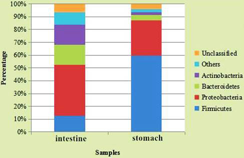
The classification of reads at the phylum level (only the top 5 enriched class categories are shown in the figure).
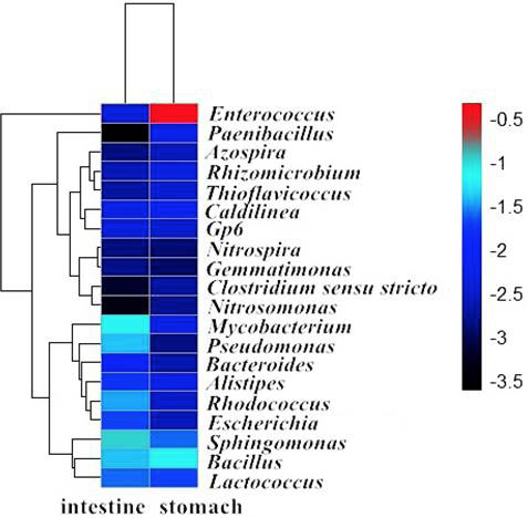
Heat map showing the relative abundance of more than 0.1% of reads at the genus level.
References
- Banskar, S., Bhute, S.S., Suryavanshi, M.V., Punekar, S. and Shouche, Y.S., 2016. Microbiome analysis reveals the abundance of bacterial pathogens in Rousettus leschenaultii guano. Scient. Rep., 6: 36948. https://doi.org/10.1038/srep36948[CrossRef]
- Bu, Y.Z., Wang, Y.M., Zhang, C., Liu, W., Zhou, H.X., Yu, Y. and Niu, H.X., 2015. Geographical distribution, roost selection, and conservation state of cave-dwelling bats in China. Mammalia, 79: 409-417. https://doi.org/10.1515/mammalia-2014-0008[CrossRef]
- Calisher, C.H., Childs, J.E., Field, H.E., Holmes, K.V. and Schountz, T., 2006. Bats: Important reservoir hosts of emerging viruses. Clin. Microbiol. Rev., 19: 531-545. https://doi.org/10.1128/CMR.00017-06[CrossRef]
- Carrillo-Araujo, M., Taş, N., Alcántara-Hernández, R.J., Gaona, O., Schondube, J.E., Medellín, R.A., Jansson, J.K. and Falcón, L.I., 2015. Phyllostomid bat microbiome composition is associated to host phylogeny and feeding strategies. Front. Microbiol., 6: 447. https://doi.org/10.3389/fmicb.2015.00447[CrossRef]
- Daniel, D.S., Ng, Y.K., Chua, E.L., Arumugam, Y., Wong, W.L. and Kumaran, J.V., 2013. Isolation and identification of gastrointestinal microbiota from the short-nosed fruit bat Cynopterus brachyoti brachyotis. Microbiol. Res., 168: 485-496. https://doi.org/10.1016/j.micres.2013.04.001[CrossRef]
- de Mandal, S., Zothansanga, Panda, A.K., Bisht, S.S. and Senthil-Kumar, N., 2015. First report of bacterial community from a Bat Guano using Illumina next-generation sequencing. Genom. Data, 4: 99-101. https://doi.org/10.1016/j.gdata.2015.04.001[CrossRef]
- Golińska, E., Tomusiak, A., Gosiewski, T., Więcek, G., Machul, A., Mikołajczyk, D., Bulanda, M., Heczko, P.B. and Strus, M., 2013. Virulence factors of Enterococcus strains isolated from patients with inflammatory bowel disease. World J. Gastroenterol., 19: 3562-3572. https://doi.org/10.3748/wjg.v19.i23.3562[CrossRef]
- Graves, S.R., Kennelly-Merrit, S.A., Tidemann, C.R., Rawlinson, P.A., Harvey, K.J. and Thornton, I.W., 1988. Antibiotic-resistance patterns of enteric bacteria of wild mammals on the Krakatau Islands and West Java, Indonesia. Phil. Trans. R. Soc. Lond. B: Biol. Sci., 322: 339-353. https://doi.org/10.1098/rstb.1988.0130[CrossRef]
- Gulraiz, T.L., Javid A., Hussain, S.M., Shahbaz, M., Irfan, M. and Daud, S., 2017. Microbial analysis of Indian flying fox (Pteropus giganteus) ejecta collected from two public parks in Lahore, Pakistan. Pakistan J. Zool., 49: 305-312. http://dx.doi.org/10.17582/journal.pjz/2017.49.1.305.312[CrossRef]
- Hanning, I. and Diaz-Sanchez, S., 2015. The functionality of the gastrointestinal microbiome in non-human animals. Microbiome, 3: 51. https://doi.org/10.1186/s40168-015-0113-6[CrossRef]
- Hatta, Y., Omatsu, T., Tsuchiaka, S., Katayama, Y., Taniguchi, S., Masangkay, J.S., Puentespina, Jr. R., Eres, E., Cosico, E., Une, Y., Yoshikawa, Y., Maeda, K., Kyuwa, S. and Mizutani, T., 2016. Detection of Campylobacter jejuni in rectal swab samples from Rousettus amplexicaudatus in the Philippines. J. Vet. Med. Sci., 78: 1347-1350. https://doi.org/10.1292/jvms.15-0621[CrossRef]
- He, B., Li, Z., Yang, F., Zheng, J., Feng, Y., Guo, H., Li, Y., Wang, Y., Su, N., Zhang, F., Fan, Q. and Tu, C., 2013. Virome profiling of bats from Myanmar by metagenomic analysis of tissue samples reveals more novel mammalian viruses. PLoS One, 8: e61950. https://doi.org/10.1371/journal.pone.0061950[CrossRef]
- Heard, D.J., de Young, J.L., Goodyear, B. and Ellis, G.A., 1997. Comparative rectal bacterial flora of four species of flying fox (Pteropus sp.). J. Zool. Wildl. Med., 28: 471-475.
- Hirakawa, T.F., Costa, F.A., Vilela, M.C., Rigon, M., Abensur, H. and Araújo, M.R., 2011. Lactococcus garvieae endocarditis: first case report in Latin America. Arq. Bras. Cardiol., 97: e108-110. https://doi.org/10.1590/S0066-782X2011001400016[CrossRef]
- Huang, H.X., Wang, H. and Zhang, W.M., 2010. Potential pathogenicity of Bacillus amyloliquefaciens to infants. J. Clin. Pediatr., 28: 190-192.
- Human Microbiome Project Consortium, 2012. Structure, function and diversity of the healthy human microbiome. Nature, 486: 207-214. https://doi.org/10.1038/nature11234[CrossRef]
- Hu, J., He, X.H., Li, D.P. and Liu, Q., 2007. Progress in research of Sphingomonas. Chin. J. appl. Environ. Biol., 13: 431-437.
- Jagielski, T., Minias, A., van Ingen, J., Rastogi, N., Brzostek, A., Żaczek, A. and Dziadek, J., 2016. Methodological and clinical aspects of the molecular epidemiology of Mycobacterium tuberculosis and other Mycobacteria. Clin. Microbiol. Rev., 29: 239-290. https://doi.org/10.1128/CMR.00055-15[CrossRef]
- Kaewpoowat Q., Permpalung N. and Sentochnik D.E., 2013. Emerging Escherichia pathogen. J. clin. Microbiol., 51: 2785-2786. https://doi.org/10.1128/JCM.00983-13[CrossRef]
- Kim, Y.S., Kim, J. and Park, S.J., 2015. High-throughput 16S rRNA gene sequencing reveals alterations of mouse intestinal microbiota after radiotherapy. Anaerobe, 33: 1-7. https://doi.org/10.1016/j.anaerobe.2015.01.004[CrossRef]
- Kunz, T.H., Braun de Torrez, E., Bauer, D., Lobova, T. and Fleming, T.H., 2011. Ecosystem services provided by bats. Annls. N.Y. Acad. Sci., 1223: 1-38. https://doi.org/10.1111/j.1749-6632.2011.06004.x[CrossRef]
- Lane, D.J., Pace, B., Olsen, G.J., Stahl, D.A., Sogin, M.L. and Pace, N.R., 1985. Rapid determination of 16S ribosomal RNA sequences for phylogenetic analyses. Proc. natl. Acad. Sci. USA, 82: 6955-6959. https://doi.org/10.1073/pnas.82.20.6955[CrossRef]
- Liu, W., Wang, Y.X., He, X.H. and Niu, H.X., 2011. Distribution and analysis of the importance of underground habitats of cave-dwelling bats in the south of Taihang Mountain. Acta. Theriol. Sin., 31: 371-379.
- Liu, Z.X., Zhang, Y.X. and Zhang, L.B., 2013. Research perspectives and achievements in taxonomy and distribution of bats in China. Zool. Res., 34: 687-693.
- Mühldorfer, K., 2013. Bats and bacterial pathogens: A review. Zoon. Publ. Hlth., 60: 93-103. https://doi.org/10.1111/j.1863-2378.2012.01536.x[CrossRef]
- Mühldorfer, K., Speck, S., Kurth, A., Lesnik, R., Freuling, C., Müller, T., Kramer-Schadt, S. and Wibbelt, G., 2011. Diseases and causes of death in European bats: dynamics in disease susceptibility and infection rates. PLoS One, 6: e29773. https://doi.org/10.1371/journal.pone.0029773[CrossRef]
- Mühldorfer, K., Wibbelt, G., Haensel, J., Riehm, J. and Speck, S., 2010. Yersinia species isolated from bats, Germany. Emerg. Infect. Dis., 16: 578-580. https://doi.org/10.3201/eid1603.091035[CrossRef]
- Peterson, J.W., 1996. Bacterial pathogenesis, Chapter 7. In: Medical microbiology, 4th edition (ed. S. Baron). University of Texas Medical Branch at Galveston, Galveston, TX.
- Prem-Anand, A.A. and Sripathi, K., 2004. Digestion of cellulose and xylan by symbiotic bacteria in the intestine of the Indian flying fox (Pteropus giganteus). Comp. Biochem. Physiol. A: Mol. Integr. Physiol., 139: 65-69. https://doi.org/10.1016/j.cbpb.2004.07.006[CrossRef]
- Simmons N.B., 2005. Order Chiroptera. In: Mammal species of the world: A taxonomic and geographic reference, 3rd edition (eds. D.E. Wilson and D.M. Reeder). Johns Hopkins University Press, Baltimore, Maryland, pp. 312-529.
- Smith A.T. and Xie, Y., 2008. A guide to the mammals of China. Princeton University Press, Princeton, pp. 338.
- Valdez, H. and Salata, R.A., 1999. Bat-associated histoplasmosis in returning travelers: Case presentation and description of a cluster. J. Travel Med., 6: 258-260. https://doi.org/10.1111/j.1708-8305.1999.tb00529.x[CrossRef]
- Veikkolainen, V., Vesterinen, E.J., Lilley, T.M. and Pulliainen, A.T., 2014. Bats as reservoir hosts of human bacterial pathogen, Bartonella mayotimonensis. Emerg. Infect. Dis., 20: 960-967. https://doi.org/10.3201/eid2006.130956[CrossRef]
- Welkos, S., Bozue, J., Twenhafel, N. and Cote, C., 2015. Animal models for the pathogenesis, treatment, and prevention of infection by Bacillus anthracis. Microbiol. Spectr., 3: TBS-0001-2012. https://doi.org/10.1128/microbiolspec.TBS-0001-2012[CrossRef]
- You, Y., Wang, M.H., Dai, Q.Y. and Nie, S.Z., 2013. Correlation between genital tract Mycoplasma or Chlamydia Infections and infertility. Chin. J. Nosocomiol., 23: 5620-5626.
Services
Article usage statistics
Loading…