Metabolic and Stress Plasticity in Fish: A Review of Fasting-Refeeding Physiology
Affiliations & Notes ⌄
1Department of Biology, College of Science, Jazan University, P.O. Box. 114, Jazan 45142, Saudi Arabia. *Correspondence | Mohsen A. Khormi, Department of Biology, College of Science, Jazan University, P.O. Box. 114, Jazan 45142, Saudi Arabia
Short-term fasting enhances glucose utilization, stress tolerance, and antioxidant defenses.
Ultimately, the study of fasting-refeeding cycles highlights the interplay between energy metabolism, endocrine regulation, and oxidative balance.
The most significant gap identified in fasting–refeeding physiology is the most significant gap lies in connecting mechanistic insights from controlled laboratory studies with ecologically realistic and commercially relevant aquaculture systems.
Addressing this gap through multi-omics and long-term, applied trials will be essential for translating fasting–refeeding physiology into sustainable aquaculture strategies.
Selected from the article's own text.
Abstract
Fasting-refeeding cycles represent a critical adaptive strategy that influences metabolism, physiology, and stress resistance. Fish exhibit remarkable tolerance to fasting compared with other vertebrates, maintaining homeostasis through coordinated metabolic, endocrine, and oxidative mechanisms. During fasting, carbohydrate reserves are rapidly mobilized, followed by lipid oxidation and eventually protein catabolism under prolonged deprivation. These shifts are regulated by hormones such as insulin, glucagon, and cortisol, which orchestrate energy redistribution. Short-term fasting often enhances glucose efficiency, stress resistance, and antioxidant protection, whereas extended starvation can impair tissue integrity, weaken defenses, and disrupt metabolic balance. Refeeding restores biochemical parameters and supports recovery, highlighting the interplay between energy metabolism, osmotic regulation, and adaptive resilience. This review synthesizes findings across diverse fish species and experimental conditions to evaluate the physiological responses to fasting-refeeding, with emphasis on metabolic and stress-related adaptations. The insights presented have direct implications for optimizing feeding strategies, improving fish welfare, and enhancing sustainability in aquaculture, while also contributing to a broader understanding of evolutionary physiological plasticity in fish. The single most important aquaculture implication is that short, controlled fasting before handling or transport can reduce stress and mortality while preserving growth performance, offering farmers a practical strategy to improve welfare and production outcomes.
Fasting in fish challenges metabolism and physiology, yet fish exhibit extraordinary resilience to food deprivation compared with other animals ; . Adaptive mechanisms preserve homeostasis, redistribute energy, and sustain vital functions. Understanding metabolic and oxidative responses is essential for aquaculture. Fish survive starvation by regulating biochemical and physiological mechanisms, which reflect fish condition and are influenced by biotic and abiotic factors such as age, sex, water temperature, seasonal variation, and diet .
Energy metabolism in fish is sequential and coordinated. Carbohydrate reserves are mobilized first through glycogenolysis, gluconeogenesis, and glycolysis, controlled by insulin and glucagon ; . Short-term fasting enhances glucose efficiency and stress resistance (Peng et al., 2026; , whereas prolonged starvation depletes glycogen, leading to hypoglycemia or fluctuations in glucose levels ; ; . Once carbohydrate reserves are exhausted, lipids become the primary energy source. Triglycerides fuel β-oxidation and gluconeogenesis, usually lowering plasma levels ; , though some species show increases . Protein reserves are preserved initially but are mobilized during extended fasting, causing plasma protein decline; refeeding restores these levels, aided by glycogen sparing ; . Together, carbohydrate, lipid, and protein metabolism interact to maintain osmotic balance and support recovery.
Cortisol orchestrates metabolic shifts, promoting gluconeogenesis and immune modulation ; . Its response varies rising , falling (Peng et al., 2026), or stable . Chronic elevation may impair tissues , showing both adaptive and harmful effects .
Fasting also alters oxidative balance. Short-term fasting activates antioxidant defences, including key enzymes such as superoxide dismutase (SOD) and catalase (CAT), enhancing protection against reactive oxygen species (ROS) ; . In contrast, prolonged deprivation may lead to oxidative damage .
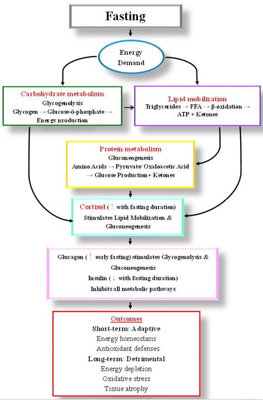
The indicators discussed (e.g. plasma glucose and glycogen reserves, cortisol levels, and antioxidant enzyme activity) are widely applicable across species, integrative of multiple physiological systems (metabolic, endocrine, oxidative), and sensitive to both short-term fasting and prolonged deprivation and can be used to assess the impact of fasting-refeeding strategies. Accordingly, it is useful to study the physiological changes that occur in fish under fasting-refeeding strategies. This review aims to evaluate the physiological responses of fish to fasting-refeeding cycles, with emphasis on metabolic, and stress-related adaptations. By synthesizing findings across diverse species, it highlights the impact of fasting-refeeding strategies on plasma metabolites, enzyme activity, and antioxidant defences. While numerous studies have experimentally investigated specific aspects of fasting or refeeding in individual fish species, there is currently no comprehensive review that integrates these findings across multiple species. By addressing this gap, the present review synthesizes systemic and molecular evidence and highlights the effects of fasting–refeeding strategies on plasma metabolites, enzyme activity, and antioxidant defences. Figure 1 illustrates the key physiological responses involved in metabolic adaptation to fasting, as discussed in the text.
Energy metabolism
Energy metabolism in fish is coordinated through tightly regulated pathways that ensure adequate ATP supply during both fed and fasting states. These metabolic processes rely on the dynamic utilization of carbohydrates, lipids, and proteins, which collectively maintain cellularenergy balance and support vital physiological functions.
Fasting phases in fish can be classified based on physiological indicators rather than arbitrary durations. Short-term fasting corresponds to periods in which glycogen stores are partially depleted, plasma metabolites remain within homeostatic range, and cortisol elevation is moderate, reflecting adaptive metabolic adjustments ; . In contrast, long-term fasting involves extensive glycogen depletion, significant lipid and protein mobilization, and pronounced endocrine and oxidative stress responses ; . This physiology-based classification facilitates interspecies comparisons and provides a reproducible framework for linking metabolic and hormonal changes to defined fasting phases. Table 1 summarizes the impacts of different fasting-refeeding on metabolic and stress parameters of fish.
Carbohydrate metabolism
Carbohydrate metabolism provides rapid and readily accessible energy through glucose oxidation and gluconeogenic pathways. Glucose serves as the primaryenergy source for cellular function, and during
Table 1: Metabolic responses to fasting-refeeding cycles.
Fasting duration
Refeeding duration
Species
Specific findings during fasting
and refeeding
Key context / proposed explanation
References
A. Hypoglycaemic responders
1, 2, 3, 4 weeks
Refed to end of experiment (13 weeks total)
Nile tilapia (O. niloticus)
Fasted fish exhibited reduced plasma glucose.
After refeeding, glucose in 1–3-week fasted fish matched controls.
4-week fasted group had lower glucose even after prolonged refeeding.
Progressive shift from glycolysis to gluconeogenesis and glycogenolysis as fasting continues.
2-3 days
Not specified
Blunt snout bream (Megalobrama amblycephala)
Fasting stimulated glycolysis and redirected amino acid catabolism toward energy production. Alleviated ammonia nitrogen stress.
Unique short-term response where glycolysis is activated, possibly for rapid energy from remaining glycogen.
fasting, its demand is efficiently met through glycogenolysis to maintain blood glucose homeostasis ; . In fish, glycaemia is tightly regulated by the pancreatic hormones’ insulin and glucagon . Insulin promotes hepatic glycogen storage, while glucagon facilitates the release of glucose from the liver when plasma levels decline. Additionally, hepatic gluconeogenesis and reduced glycolytic activity may also contribute to glucose regulation.
Glucose responses to starvation vary notably among fish. For instance, multiple studies have demonstrated that fasting followed by refeeding had no significant impact on glycaemia ; ; . This indicates that some species maintain glucose homeostasis during short-term fasting. While other studies reported declines in glucose levels ; ; . Suggesting downregulation of gluconeogenesis and glycogenolysis in response to limited nutrient availability. Similarly, Peng et al. (2026) reported that fasting stimulated glycolysis and redirected amino acid catabolism toward energy production, thereby improving resistance to transport-induced ammonia stress. In contrast, fasting increased glucose levels in S. scherzeri . These observations emphasize the importance of distinguishing between adaptive responses during short-term fasting and potential metabolic disruptions during prolonged fasting.
At the molecular level, Ntantali et al. (2023) demonstrated that fasting in sea bass altered the expression of key glycolytic genes, suggesting that metabolic responses extend beyond systemic blood parameters. In crustaceans, Tian et al. (2024) showed that alternate-day fasting elevated uridine diphosphate N-acetylglucosamine levels a marker of nucleotide sugar metabolism, indicating the presence of adaptive hyperglycemia under metabolic stress. These molecular adaptations underscore the complexity of fasting responses. While temporary glucose suppression may offer protective benefits during infections, extended fasting can ultimately disrupt metabolic stability.
During starvation, fish regulate glucose metabolism by suppressing the activity of enzymes involved in glycolysis (pyruvate kinase, PK and phosphofructokinase) and the pentose phosphate pathway, while enhancing those linked to gluconeogenesis to maintain blood glucose homeostasis . Interestingly, glucokinase (GK) activity increased significantly during starvation, a pattern that contrasts with observations in Oncorhynchus mykiss , Sparus aurata , and hybrid grouper . The elevated GK activity during fasting may be attributed to its role as the initial rate-limiting enzyme in the glycolytic pathway, the upregulation of GK during fasting as a preparative adaptation for refeeding, ensuring that the liver can efficiently handle the postprandial surge in glucose. GK induction during fasting does not necessarily indicate active glycolysis but rather reflects anticipatory regulation of hepatic glucose metabolism.
As a key substrate for glycogen synthesis, glucose is essential for sustaining physiological functions. While glycolysis facilitates glucose breakdown and utilization, gluconeogenesis supports its synthesis and helps elevate blood glucose levels, indicating that glucose is not entirely depleted during short-term fasting.
Lipid metabolism
Lipid metabolism plays a vital role in maintaining energy balance and physiological stability, particularly under nutritional conditions. During fasting, lipid stores in adipose tissue, liver, and muscle serve as the primary energy source, compensating for reduced carbohydrate. Key processes such as lipolysis, the release of free fatty acids, and β-oxidation are upregulated to produce ATP, while glycerol from triglyceride breakdown contributes to gluconeogenesis. Once refeeding begins, lipogenesis and lipid assimilation pathways are reactivated to replenish energy and restore metabolic equilibrium. These dynamic shifts in lipid mobilization, transport, and storage are essential for promoting growth, osmotic balance, and enhancing resilience to nutritional stress ; .
Cholesterol, is a key structural lipid, plays an essential role in the synthesis of steroid hormones such as cortisol, which governs various physiological functions including energy metabolism . Cholesterol may be synthesized or mobilized during fasting depending on species-specific metabolic responses. Variation in cholesterol response as reflecting a balance between endocrine precursor functions and structural/functional demands, with the relative importance of each pathway differing by species.
Several studies reported a decline in cholesterol levels under fasting , 2021, 2022). Messina et al. (2023) similarly found that fasted fish maintained lower cholesterol levels that returned to baseline only after 14-days of refeeding, in agreement with extended fasting–refeeding trials in trout and sturgeon . Conversely, fasting increases cholesterol levels in climbing perch (A. testudineus) and fat and lean pacu (P. mesopotamicus) due to higher energetic and stress-related demands. Supporting this, elevated cortisol was observed in fasted pacu, experimentally infected with bacteria, suggesting a role for cortisol in promoting gluconeogenesis Gimbo et al. (2015).
In contrast, several species show stable cholesterol concentrations during fasting ; ; . Likewise, no significant changes were detected during fasting–refeeding cycles in ; . Collectively, these findings highlight marked species-specific variability yet emphasize that cholesterol is tightly linked to dietary intake rather than solely to lipid mobilization . Moreover, as a precursor for cortisol, cholesterol plays a critical role in supporting gluconeogenesis and enhancing stress resilience, linking its metabolic changes directly to physiological adaptation during nutritional deprivation . The observed variability in cholesterol responses across fish species may reflect differences in ecological and physiological traits. Benthic versus pelagic lifestyles, temperate versus tropical habitats, dietary lipid composition, and acclimation temperature all influence lipid metabolism and cortisol regulation. Metabolic flexibility, including the capacity to switch between carbohydrate, lipid, and protein catabolism, further modulates these responses. Recognizing these factors provides a predictive framework for interpreting species-specific outcomes and highlights critical gaps in understanding the mechanisms underlying fasting-induced lipid adjustments.
During periods of starvation, triglycerides (TG) serve as the principal lipid reserves mobilized to fulfill energy requirements. The breakdown of TG releases glycerol, which acts as a substrate for gluconeogenesis, thereby sustaining glucose availability for essential physiological functions .
During fasting, fish shift toward lipid catabolism. Triacylglycerols are mobilized by adipose triglyceride lipase (ATGL) and hormone-sensitive lipase (HSL), releasing free fatty acids (FFAs) that enter mitochondria via the carnitine palmitoyl transferase (CPT) system (CPT1/CPT2) for β-oxidation, with peroxisomal acyl-CoA oxidase (ACOX) supporting long-chain fatty-acid breakdown . Lipoprotein lipase (LPL) regulates uptake of circulating lipids, while lipogenic enzymes (ACC, FAS, DGAT) are suppressed. Lipophagy further contributes to lipid-droplet mobilization ; . Overall, fasting upregulates lipolysis and fatty-acid oxidation while downregulating lipogenesis to maximize ATP production from stored lipids.
The response of circulating TG to fasting varies across fish species and experimental conditions. Numerous studies have documented significant declines in TG and lipoprotein concentrations in fast fish, underscoring the critical role of lipid catabolism in energy metabolism during nutrient deprivation ; . These observations suggest that lipids function as a primary energy buffer, enabling fish to endure fasting periods before resorting to protein catabolism. In contrast, Norouzi et al. (2020) observed elevated plasma TG concentrations after fasting, possibly reflecting increased lipid mobilization to meet heightened energy demands.
Protein metabolism
Although protein catabolism is often described as a tertiary response to fasting, evidence indicates that energy substrate utilization in fish is not strictly sequential. Glycogenolysis, lipolysis, and proteolysis may occur concurrently, with their relative contribution depending on species-specific metabolism, nutritional condition, and environmental context ; . Species with limited lipid reserves or high metabolic rates may initiate protein catabolism early, whereas lipid-rich species rely longer on fat oxidation, thereby sparing proteins ; .
During fasting, fish enhance proteolysis to supply amino acids for energy metabolism. Proteases such as cathepsins and components of the ubiquitin–proteasome system increase to mobilize tissue proteins . Concurrently, amino acid–catabolizing enzymes including alanine aminotransferase (ALT) and aspartate aminotransferase (AST) rise to support transamination and gluconeogenic substrate production . Together, these adjustments reflect a metabolic shift toward protein catabolism when other energy reserves decline.
Extended fasting for 45-days did not lead to significant changes in total protein, except for a marked decline at the 30-day point compared to fed group . This temporary reduction suggests a threshold beyond which protein catabolism may begin to impact systemic reserves. Importantly, refeeding facilitated a recovery of protein levels, indicating that protein stores can be replenished effectively and return to levels comparable to those in continuously fed fish. While, Nebo et al. (2017) reported no significant changes during fasting, which may be attributed to the mobilization of liver glycogen, a key mechanism for maintaining blood glucose and stabilizing other serum parameters. However, refeeding restored liver glycogen, underscoring its role in metabolic recovery. Similar patterns have been observed in traira Hoplias malabaricus , and sunshine bass Morone chrysops × Morone saxatilis , where liver glycogen stores declined during fasting.
Plasma osmolarity remained stable during 14-days of fasting but increased following acute stress, consistent with observations in fasted gilthead seabream , underscoring the prioritization of homeostasis. A moderate decline in blood glucose was noted during fasting, while hyperglycemia is a classical secondary stress response, reflecting glucose mobilization triggered by cortisol release . The concurrent rise in plasma cortisol, elevated osmolarity, and hyperglycemia in confined fish collectively indicated a mild but typical physiological stress response.
The pronounced interspecific variability in metabolic and hormonal responses to fasting–refeeding likely arises from a combination of phylogenetic constraints and ecological adaptation. Species occupying distinct ecological niches, such as ambush predators versus continuous foragers, or those inhabiting environments with strong seasonal fluctuations in food availability, may differ in baseline metabolic rate, energy storage strategies, and reliance on cortisol-mediated energy mobilization. Furthermore, evolutionary exposure to natural fasting periods may confer enhanced metabolic flexibility and stress tolerance. Future studies should adopt comparative frameworks that integrate phylogeny, ecological traits, and standardized experimental conditions to disentangle these interacting drivers of physiological variability
Beyond changes in enzyme activity, fasting-induced metabolic adjustments in fish are regulated by conserved nutrient-sensing and transcriptional pathways. AMP-activated protein kinase (AMPK) functions as a central cellular energy sensor, promoting catabolic processes such as fatty-acid oxidation while suppressing anabolic pathways through inhibition of mTOR signaling during nutrient deprivation ; . In parallel, peroxisome proliferator-activated receptors (PPARs) play a key role in lipid mobilization and β-oxidation, thereby supporting energy supply during prolonged fasting ; . Fasting also activates FOXO transcription factors, which enhance gluconeogenesis, antioxidant defenses, and cellular stress resistance . Although these signaling pathways appear broadly conserved across teleosts, their relative contribution and sensitivity vary among species, likely reflecting differences in metabolic rate, ecological niche, and evolutionary history. Emerging evidence further suggests that epigenetic mechanisms, including DNA methylation and histone modifications, may contribute to fasting memory and long-term metabolic plasticity in fish, representing an important avenue for future research .
Stress response
Cortisol regulation
Cortisol is the major corticosteroid in teleosts and a key catabolic hormone that increases under stress . It plays a vital role in maintaining physiological homeostasis and its plasma concentration is widely used as stress biomarker . Activation of the hypothalamic–pituitary–interrenal (HPI) axis during stress leads to the release of neuroendocrine hormones, including cortisol, through a cascade initiated by corticotropin-releasing hormone and followed by the secretion of adrenocorticotropic hormone (Dar et al., 2019). The activation of the HPI axis during fasting is tightly linked to the energetic state of the fish. Individuals with sufficient lipid reserves may rely primarily on lipid mobilization, resulting in attenuated or delayed cortisol responses, whereas fish with depleted energy stores exhibit heightened HPI axis sensitivity and elevated cortisol secretion ; . In this context, cortisol functions not only as an energy-mobilizing hormone that promotes gluconeogenesis and lipolysis, but also as a mediator of stress when food deprivation is combined with social competition, confinement, or high stocking density ; . Experimental studies have shown that fasting under socially or environmentally constrained conditions elicits stronger cortisol responses than fasting alone, indicating that endocrine activation reflects both metabolic demand and perceived stress ; . Therefore, cortisol elevation during fasting should be interpreted as an integrated physiological response shaped by internal energy status and external environmental pressures, rather than a purely metabolic signal.
Cortisol regulates diverse physiological and biochemical processes, including growth, metabolism, and immunity . During starvation, known stressor, cortisol secretion increases as part of an adaptive response aimed at mobilizing energy reserves and inducing protective proteins against oxidative stress (OS) . The rapid elevation of cortisol represents a fundamental stress response that promotes energy utilization and supports the re-establishment of internal homeostasis .
In response to environmental or metabolic challenges, cortisol stimulates hepatic gluconeogenesis to elevate blood glucose levels, thereby meeting increased energy demands . Supporting this, Wang et al. (2019) found that phosphoenolpyruvate carboxykinase activity increased significantly after 7-days of fasting, while hexokinase and pyruvate kinase activities decreased. These results suggest that during starvation, Nile tilapia sustain energy balance by enhancing gluconeogenic enzyme activity while downregulating key glycolytic enzymes, reflecting a metabolic shift directed by cortisol-mediated adaptation.
Previous studies have reported variable cortisol responses to fasting across fish species, suggesting that both the duration of fasting and experimental conditions influence endocrine regulation. Peng et al. (2026) found that cortisol levels in the fasting group were decreased, and accompanied by downregulation of isoproterenol, suggesting a reduced sympathetic response. Wang et al. (2019) observed that serum cortisol levels initially decreased and then increased as fasting duration extended, indicating that short-term fasting might transiently suppress cortisol production. Conversely, Liu et al. (2022) reported that cortisol levels increased after 7-days of starvation, consistent with previous observations in several fish species, including Garra gotyla and Salmo salar . Notably, refeeding for 7-days restored cortisol concentrations to normal, demonstrating that cortisol elevation during fasting is a reversible adaptive response. Also, marine species such as gilthead sea bream (Sparus auratus) starved for 14-days and Senegalese sole (Solea senegalensis) starved for 21-days showed pronounced cortisol elevation, reflecting enhanced gluconeogenic and amino acid metabolism for energy supply. In contrast, Pottinger et al. (2003) observed no significant changes in plasma cortisol during long-term fasting of rainbow trout, indicating that energy mobilization may occur without endocrine involvement of cortisol, growth hormone, or somatolactin. Similarly, Mørkøre et al. (2008) found improved resistance to acute stress in long-term starved fish. These differences indicate that species, age, and condition modulate endocrine regulation during fasting, with well-conditioned cold-water fish being less reliant on cortisol-driven energy mobilization.
During short-term fasting and transient cortisol elevation appear to be adaptive, promoting gluconeogenesis and mobilization of energy reserves while simultaneously supporting protective functions such as oxidative stress defense and osmoregulatory integrity ; . In this context, cortisol may enhance resilience to acute stressors and maintain barrier function, thereby exerting a positive effect on disease resistance. Prolonged or intense cortisol elevation is often associated with sustained cortisol secretion that reallocates energy away from growth and immunity toward essential survival processes ; . Chronic elevation has been linked to intestinal atrophy and reduced absorptive capacity , which may compromise nutrient uptake and immune competence. This immunosuppressive effect can reduce disease resistance, particularly in species or conditions where prolonged cortisol signaling dominates metabolic regulation. Importantly, the balance between positive and negative outcomes is modulated by species, age, and physiological condition.
Oxidative stress and antioxidant responses
Stress in fish triggers ROS production, leading to OS in tissues such as muscle and liver. In response, fish activate their antioxidant defense to scavenge oxygen-free radicals and maintain redox balance . Both SOD and CAT are recognized as key antioxidant enzymes in the fish antioxidant defense . SOD is considered the primary and major first-line defense against OS , as it catalyzes the conversion of the superoxide anion (O2−) into oxygen (O2) and hydrogen peroxide (H2O2), which is subsequently converted into water (H2O) by CAT and glutathione peroxidase (GPx) . The activation of SOD requires coordination with the activation of cytosolic GPx and/or CAT activities to protect cells from OS. CAT eliminates the H2O2 produced by SOD, thereby helping to mitigate the effects of OS . Antioxidant enzyme, glutathione reductase (GR) catalyzes the reduction of glutathione disulfide to glutathione, and glutathione S-transferase (GST) facilitates detoxification by conjugating glutathione to target molecules . To maintain homeostasis and protect fish from free radicals, the body’s antioxidant defenses neutralize ROS effects . The accumulation of ROS leads to excessive concentrations that increase OS . Excessive ROS production may cause loss of cellular function and even trigger apoptosis or necrosis. When exposed to stress, fish regulate ROS production and activate antioxidant defenses to maintain proper ROS, thereby controlling OS .
Fasting in fish consistently elevates ROS, which stimulates both enzymatic (SOD, CAT, GPx, GST, GR) and non-enzymatic antioxidant defenses to maintain redox balance and limit autophagy induction ; . Multiple studies summarized in Table 2 demonstrate that fasting in fish generally elevates ROS production, which in turn stimulates antioxidant defenses. Short-term fasting enhances antioxidant activity and immune defense , increased hepatic SOD activity was reported in sea bream (S. aurata) and in D. dentex, where CAT, SOD and GPx activities were elevated, while GR activity was significantly depressed . Similarly, L. rohita showed pronounced increases in SOD and CAT after 1 week, along with antioxidant gene overexpression , while mirror carp displayed enhanced intestinal SOD, CAT, and total antioxidant capacity (T-AOC) during short-term starvation . In tilapia, SOD and CAT expression peaked after 14 days of fasting, highlighting the up-regulation of antioxidant defenses .
However, prolonged starvation depletes antioxidant capacity, reduces T-AOC, and promotes oxidative damage. In carp, SOD, GPx, and CAT activities declined after 30 days , while long-term starvation in mirror carp reduced intestinal SOD, CAT, and T-AOC, leading to insufficient defense against lipid peroxidation . Moreover, OS persisted even after 60 days of refeeding in trout and sturgeon .
Refeeding generally lowers ROS and down-regulates antioxidant expression, restoring redox homeostasis, though recovery time varies by species and fasting duration. For instance, antioxidant enzyme activities declined after 1 week of refeeding in L. rohita , and in O. niloticus, SOD and CAT expression decreased after 21 days of refeeding . In A. latus, CAT, GR, and GST normalized only after 80 days of refeeding , whereas in rainbow trout and sturgeon OS persisted despite extended refeeding . Collectively, these findings suggest that short-term fasting enhances antioxidant defenses, but prolonged deprivation leads to antioxidant depletion and OS, with recovery upon refeeding being species- and duration-dependent.
Although oxidative stress during fasting is often framed as a negative outcome, increasing evidence indicates that mild and transient oxidative stress may act as a hormetic signal, inducing adaptive upregulation of endogenous antioxidant defenses. In fish, short-term food deprivation is frequently associated with elevated ROS accompanied by increased activities of antioxidant enzymes such as SOD, GR, and GPx, reflecting an adaptive and reversible response rather than pathological damage ; ; . Such early antioxidant activation suggests that fasting-induced oxidative challenges can enhance cellular protection mechanisms ; . Moreover, fasting has been shown to increase tolerance to secondary stressors, supporting the concept of cross-tolerance; for example, moderate fasting improved resistance to acute cold stress in zebrafish, likely through coordinated metabolic and stress-response signaling pathways . Together, these findings support a hormetic framework in which short-term fasting induces beneficial redox adaptations, whereas prolonged fasting overwhelms antioxidant capacity, leading to oxidative damage and impaired physiological function.
Implications for aquaculture
The insights gained from this review provide a physiology-based framework that can guide species- and life stage-specific feeding protocols in aquaculture. Short-term fasting, defined by the depletion of hepatic glycogen, is often used tactically before transport or sampling to reduce stress and mortality, as reported in a range of cultured species ; . Prolonged fasting, characterized by lipid mobilization, underpins skip-feeding or alternate-day feeding protocols in juveniles and grow-out stages of species such as Nile tilapia, with evidence for improvements in feed conversion
Table 2: Stress responses to fasting-refeeding cycles, categorized by antioxidant response pattern.
Fasting duration
Refeeding duration
Species
Specific findings during fasting and refeeding
Key context / proposed explanation
References
A. Antioxidant up-regulators (adaptive response)
46 days
Not specified
Sea bream (S. aurata)
Increased hepatic SOD and MDA.
Initial upregulation of SOD is compensatory, but prolonged fasting leads to oxidative damage (high MDA)
3, 7, 14, 21, 28 days
Not specified
Sparus macrocephalus
SOD increased from day 3. GPx increased after 3 days and remained high. MDA increased from day 21.
Antioxidant enzymes upregulated early as protective measure.
7 days
7 days
L. rohita
SOD and CAT activities in liver/gills increased after fasting, with gene overexpression. Declined after refeeding.
Clear, reversible oxidative challenge inducing a coordinated gene-expression and enzymatic response.
30 days
30 days
Hybrid grouper
SOD, GPX, and CAT enhanced during first 15 days of starvation but declined after 30 days.
Capacity for antioxidant upregulation may be time-limited; prolonged fasting depletes this capacity.
21 days
21 days
Nile tilapia (O. niloticus)
SOD and CAT expression were upregulated during fasting and lower during refeeding.
Gene-level confirmation of an inducible, reversible antioxidant defence.
3 or 7 days
Not specified
Rice flower carp (C. carpio)
Fasting increased MDA, SOD, CAT, GST, GR, and GPX. Autophagy marker LC3B increased at 7 days.
Short-term fasting induces a full-spectrum oxidative and autophagic response, likely a protective effect.
B. Antioxidant suppressors
72 days
2 months
Rainbow trout & Sturgeon
Fasting decreased SOD, CAT, GPX and GR in both species. SOD recovered upon refeeding in both; CAT only in trout.
Very prolonged fasting may suppress overall antioxidant machinery, indicating severe physiological stress. Recovery varies by enzyme and species.
Furné et al. (2009)
1, 2, 3, 4 weeks
Not specified
Songpu mirror carp
SOD and CAT in intestines increased during short-term fasting (1-3 weeks) and decreased during 4-weeks fasting.
Increased by short fasting but demonstrates subsequent suppression with extended fasting.
C. Mixed/modulated response
5 weeks
3 weeks
D. dentex
Increased SOD, CAT, GPX but decreased GR activity after fasting. MDA increased.
Complex enzyme-specific responses; decreased GR (regenerates glutathione) may limit the entire GPX system, contributing to damage.
2, 4, 8 days
8, 16, 32 days refeed
Yellowfin seabream (A. latus)
2-day fast/8-day refeed group had higher CAT and GST. Levels returned to normal after 80 days refeeding. GR not changed.
Short fasting can induce a mild oxidative stress that is resolved with adequate refeeding time.
2 or 3 weeks
5 weeks
S. hasta & A. latus
GST, GPx, SOD higher in 2-week fasted fish; 3-week group showed intermediate values. Thiobarbituric acid increased in liver.
Non-linear response, antioxidant induction may peak and then wane as fasting severity increases, with damage accruing.
2-3 days
Not specified
Blunt snout bream (M. amblycephala)
Decrease in SOD level. Decreased cortisol level.
Short-term fasting may reduce metabolic rate and baseline stress (cortisol), possibly explaining SOD decrease.
efficiency and water quality when matched to metabolic capacity and environmental conditions . Long-term fasting corresponds to pronounced protein catabolism and is associated with reduced growth performance and compromised welfare . For example, intermittent feeding regimes have been shown to enhance compensatory growth and feed efficiency in tilapia and seabream when applied during early life stages, whereas extended fasting can impair growth trajectories in fast-growing juveniles .
Importantly, potential long-term risks of repeated fasting cycles should be considered, as chronic or routine application may inadvertently select for undesirable traits such as reduced growth efficiency, altered stress reactivity, or leaner body composition in breeding nuclei and production stocks. To mitigate this risk, intermittent fasting should be used primarily as a management tool for logistical events (e.g., transport, handling) or temporary water quality challenges, while broodstock and seed production should be maintained on optimized daily rations, with multi-generation monitoring of growth performance, stress biomarkers, and body composition to detect correlated responses. Integrating these physiological indicators with practical feeding strategies will help optimize feeding regimes and minimize unintended selection pressures in aquaculture.
Conclusion
Fasting-refeeding cycles in fish reveal the remarkable physiological plasticity that underpins their survival in fluctuating environments. By coordinating metabolic, immune, and oxidative responses, fish are able to redistribute energy, preserve homeostasis, and sustain vital functions during periods of food deprivation. Short-term fasting enhances glucose utilization, stress tolerance, and antioxidant defenses. In contrast, prolonged starvation triggers glycogen depletion, lipid mobilization, protein catabolism, and variable cortisol responses, which may compromise tissue integrity. Refeeding restores metabolic balance, plasma proteins, and antioxidant capacity, showing fish resilience and recovery.
Understanding these adaptive mechanisms can inform aquaculture practices by guiding optimal fasting durations and feeding schedules to promote growth, health, and stress resistance while minimizing metabolic disruption. Ultimately, the study of fasting-refeeding cycles highlights the interplay between energy metabolism, endocrine regulation, and oxidative balance. This review also identifies opportunities for future research, including exploring molecular mechanisms and species-specific adaptations, providing a valuable framework for advancing both fundamental and applied fish physiology research.
The most significant gap identified in fasting–refeeding physiology is the most significant gap lies in connecting mechanistic insights from controlled laboratory studies with ecologically realistic and commercially relevant aquaculture systems. While molecular and endocrine pathways are relatively well characterized under controlled conditions, there is a shortage of studies linking these mechanisms to realistic environmental or aquaculture scenarios. Multi-omics approaches and long-term, multi-generation trials are particularly lacking, limiting translation to robust, evidence-based aquaculture practices. Addressing this gap through multi-omics and long-term, applied trials will be essential for translating fasting–refeeding physiology into sustainable aquaculture strategies.
Acknowledgement
The author gratefully acknowledges Jazan University, Saudi Arabia for endless support.
Novelty Statement
This review provides the first integrated synthesis of metabolic, endocrine, and oxidative stress responses to fasting–refeeding cycles across diverse fish species. Unlike previous studies that examined isolated parameters or single species, our work establishes a comparative, physiology-based framework that classifies fasting phases according to biochemical and hormonal indicators rather than arbitrary durations. By linking carbohydrate, lipid, and protein metabolism with cortisol regulation and antioxidant defenses, the review highlights systemic plasticity and interspecific variability in adaptive responses. Importantly, it identifies the aquaculture implications of controlled fasting as a practical welfare strategy, bridging fundamental physiology with applied production outcomes. This dual focus on mechanistic pathways and translational relevance distinguishes the present synthesis from earlier fragmented reports and advances understanding of evolutionary and applied aspects of fasting–refeeding physiology in fish.
Data availability statement
No data analyzed, this is reviewing article
Generative AI and AI-assisted technology statement
No generative AI or AI-assisted technologies were employed. Only paraphrasing/rephrasing text were used. All processes of literature selection, data interpretation, and conclusion development were conducted and verified solely by the author.
Conflicts of interest
The author has declared no conflict of interest.
Figures
Figure 1:.
Physiological responses of metabolic adaptation to fasting in fish.
References
Abdel-Tawwab M, Khattab Y, Ahmad M, Shalaby A, (2006). Compensatory Growth, Feed Utilization, Whole-Body Composition, and Hematological Changes in Starved Juvenile Nile Tilapia, Oreochromis niloticus (L.). Journal of Applied Aquaculture. 18: 17-36. https://doi.org/10.1300/J028v18n03_02[CrossRef]
Alzaid A, Castro R, Wang T, Secombes CJ, Boudinot P, Macqueen DJ, Martin SA (2016). Cross talk between growth and immunity: Coupling of the IGF axis to conserved cytokine pathways in rainbow trout. Endocrinology, 157(5): 1942-1955. https://doi.org/10.1210/en.2015-2024[CrossRef]
Arjona FJ, Vargas-Chacoff L, Ruiz-Jarabo I, Gonçalves O, Páscoa I, Martín del Río MP, Mancera JM (2009). Tertiary stress responses in Senegalese sole (Solea senegalensis Kaup, 1858) to osmotic challenge: Implications for osmoregulation, energy metabolism and growth. Aquaculture, 287(3): 419-426. https://doi.org/10.1016/j.aquaculture.2008.10.047[CrossRef]
Assis YPAS, de Assis Porto L, de Melo NFAC, Palheta GDA, Luz RK, Favero GC (2020). Feed restriction as a feeding management strategy in Colossoma macropomum juveniles under recirculating aquaculture system (RAS). Aquaculture, 529: 735689. https://doi.org/10.1016/j.aquaculture.2020.735689[CrossRef]
Bar N (2014). Physiological and hormonal changes during prolonged starvation in fish. Can. J. Fish. Aquat. Sci., 71: 1447-1458. https://doi.org/10.1139/cjfas-2013-0175[CrossRef]
Barcellos LJG, Marqueze A, Trapp M, Quevedo RM, Ferreira D (2010). The effects of fasting on cortisol, blood glucose and liver and muscle glycogen in adult jundiá Rhamdia quelen. Aquaculture, 300(1): 231-236. https://doi.org/10.1016/j.aquaculture.2010.01.013[CrossRef]
Caruso G, Maricchiolo G, Micale V, Genovese L, Caruso R, Denaro MG, (2010). Physiological responses to starvation in the European eel (Anguilla anguilla): effects on haematological, biochemical, non-specific immune parameters and skin structures. Fish physiology and biochemistry. 36 (1): 71-83. https://doi.org/10.1007/s10695-008-9290-6[CrossRef]
Caruso G, Denaro MG, Caruso R, Mancari F, Genovese L, Maricchiolo G (2011). Response to short term starvation of growth, haematological, biochemical and non-specific immune parameters in European sea bass (Dicentrarchus labrax) and blackspot sea bream (Pagellus bogaraveo). Mar. Environ. Res., 72(1-2): 46-52. https://doi.org/10.1016/j.marenvres.2011.04.005[CrossRef]
Caruso G, Denaro MG, Caruso R, Genovese L, Mancari F, Maricchiolo G, (2012). Short fasting and refeeding in red porgy (Pagrus pagrus, Linnaeus 1758): response of some haematological, biochemical and non specific immune parameters. Marine environmental research. 81: 18-25. https://doi.org/10.1016/j.marenvres.2012.07.003[CrossRef]
Chen G, Qian J, Liu H, Tan B, Dong X, Yang Q, Chi S, Zhang S (2022). Metabolic responses of hybrid grouper (♀ Epinephelus fuscoguttatus × ♂ E. lanceolatus) induced by different feeding modes: Effects on growth, serum biochemical indices, intestinal digestion and hepatic metabolism of glucose and lipid. Aquacult. Res., 53: 2708–2723. https://doi.org/10.1111/are.15787[CrossRef]
Cheng J, Luo J, Xu Z, Liu Z, Bao L, Xue L (2024). ROS-Induced autophagy of skeletal muscle confers resistance of rice flower carp (Cyprinus carpio) to short-term fasting. Genes, 15(7). https://doi.org/10.3390/genes15070840[CrossRef]
Conde-Sieira M, Soengas JL (2016). Nutrient sensing systems in fish: Impact on food intake regulation and energy homeostasis. Front. Neurosci., 10: 603. https://doi.org/10.3389/fnins.2016.00603[CrossRef]
Cong M, Wu H, Cao T, Ji C, Lv J (2019). Effects of ammonia nitrogen on gill mitochondria in clam Ruditapes philippinarum. Environ. Toxicol. Pharmacol., 65: 46-52. https://doi.org/10.1016/j.etap.2018.12.003[CrossRef]
Costas B, Aragão C, Ruiz-Jarabo I, Vargas-Chacoff L, Arjona FJ, Mancera JM, Dinis MT, Conceição LE (2012). Different environmental temperatures affect amino acid metabolism in the eurytherm teleost Senegalese sole (Solea senegalensis Kaup, 1858) as indicated by changes in plasma metabolites. Amino Acids, 43(1): 327-335. https://doi.org/10.1007/s00726-011-1082-0[CrossRef]
Cui X, Huang X, Chen X, Li H, Wu Y, Yang Z, Liu Z, Feng R, Xu J, Wei C, Ding Z, Cheng H (2024). Influence of starvation on biochemical, physiological, morphological, and transcriptional responses associated with glucose and lipid metabolism in the liver of javelin goby (Synechogobius hasta). Animals, 14(18): 2734. https://doi.org/10.3390/ani14182734[CrossRef]
Dai Y, Shen Y, Guo J, Yang H, Chen F, Zhang W, Wu W, Xu X, Li J (2024). Glycolysis and gluconeogenesis are involved of glucose metabolism adaptation during fasting and re-feeding in black carp (Mylopharyngodon piceus). Aquacult. Fish., 9(2): 226-233. https://doi.org/10.1016/j.aaf.2022.04.003[CrossRef]
Dar SA, Srivastava PP, Varghese T, Nazir MI, Gupta S, Krishna G (2019). Temporal changes in superoxide dismutase, catalase, and heat shock protein 70 gene expression, cortisol and antioxidant enzymes activity of Labeo rohita fingerlings subjected to starvation and refeeding. Gene, 692: 94-101. https://doi.org/10.1016/j.gene.2018.12.058[CrossRef]
Davis KB, Gaylord TG (2011). Effect of fasting on body composition and responses to stress in sunshine bass. Comparative biochemistry and physiology. Part A, Mol. Integr. Physiol., 158(1): 30-36. https://doi.org/10.1016/j.cbpa.2010.08.019[CrossRef]
Elbialy ZI, Gamal S, Al H, II, Shukry M, Salah AS, Aboshosha AA, Assar DH (2022). Exploring the impacts of different fasting and refeeding regimes on Nile tilapia (Oreochromis niloticus L.): growth performance, histopathological study, and expression levels of some muscle growth-related genes. Fish Physiol. Biochem., 48(4): 973-989. https://doi.org/10.1007/s10695-022-01094-0[CrossRef]
Fan X, Hou T, Zhang S, Guan Y, Jia J, Wang Z (2020). The cellular responses of autophagy, apoptosis, and 5-methylcytosine level in zebrafish cells upon nutrient deprivation stress. Chemosphere, 241: 124989. https://doi.org/10.1016/j.chemosphere.2019.124989[CrossRef]
Fatima S, Izhar S, Usman Z, Rashid F, Kanwal Z, Jabeen, Latif A (2017). Effects of high stocking density on condition factor and profile of free thyroxine and cortisol in Catla catla (Hamilton, 1822) and Labeo rohita (Hamilton, 1822). Turk. J. Fish. Aquat. Sci., 18.
Favero GC, dos Santos FAC, da Costa Júlio GS, Pedras PPC, Ferreira AL, de Souza e Silva W, Ferreira NS, do Carmo Neves L, Luz RK (2021). Effects of short feed restriction cycles in Piaractus brachypomus juveniles. Aquaculture, 536: 736465. https://doi.org/10.1016/j.aquaculture.2021.736465[CrossRef]
Favero GC, Gimbo RY, Franco Montoya LN, Zanuzzo FS, Urbinati EC (2018). Fasting and refeeding lead to more efficient growth in lean pacu (Piaractus mesopotamicus). Aquacult. Res., 49(1): 359–366. https://doi.org/10.1111/are.13466[CrossRef]
Fernández-Muela M, Bermejo-Poza R, Cabezas A, Pérez C, Gonzalez de Chavarri E, Díaz M, Torrent F, Villarroel M, Fuente J, (2023). Effects of Fasting on Intermediary Metabolism Enzymes in the Liver and Muscle of Rainbow Trout. Fishes. 8: 53. https://doi.org/10.3390/fishes8010053[CrossRef]
Furné M, García-Gallego M, Hidalgo MC, Morales AE, Domezain A, Domezain J, Sanz A (2009). Oxidative stress parameters during starvation and refeeding periods in Adriatic sturgeon (Acipenser naccarii) and rainbow trout (Oncorhynchus mykiss). Aquacult. Nutr., 15: 587-595. https://doi.org/10.1111/j.1365-2095.2008.00626.x[CrossRef]
Furné M, Morales AE, Trenzado CE, García-Gallego M, Carmen Hidalgo M, Domezain A, Sanz Rus A (2012). The metabolic effects of prolonged starvation and refeeding in sturgeon and rainbow trout. Journal of comparative physiology. B, Biochemical, systemic, and environmental physiology. 182 (1): 63-76. https://doi.org/10.1007/s00360-011-0596-9[CrossRef]
Gimbo RY, Fávero GC, Franco Montoya LN, Urbinati EC (2015). Energy deficit does not affect immune responses of experimentally infected pacu (Piaractus mesopotamicus). Fish Shellfish Immunol., 43(2): 295-300. https://doi.org/10.1016/j.fsi.2015.01.005[CrossRef]
Godavarthy P, Sunila Kumari Y, Bikshapathy E (2012). Starvation induced cholesterogenesis in hepatic and extra hepatic tissues of climbing Perch, Anabas testudineus (Bloch). Saudi J. Biol. Sci., 19(4): 489-494. https://doi.org/10.1016/j.sjbs.2012.07.004[CrossRef]
Grigorakis K, Alexis MN (2005). Effects of fasting on the meat quality and fat deposition of commercial-size farmed gilthead sea bream (Sparus aurata L.) fed different dietary regimes. Aquacult. Nutr., 11: 341-344. https://doi.org/10.1111/j.1365-2095.2005.00351.x[CrossRef]
Gross DN, Wan M, Birnbaum MJ (2009). The role of FOXO in the regulation of metabolism. Curr. Diabetes Rep., 9(3): 208-214. https://doi.org/10.1007/s11892-009-0034-5[CrossRef]
Karatas T, Onalan S, Yildirim S (2021). Effects of prolonged fasting on levels of metabolites, oxidative stress, immune-related gene expression, histopathology, and DNA damage in the liver and muscle tissues of rainbow trout (Oncorhynchus mykiss). Fish Physiol. Biochem., 47(4): 1119-1132. https://doi.org/10.1007/s10695-021-00949-2[CrossRef]
Kim Y-O, Oh S-Y, Kim T (2021). Effects of the feeding rate on growth performance, body composition, and hematological properties of juvenile mandarin fish siniperca scherzeri in a recirculating aquaculture system. Sustainability, 13: 8257. https://doi.org/10.3390/su13158257[CrossRef]
Kim YO, Oh SY, Kim T (2022). Effect of fasting and refeeding on juvenile leopard mandarin fish Siniperca scherzeri. Animals, 12(7). https://doi.org/10.3390/ani12070889[CrossRef]
Kim Y-O, Oh S-Y, Lee SM (2020). Influence of different feeding frequency on the growth and body composition of juvenile mandarin fish siniperca scherzeri reared in a recirculating aquaculture system (RAS). Korean J. Fish. Aquat. Sci., 53: 538-543.
Kirchner S, Seixas P, Kaushik S, Panserat S (2005). Effects of low protein intake on extra-hepatic gluconeogenic enzyme expression and peripheral glucose phosphorylation in rainbow trout (Oncorhynchus mykiss). Comparative biochemistry and physiology. Part B, Biochem. Mol. Biol., 140(2): 333-340. https://doi.org/10.1016/j.cbpc.2004.10.019[CrossRef]
Kloska A, Węsierska M, Malinowska M, Gabig-Cimińska M, Jakóbkiewicz-Banecka J (2020). Lipophagy and lipolysis status in lipid storage and lipid metabolism diseases. Int. J. Mol. Sci., 21(17): 6113. https://doi.org/10.3390/ijms21176113[CrossRef]
Leaver M, Ezaz T, Fontagné-Dicharry S, Tocher D, Boukouvala E, Krey G (2007). Multiple peroxisome proliferator-activated receptor subtypes from Atlantic salmon (Salmo salar). J. Mol. Endocrinol., 38: 391-400. https://doi.org/10.1677/JME-06-0043[CrossRef]
Liu B, Guo H-Y, Liu B-S, Zhang N, Yang J-W, Guo L, Jiang S-G, Zhang D-C (2022). Starvation and refeeding influence the growth, biochemical index, intestinal microbiota, and transcriptomic profiles of golden pompano Trachinotus ovatus (Linnaeus 1758). Front. Mar. Sci., 9. https://doi.org/10.3389/fmars.2022.998190[CrossRef]
Liu X, Shi H, He Q, Lin F, Wang Q, Xiao S, Dai Y, Zhang Y, Yang H, Zhao H (2020). Effect of starvation and refeeding on growth, gut microbiota and non-specific immunity in hybrid grouper (Epinephelus fuscoguttatus♀×E. lanceolatus♂). Fish Shellf. Immunol., 97: 182-193. https://doi.org/10.1016/j.fsi.2019.11.055[CrossRef]
Lu DL, Ma Q, Wang J, Li LY, Han SL, Limbu SM, Li DL, Chen LQ, Zhang ML, Du ZY (2019). Fasting enhances cold resistance in fish through stimulating lipid catabolism and autophagy. J. Physiol., 597(6): 1585-1603. https://doi.org/10.1113/JP277091[CrossRef]
Lu G, Wang J, Luo M, Wang C (2025). Epigenetic mechanisms and applications in aquaculture and fisheries, with perspectives on technology and analysis. Aquacult. Fish., https://doi.org/10.1016/j.aaf.2025.10.002[CrossRef]
McCue MD (2010). Starvation physiology: Reviewing the different strategies animals use to survive a common challenge. Compar. Biochem. Physiol. A, Mol. Integr. Physiol., 156(1): 1-18. https://doi.org/10.1016/j.cbpa.2010.01.002[CrossRef]
Messina M, Iacumin L, Pascon G, Tulli F, Tibaldi E, Cardinaletti G (2023). Effect of feed restriction and refeeding on body condition, digestive functionality and intestinal microbiota in rainbow trout (Oncorhynchus mykiss). Fish Physiol. Biochem., 49: 169–189. https://doi.org/10.1007/s10695-023-01170-z[CrossRef]
Metón I, Caseras A, Fernández F, Baanante IV (2004). Molecular cloning of hepatic glucose-6-phosphatase catalytic subunit from gilthead sea bream (Sparus aurata): response of its mRNA levels and glucokinase expression to refeeding and diet composition. Compar. Biochem. Physiol. B, Biochem. Mol. Biol., 138(2): 145-153. https://doi.org/10.1016/j.cbpc.2004.03.004[CrossRef]
Mishra R, Gamango B, Jamwal A, Kumar R, Sharma A, Rathor P (2024). Effect of fasting and re-feeding cycles on growth, glucose level, glycogen level, and digestive enzyme activity of Nile tilapia juveniles (Oreochromis niloticus) for cost-effective aquaculture. Aquacult. Int., 33(1): 61. https://doi.org/10.1007/s10499-024-01764-1[CrossRef]
Mommsen T, Vijayan M, Moon T (1999). Cortisol in teleost Dynamics, mechanisms of action, and metabolic regulation. Rev. Fish Biol. Fish., 9: 211-268. https://doi.org/10.1023/A:1008924418720[CrossRef]
Morales AE, Pérez-Jiménez A, Carmen Hidalgo M, Abellán E, Cardenete G (2004). Oxidative stress and antioxidant defenses after prolonged starvation in Dentex dentex liver. Compar. Biochem. Physiol. Part C Toxicol. Pharmacol., 139(1): 153-161. https://doi.org/10.1016/j.cca.2004.10.008[CrossRef]
Morash AJ, McClelland GB (2011). Regulation of carnitine palmitoyltransferase (CPT) I during fasting in rainbow trout (Oncorhynchus mykiss) promotes increased mitochondrial fatty acid oxidation. Physiol. Biochem. Zool., 84 (6): 625-633. https://doi.org/10.1086/662552[CrossRef]
Morshedi V, Kochanian P, Bahmani M, Yazdani MA, Pourali HR, Ashouri G, Pasha-Zanoosi H, (2017). Cyclical short-term starvation and refeeding provokes compensatory growth in sub-yearling Siberian sturgeon, Acipenser baerii Brandt, 1869. Animal Feed Science and Technology. 232: 207-214. https://doi.org/10.1016/j.anifeedsci.2016.10.005[CrossRef]
Mørkøre T, Mazo T PI, Tahirovic V, Einen O (2008). Impact of starvation and handling stress on rigor development and quality of Atlantic salmon (Salmon salar L). Aquaculture, 277(3): 231-238. https://doi.org/10.1016/j.aquaculture.2008.02.036[CrossRef]
Nebo C, Gimbo R, Kojima J, Overturf K, Dal-Pai-Silva M, Portella M (2017). Depletion of stored nutrients during fasting in Nile tilapia (Oreochromis niloticus) juveniles. J. Appl. Aquacult., 30(1): 157–173. https://doi.org/10.1080/10454438.2017.1420516[CrossRef]
Nordberg J, Arnér ES (2001). Reactive oxygen species, antioxidants, and the mammalian thioredoxin system. Free Radic. Biol. Med., 31(11): 1287-1312. https://doi.org/10.1016/S0891-5849(01)00724-9[CrossRef]
Norouzi M, Kochanian P, Salati A (2020). Changes in growth, orexigenic hormones and blood metabolites following short‐term food deprivation and refeeding in juvenile Asian sea bass (Lates calcarifer). Aquacult. Res., 52. https://doi.org/10.1111/are.14958[CrossRef]
Ntantali O, Malandrakis E, Abbink W, Bastiaansen J, Chatzoglou E, Karapanagiotidis I, Golomazou E, Panagiota P (2023). Effects of short-term intermittent fasting on growth performance, fatty acids profile, glycolysis and cholesterol synthesis gene expression in European seabass Dicentrarchus labrax. Fishes, 8: 582. https://doi.org/10.3390/fishes8120582[CrossRef]
Pascual P, Pedrajas JR, Toribio F, López-Barea J, Peinado J (2003). Effect of food deprivation on oxidative stress biomarkers in fish (Sparus aurata). Chemico-Biol. Interact., 145(2): 191-199. https://doi.org/10.1016/S0009-2797(03)00002-4[CrossRef]
Peng L, You J, Liu R, Long Y, Song G, Benjakul S, Xiong S, Rahman Z, Huang Q, Chen S, Yin T (2026). Fasting influences the muscle quality of fish during transportation by regulating the balance between energy metabolism and ammonia nitrogen stress. J. Adv. Res., 79: 75-88. https://doi.org/10.1016/j.jare.2025.03.047[CrossRef]
Pérez-Jiménez A, Cardenete G, Hidalgo MC, García-Alcázar A, Abellán E, Morales AE, (2012). Metabolic adjustments of Dentex dentex to prolonged starvation and refeeding. Fish physiology and biochemistry. 38 (4): 1145-1157. https://doi.org/10.1007/s10695-011-9600-2[CrossRef]
Peragón J, Barroso JB, García-Salguero L, Aranda F, de la Higuera M, Lupiáñez JA (1999). Selective changes in the protein-turnover rates and nature of growth induced in trout liver by long-term starvation followed by re-feeding. Mol. Cell. Biochem., 201(1-2): 1-10. https://doi.org/10.1023/A:1006953917697[CrossRef]
Pfalzgraff T, Lund I, Skov PV (2022). Prolonged cortisol elevation alters whole body and tissue metabolism in rainbow trout (Oncorhynchus mykiss). Compar. Biochem. Physiol. Part A, Mol. Integr. Physiol., 263: 111098. https://doi.org/10.1016/j.cbpa.2021.111098[CrossRef]
Polakof S, Arjona FJ, Sangiao-Alvarellos S, Martín del Río MP, Mancera JM, Soengas JL (2006). Food deprivation alters osmoregulatory and metabolic responses to salinity acclimation in gilthead sea bream Sparus auratus. J. Compar. Physiol. B, Biochem. Syst. Environ. Physiol., 176(5): 441-452. https://doi.org/10.1007/s00360-006-0065-z[CrossRef]
Polakof S, Panserat S, Soengas JL, Moon TW (2012). Glucose metabolism in fish: A review. J. Compar. Physiol. B, Biochem. Syst. Environ. Physiol., 182 (8): 1015-1045. https://doi.org/10.1016/j.metabol.2012.02.006[CrossRef]
Porto L, Assis Y, Amorim M, Oliveira P, Paschoalini A, Bazzoli N, Kennedy L, Favero G (2024). Effects of prolonged fasting and refeeding on metabolic, physiological, tissue, and growth performance adjustments in Colossoma macropomum. Fishes, 9(2): 71. https://doi.org/10.3390/fishes9020071[CrossRef]
Pottinger TG (2008). The stress response in fish mechanisms, effects and measurements. In: Branson EJ, ed. Fish welfare. Oxford: Blackwell Publishing Ltd, 32-48. https://doi.org/10.1002/9780470697610.ch3[CrossRef]
Pottinger TG, Rand-Weaver M, Sumpter JP (2003). Overwinter fasting and re-feeding in rainbow trout: Plasma growth hormone and cortisol levels in relation to energy mobilisation. Compar. Biochem. Physiol. Part B, Biochem. Mol. Biol., 136(3): 403-417. https://doi.org/10.1016/S1096-4959(03)00212-4[CrossRef]
Qosimah D, Widyaputri T, Ataullah MF, Fu’ada S, Najib Z, Farhan A, Putri FZA, Ratuyustiarany AJVW (2025). Short-term fasting enhances the resistance of common carp (Cyprinus carpio) to Aeromonas hydrophila: Impacts on gut microbiota, glucose, and oxidative stress. Vet. World, 18: 1955-1963. https://doi.org/10.14202/vetworld.2025.1955-1963[CrossRef]
Ranganathan G, Unal R, Pokrovskaya I, Yao-Borengasser A, Phanavanh B, Lecka-Czernik B, Rasouli N, Kern PA (2006). The lipogenic enzymes DGAT1, FAS, and LPL in adipose tissue: Effects of obesity, insulin resistance, and TZD treatment. J. Lipid Res., 47(11): 2444-2450. https://doi.org/10.1194/jlr.M600248-JLR200[CrossRef]
Řehulka J, Minařík B, Rehulkova E (2004). Red blood cell indices of rainbow trout Oncorhynchus mykiss (Walbaum) in aquaculture. Aquacult. Res., 35: 529-546. https://doi.org/10.1111/j.1365-2109.2004.01035.x[CrossRef]
Rios FS, Moraes G, Oba ET, Fernandes MN, Donatti L, Kalinin AL, Rantin FT (2006). Mobilization and recovery of energy stores in traíra, Hoplias malabaricus Bloch (Teleostei, Erythrinidae) during long-term starvation and after re-feeding. J. Compar. Physiol. B, Biochem. Syst. Environ. Physiol., 176(7): 721-728. https://doi.org/10.1007/s00360-006-0098-3[CrossRef]
Roa F, Silva S, Hoshiba M, Savay-da-Silva L, Barros A, de Abreu J, (2019). Production performance of tambaqui juveniles subjected to short feed-deprivation and refeeding cycles. Boletim do Instituto de Pesca. 45 (4): e466. https://doi.org/10.20950/1678-2305.2019.45.4.466[CrossRef]
Rosengren M, Thörnqvist PO, Winberg S, Sundell K (2018). The brain-gut axis of fish: Rainbow trout with low and high cortisol response show innate differences in intestinal integrity and brain gene expression. Gen. Compar. Endocrinol., 257: 235-245. https://doi.org/10.1016/j.ygcen.2017.09.020[CrossRef]
Ross FA, MacKintosh C, Hardie DG (2016). AMP-activated protein kinase: a cellular energy sensor that comes in 12 flavours. FEBS J., 283(16): 2987-3001. https://doi.org/10.1111/febs.13698[CrossRef]
Sadoul B, Geffroy B (2019). Measuring cortisol, the major stress hormone in fishes. J. Fish Biol., 94(4): 540-555. https://doi.org/10.1111/jfb.13904[CrossRef]
Sakyi ME, Cai J, Tang J, Abarike ED, Xia L, Li P, Kuebutornye FKA, Zou Z, Liang Z, Jian J (2020). Effects of starvation and subsequent re-feeding on intestinal microbiota, and metabolic responses in Nile tilapia, Oreochromis niloticus. Aquacult. Rep., 17: 100370. https://doi.org/10.1016/j.aqrep.2020.100370[CrossRef]
Schreck CB (2010). Stress and fish reproduction: The roles of allostasis and hormesis. Gen. Compar. Endocrinol., 165(3): 549-556. https://doi.org/10.1016/j.ygcen.2009.07.004[CrossRef]
Schweiger M, Schreiber R, Haemmerle G, Lass A, Fledelius C, Jacobsen P, Tornqvist H, Zechner R, Zimmermann R (2006). Adipose triglyceride lipase and hormone-sensitive lipase are the major enzymes in adipose tissue triacylglycerol catabolism. J. Biol. Chem., 281(52): 40236-40241. https://doi.org/10.1074/jbc.M608048200[CrossRef]
Sharma J, Dar S, Sayani AN, Langer S (2017). Effect of Stressors on Haematological and Hormonal Parameters of Garra gotyla gotyla. Int. J. Curr. Microbiol. Appl. Sci., 6: 357-369. https://doi.org/10.20546/ijcmas.2017.605.041[CrossRef]
Silva WdSe, Hisano H, Mattioli CC, Torres IFA, Paes-Leme FdO, Luz RK (2019). Effects of cyclical short-term fasting and refeeding on juvenile Lophiosilurus alexandri, a carnivorous Neotropical catfish. Aquaculture, 505: 12-17. https://doi.org/10.1016/j.aquaculture.2019.02.047[CrossRef]
Su Y, Liu X, Yi H, Bi S, Chen X, Lai H, Liu S, Zeng Z, Chen Q, Li G (2022). Effects of starvation and refeeding on growth, biochemical composition, enzyme activities and intestine structure of lined seahorses, Hippocampus erectus. Aquaculture, 548: 737733. https://doi.org/10.1016/j.aquaculture.2021.737733[CrossRef]
Tamadoni R, Bahabadi MN, Morshedi V, Bagheri D, Mozanzadeh MT (2020). Effect of short-term fasting and re-feeding on growth, digestive enzyme activities and antioxidant defence in yellowfin seabream, Acanthopagrus latus (Houttuyn, 1782). Aquacult. Res., 51: 1437–1445. https://doi.org/10.1111/are.14489[CrossRef]
Tian Y, Zheng D, Xu H, Xu Z, Zhou T, Huang Y, (2024). Effects of intermittent fasting on growth, metabolism and stress resistance in freshwater crayfish (Procambarus clarkii). Aquaculture Reports. 38: 102275. https://doi.org/https://doi.org/10.1016/j.aqrep.2024.102275[CrossRef]
Tort L (2011). Stress and immune modulation in fish. Dev. Compar. Immunol., 35(12): 1366-1375. https://doi.org/10.1016/j.dci.2011.07.002[CrossRef]
Torfi Mozanzadeh M, Safari O, Ghaedi A, Najafabadi M, Pagheh E, Oosooli R, Mehrjooyan S, Hoseini S, Saghavi H, Monem J, (2021). Effects of a single-phase fasting period and subsequent re-feeding on compensatory growth, digestive enzyme activities, and antioxidant capacity of Sobaity (Sparidentex hasta) and Yellowfin seabream (Acanthopagrus latus). Annals of Animal Science. 000010247820210070. https://doi.org/10.2478/aoas-2021-0070[CrossRef]
Waagbø R, Jørgensen SM, Timmerhaus G, Breck O, Olsvik PA (2017). Short-term starvation at low temperature prior to harvest does not impact the health and acute stress response of adult Atlantic salmon. PeerJ, 5: e3273. https://doi.org/10.7717/peerj.3273[CrossRef]
Wang J, Du JJ, Jiang B, He RZ, Li AX (2019). Effects of short-term fasting on the resistance of Nile tilapia (Oreochromis niloticus) to Streptococcus agalactiae infection. Fish Shellf. Immunol., 94: 889-895. https://doi.org/10.1016/j.fsi.2019.09.055[CrossRef]
Weber JM, Choi K, Gonzalez A, Omlin T (2016). Metabolic fuel kinetics in fish: Swimming, hypoxia and muscle membranes. J. Exper. Biol., 219(Pt 2): 250-258. https://doi.org/10.1242/jeb.125294[CrossRef]
Won ET, Borski RJ (2013). Endocrine regulation of compensatory growth in fish. Front. Endocrinol., 4: 74. https://doi.org/10.3389/fendo.2013.00074[CrossRef]
Xu Z, Regenstein JM, Xie D, Lu W, Ren X, Yuan J, Mao L (2018). The oxidative stress and antioxidant responses of Litopenaeus vannamei to low temperature and air exposure. Fish Shellf. Immunol., 72: 564-571. https://doi.org/10.1016/j.fsi.2017.11.016[CrossRef]
Yarmohammadi M, Pourkazemi M, Kazemi R, Pourdehghani M, Saber M, Azizzadeh L (2015). Effects of starvation and re-feeding on some hematological and plasma biochemical parameters of juvenile Persian sturgeon ,Acipenser persicus Borodin, 1897. Caspian J. Environ. Sci., 132.
Yu BP (1994). Cellular defenses against damage from reactive oxygen species. Physiol. Rev., 74(1): 139-162. https://doi.org/10.1152/physrev.1994.74.1.139[CrossRef]
Zhang XD, Wu TX, Cai LS, Zhu YF (2007). Influence of fasting on muscle composition and antioxidant defenses of market-size Sparus macrocephalus. J. Zhejiang Univ. Sci. B, 8(12): 906-911. https://doi.org/10.1631/jzus.2007.B0906[CrossRef]
Zhao Z, Xie J, Liu B, Ge X, Song C, Ren M, Zhou Q, Miao L, Zhang H, Shan F, Yang Z (2017). The effects of emodin on cell viability, respiratory burst and gene expression of Nrf2-Keap1 signaling molecules in the peripheral blood leukocytes of blunt snout bream (Megalobrama amblycephala). Fish Shellf. Immunol., 62: 75-85. https://doi.org/10.1016/j.fsi.2017.01.006[CrossRef]
Zhao Z, Zhang X, Zhao F, Zhou Z, Zhao F, Wang J, Liu T, Yang X, Zhang X, Li Z (2022). Stress responses of the intestinal digestion, antioxidant status, microbiota and non-specific immunity in Songpu mirror carp (Cyprinus carpio L.) under starvation. Fish Shellf. Immunol., 120: 411-420. https://doi.org/10.1016/j.fsi.2021.12.008[CrossRef]
Zheng J-L, Zhu Q-L, Shen B, Zeng L, Zhu A-Y, Wu C-W (2016). Effects of starvation on lipid accumulation and antioxidant response in the right and left lobes of liver in large yellow croaker Pseudosciaena crocea. Ecol. Indicat., 66: 269-274. https://doi.org/10.1016/j.ecolind.2016.01.037[CrossRef]