Research Article · Volume 10, Issue 2, 18 January 2022 · Open Access
Mandible Size and Shape of the Red Fox (Vulpes vulpes) and Golden Jackal (Canis aureus)
· ·
Affiliations & Notes ⌄
1Department of Basic Sciences of Veterinary Medicine, Veterinary Faculty, University of Sarajevo, Sarajevo, Bosnia and Herzegovina
2Department of Anatomy, Faculty of Veterinary Medicine, Istanbul University-Cerrahpasa, Istanbul, Turkey. *Correspondence | Nedžad Hadžiomerović, Department of Basic Sciences of Veterinary Medicine, Veterinary Faculty, University of Sarajevo, Sarajevo, Bosnia and Herzegovina
The present study revealed that the mandible of the jackal was longer and of massive bone in comparison with fox mandibles, which is clearly indicated by the measurements in Table 1.
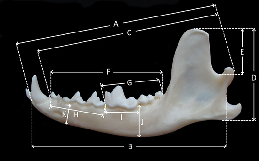
The most significant difference was the total length (A), the indentation between the condylar and the angular process (B), and the measurement from the aboral alveolus of the canine tooth to the condylar process (C).
The coronoid process (E) and the ramus mandibulae (D) were larger in jackals with height parameters of 19.4 mm and 42.6 mm, while the fox coronoid and ramus heights were 16.7 mm and 35.6 mm, respectively.
The present study provides morphometric and geometric data on red fox and golden jackal mandibles, which can be a significant resource in the taxonomy of carnivores.
Selected from the article's own text.
Abstract
The carnivore mandible as a lower part of the facial skeleton has a unique morphological characteristic and importance for the taxonomy. This study aimed to investigate size and shape differences of the mandible of the red fox and golden jackal. The research included 17 adult red fox and golden jackal mandibles from B&H. The morphometric analysis was performed on the 11 measurements and geometric analysis evaluates shape variation between two species with ten landmarks. The study revealed that the mandible of the jackal was longer and of massive bone compared to fox mandibles. The most significant difference was the total length, the indentation between the condylar and the angular process, and the measurement from the aboral alveolus of the canine tooth to the condylar process. The molar teeth parameters show similar values, especially the carnassial teeth. PC1 and PC2, which account for the highest variation, account for 68.8% of the total variation. The present study provided morphometric and geometric data on red fox and golden jackal mandibles, which can be a significant resource in the taxonomy of carnivores.
The red fox (Vulpes vulpes) and golden jackal (Canis aureus) both belong to the wild canid species present in Bosnia and Herzegovina (B&H). The golden jackal resembles a small wolf, with a more slender body, golden/yellowish fur and a long pointed muzzle. The European population ranges from the Balkans towards the Alps and, according to the data from 2014, the population in B&H was estimated to amount to 200-300 individuals ; . In 4 years the population increased substantially especially in the northern parts of B&H . The red fox has a smaller, long body, short legs, long ears and a long bushy tail. They adapt to a wide range of habitats, from tundra to desert, with huge urban populations in many European cities . Cranial morphometry is crucial for taxonomy and has a major impact on the management and maintenance of the species. Several studies have been conducted regarding the craniometrics of carnivores, including dog ; , wolf ; ; ; , fox ; ; , golden jackal ; and lynx ; . Most of these studies include the mandible as the lower part of the facial skeleton. The carnivore mandible is characterized by strong teeth, a deep masseteric fossa, several mental foramina and a unique angular process . Some recent investigations have used geometric analysis to reveal the shape differences rather than the size differences between the samples. This method focuses on analysis of data from the position of the homologous landmarks which offer more appropriate data when compared with traditional morphometric studies ; Gundemir et al., 2021).
The significant diversity of the carnivores’ size, diet, behavior, and locomotion is reflected in their skull shape and size . In addition, geographical distribution reveals variations in the carnivores’ skull morphometry . The study aimed to investigate the morphometric aspects of the red fox and golden jackal mandibles from B&H, and to reveal their shape differences using the geometric morphometric method.
MATERIALS AND METHODS
Animals
The mandibles of 11 jackals and 6 red foxes were used in this study. The study materials were obtained from the Osteology Museum at the Department of Anatomy of the Veterinary Faculty, University of Sarajevo. The mandibles were collected between 2015 and 2017 after autopsy, so no living animal was killed to obtain any of the materials used in the study. Therefore, the study did not present any ethical concerns. The muscles, fascia and other structures were dissected, boiled and macerated to remove all fat. In the final phase, the mandibles were bleached with 3% hydrogen peroxide .
Osteometric analysis
A total of eleven measurements were taken using an electronic digital caliper (0.1 mm) and the results were expressed as the mean and standard deviation (SD). The measurement parameters were selected according to a previous study (Von Den Driesch, 1976; ; and the description is given in Figure 1. The independent t-test was used to reveal the osteometric measurement differences between the 2 groups. SPSS 22 package program was used for statistical analysis.
Geometric analysis
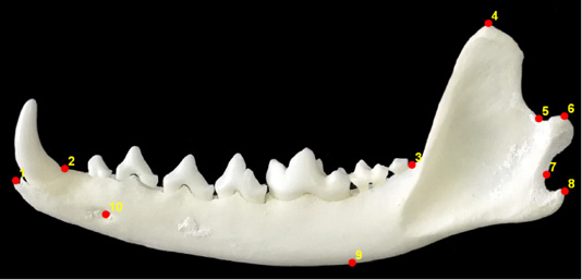
For the geometric morphometric analysis, the mandibles were photographed with a digital camera (Canon SX510 HS) from the same position and distance. The images obtained were converted from “jpg” files into a “TPS” file using the tpsUtil (Version 1.78) program. The ten landmarks shown in Figure 2 were placed using the tpsDig2 (Version 2.31) program . The data were saved as a “text” file and the MorphoJ (version 1.07a) program was used to visualize shape differences between red fox and jackal mandibles using discriminant function analysis. Principal component analysis (PCA) was performed to analyze the geometric analysis results statistically. Past (version 2.17c) statistical analysis program was used for PCA analysis. PC values explaining the highest variation were determined after the analysis. The results are given in tables. The terminology used in the study is in accordance with current anatomical terminology (NAV, 2017).
RESULTS AND DISCUSSION
The mandible consists of right and left halves, connected medially by a fibrocartilaginous symphysis (synchondrosis intermandibularis) in carnivores. Both halves are divided into a horizontal body (corpus mandibulae) and a vertical part (ramus mandibulae). The present study revealed that the mandible of the jackal was longer and of massive bone in comparison with fox mandibles, which is clearly indicated by the measurements in Table 1. The most significant difference was the total length (A), the indentation between the condylar and the angular process (B), and the measurement from the aboral alveolus of the canine tooth to the condylar process (C). The total cheek tooth row (F) was longer in jackals due to their premolars, while the molar teeth parameters show similar values, especially the carnassial teeth (I). The coronoid process (E) and the ramus mandibulae (D) were larger in jackals with height parameters of 19.4 mm and 42.6 mm, while the fox coronoid and ramus heights were 16.7 mm and 35.6 mm, respectively. The molar part of the mandible body was slightly wider in jackal with a more convex ventral margin. The two mental foramina at the level of the P1 and P3 were present in fox mandible in all specimens, while jackal mandible has single foramen in the level of the second premolar.
Table 1: The measurements (mm) of the mandibles in red fox and jackal.
Parameters
Red fox
Jackal
P value
N
Mean ± SD
N
Mean ± SD
A
22
102.1 ± 4.8
12
117.7 ± 2.4
<.001
B
22
98.3 ± 4.8
12
113.2 ± 3.6
<.001
C
22
90.6 ± 4.3
12
103.7 ± 2.2
<.001
D
22
35.6 ± 2.2
12
42.6 ± 1.4
<.001
E
22
16.7 ± 1.4
10
19.4 ± 1.07
<.001
F
22
58.03 ± 2.5
10
64.3 ± 1.5
<.001
G
22
25.7 ± 1.04
12
25.4 ± 3.1
.991
H
22
32.3 ± 1.8
10
38.9 ± 3.6
.003
I
22
14.1 ± 0.6
10
14.3 ± 1.2
.733
J
22
13.1 ± 0.7
10
16.6 ± 1.6
.001
K
22
11.3 ± 0.6
10
14.3 ± 1.6
.006
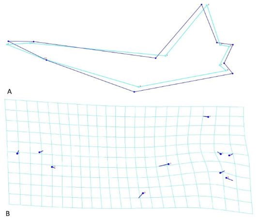
The shape variation between red fox and golden jackal mandible was determined with 10 landmarks as shown in Figure 2. The differentiation between red fox and jackal mandible was identified in several positions of the landmarks (Figure 3A). The extension and the direction of the differentiations between fox and jackal mandible were shown in Figure 3B. The ventral margin of the mandible body was more convex in the jackal at the level of landmark 9. The cheek tooth row was longer in fox mandible as shown in landmark 3. The condylar process was more massive and wider in the jackal when compared with the fox which is identified at the level of landmarks 5 and 6. Also, landmark 8 identified a stronger angular process in the jackal mandible.
PCA results are shown in Table 2. In total, 10 principal components were found. PC1 and PC2, which account for the highest variation, account for 68.8% of the total variation.
Table 2: PCA result of the mandibles in red fox and jackal.
PC
Eigenvalue
% variance
1
0,000239237
39,885
2
0,000173446
28,916
3
7,47412E-05
12,461
4
4,02184E-05
6,7051
5
3,17053E-05
5,2858
6
1,70342E-05
2,8399
7
1,27579E-05
2,1269
8
8,62367E-06
1,4377
9
2,05615E-06
0,34279
10
1,2878E-33
2,147E-28
Mandible morphology is closely associated with food habits and biomechanical performance. The carnivore mandible, along with its teeth, has been developed for tearing meat and biting with great force. The osteometric analysis of the mandible showed significant differences between carnivores, and even within the same species. The present study included 11 measurements of a total of 17 adult red fox and golden jackal mandibles from BandH. A detailed osteometric analysis of the red fox and golden jackal mandible has been reported in several studies. The total length of the red fox mandible was 9.6 ± 3.3 cm and corsac fox 9.1 ± 0.8 cm according to Munkzul et al. (2018), in the hoary fox 99.67 ± 1.14 mm according to Magalhaes et al. (2019), in golden jackals 11.2 ± 3.46 cm according to Monfared (2013). In other carnivores such as the lynx, the total length of the mandible was 102.6±5.2 mm according to Gomerčić (2010), 93.45 ± 2.98 mm according to Dayan et al. (2017) and in the wolf 190.4 ± 7.7 mm according to Okarma and Buchalczyk (1993). Our study revealed a significant difference between the total length of the fox (102.1 ± 4.8 mm) and jackal (117.7 ± 2.4 mm) mandibles, but this was similar to previous studies. Also, the total height of the vertical ramus was reported in two fox species as 3.7 ± 0.1 cm and 3.2 ± 0.2 cm in the golden jackal as 6.0 ± 0.58 cm , in the lynx as 38.92 ± 1.63 mm, 44.1 ± 3.2 mm, ; and in the wolf as 76.3 ± 3.8 mm . The present study revealed similar values as in foxes from Mongolia (35.6 ± 2.2 mm), while the total height for the jackal was 42.6 ± 1.4 mm, which was significantly smaller compared with the study of jackals from Iran. This value should be taken with caution since that study measured the maximum mandibular height from the ventral margin of the mandible, while our study measured the total height of the vertical ramus. The total cheek tooth row was longer in the red fox in comparison with the corsac fox, at 5.9 ± 0.2 and 5.0 ± 0.4 cm (Munkzul et al., 2018). Our findings were similar to the results for the red fox (58.03 ± 2.5 mm), but for the golden jackal the values were higher (64.3 ± 1.5 mm). Surprisingly, the size of the carnassial alveolus was similar in the present study in fox and jackal, and in accordance with a previous investigation of the red fox but more than double the size in the wolf, at 29.3 ± 1.2 mm .
CONCLUSIONS AND RECOMMENDATIONS
The present study provides morphometric and geometric data on red fox and golden jackal mandibles, which can be a significant resource in the taxonomy of carnivores.
Novelty Statement
The difference between the red fox and golden jackal mandible were revealed by traditional (osteometric) and developing technology (geometric) and obtained values can be a reference for future morphological and clinical studies.
AUTHOR’S CONTRIBUTION
All authors have equally contributed to this manuscript.
Conflict of interest
The authors declare that they have no conflict of interest.
Figures
Figure 1.
Parameters – jackal mandible. A. Total length; B. Length indentation between the condyle and angular process; C. Length condyl-aboral C alveolus; D. Height of the vertical ramus; E. Height of the coronoid process; F. Length of the cheektooth row M3-P1; G. Length of the molar row; H. Length of the premolar row P1-P4; I. Length of the carnassial alveolus; J. Height of the mandible behind M1; K. Height of the mandible P2-P3
Figure 2.
The points of the landmarks on red fox mandible. 1. Anterior end of the corpus mandibula; 2. Posterior edge of the canine alveolus; 3. Posterior edge of the teeth row; 4. Top of the coronoid process; 5. Level of the mandibular insicure; 6. Caudal edge of the condylar process; 7. Level between the condylar process and angular process; 8. Caudal edge of the angular process; 9. Ventral margin of the corpus mandibula at the level of posterior of the 1st molar alveolus; 10. Level of the first mental foramen
Figure 3.
Geometric morphometric analysis. A. Lineal representation of the shape differences between red fox and golden jackal; B. Landmark representation of shape differences of mandible between red fox and golden jackal
References
Aulagnier S, Haffner P, Mitchell-Jones AJ, Moutou F, Zima J (2008). Mammals of Europe, North Africa and the Middle East. Delachaux and Niestlé, Paris. pp. 100-105.
Avdić R, Hadziomerović N, Tandir F, Bejdić P, Ćutahija V (2013). Analysis of morphometric parameters of the roe deer mandible (Capreolus capreolus) and mandible of the sheep (Ovis aries). Veterinaria, 62(1-2): 1-9.
Choudhary OP, Singh I (2015). Morphometrical studies on the skull of Indian Blackbuck (Antelope cervicapra). Int. J. Morphol., 33(3): 868-876. https://doi.org/10.4067/S0717-95022015000300011[CrossRef]
Dayan MO, Gürbüz İ, Demiraslan Y, Ozgel O (2017). Craniometric measurements of the male eurasian lynx from Turkey. Anim. Vet. Sci., 5: 15-20. https://doi.org/10.11648/j.avs.20170501.13[CrossRef]
Gittleman JL (1985). Carnivore body size: Ecological and taxonomic correlates. Oecologia, 67: 540-554. https://doi.org/10.1007/BF00790026[CrossRef]
Gomerčić T, Sindičić M, Gomerčić MD, Gužvica G, Frković A, Pavlović D, Kusak J, Galov A, Huber Đ (2010). Cranial morphometry of the Eurasian lynx (Lynx lynx L.) from Croatia. Vet. Arhiv., 80(3): 393-410.
Gomes AC, Valente A (2016). Cranial and body size variation in the Iberian red fox (Vulpes vulpes silacea). Mammal. Biol., 81: 638-643. https://doi.org/10.1016/j.mambio.2016.08.005[CrossRef]
Gündemir O, Hadžiomerović N, Pazvan G, Erdikmen DO (2021). Radiometric and geometric morphometric analysis of the carpal joint area in 2-year-old thoroughbred horses. Veterinaria, 70(2): 209-217. https://doi.org/10.51607/22331360.2021.70.2.209[CrossRef]
Gürbüz I, Aytek AI, Demiraslan Y, Onar V, Ozgel O (2020). Geometric morphometric analysis of cranium of wolf (Canis lupus) and German shepherd dog (Canis lupus familiaris). Kafkas Univ. Vet. Fak. Derg., 26(4): 525-532.
Hadžiomerović N, Avdić R, Mulaomerović J, Mrvić V, Tandir F (2015). Cranial morphometry of the jackal (Canis aureus) from Neretva valley - Case report. Proceedings of the 3rd Turkish – Bosnian Scientific Days, Sarajevo 2015. pp. 41.
Ilgun R, Özüdoğru Z, Karabulut O, Can M (2021). Investigation of macroanatomical and morphometric on the skull bones of Aksaray Malakli dogs. Folia Morphol (Warsz). https://doi.org/10.5603/FM.a2021.0011[CrossRef]
Jhala YV, Moehlman PD (2004). Golden jackal (Canis aureus). In: Sillero-Zubiri C, Hoffmann M, Macdonald DW, editors. Canids: Foxes, wolves, jackals and dogs. Status survey and conservation action plan, IUCN/SSC Canid Specialist Group, Gland, Switzerland and Cambridge, UK. pp. 156-161.
Khosravi R, Kaboli M, Imani J (2012). Morphometric variations of the skull in the Gray wolf (Canis lupus) in Iran. Acta Theriol., 57: 361-369. https://doi.org/10.1007/s13364-012-0089-6[CrossRef]
König HE, Liebich HG 2004. Veterinary anatomy of domestic mammals. Stuttgart, Germany: Schattauer. pp. 69-71.
Magalhães HIR, Romão FB, de Paula YH, Luz MM, Barcelos JB, Silva Z, Carvalho-Baross RA, Ribeiro LA (2019). Morphometry of the mandibular foramen applied to local anesthesia in hoary fox (Lycalopex vetulus). Pesq. Vet. Bras., 39(4): 278-285. https://doi.org/10.1590/1678-5150-pvb-5749[CrossRef]
Meiri S, Dayan T, Simberloff D (2005). Variability and correlations in carnivore crania and dentition. Funct. Ecol., 19: 337-343. https://doi.org/10.1111/j.1365-2435.2005.00964.x[CrossRef]
Milenković M, Šipetić VJ, Blagojević J, Tattović S, Vujošević M (2010). Skull variation in Dinaric–Balkan and Carpathian gray wolf populations revealed by geometric morphometric approaches. J. Mammal., 91: 376-386. https://doi.org/10.1644/09-MAMM-A-265.1[CrossRef]
Monfared AL (2013). Macroanatomical investigation of the skull of golden jackal (Canis aureus) and its clinical application during regional anethesia. Glob. Vet., 10: 547-550.
Munkhzull T, Reading RP, Buuveibaatar B, Murdoch JD (2018). Comparative craniometric measurements of two sympatric species of vulpes in Ikh Nart nature reserve, Mongolia. Mongol. J. Biol. Sci., 16(1): 19-28.
NAV (2017). The international committee on veterinary gross anatomical nomenclature. 6th ed. (Revised version). Published by the Editorial Committee Hannover (Germany), Columbia, MO (USA), Ghent (Belgium), Sapporo (Japan). pp. 16-17.
Okarma H, Buchalczyk T (1993). Craniometrical characteristic of wolves Canis lupus from Poland. Acta Theriol., 38(3): 253-262. https://doi.org/10.4098/AT.arch.93-21[CrossRef]
Onar V (1999). A morphometric study on the skull of the german shepherd dog [Alsatian]. Anat. Histol. Embryol., 28: 253-256. https://doi.org/10.1046/j.1439-0264.1999.00202.x[CrossRef]
Rezić A, Bošković I, Lubinu P, Piria M, Florijančić T, Scandura M, Šprem N (2017). Dimorphism in the skull form of golden jackals (Canis aureus Linnaeus, 1758) in the Western Balkans: A geometric morphometric approach. Pakistan J. Zool., 49: 989-997. https://doi.org/10.17582/journal.pjz/2017.49.3.989.997[CrossRef]
Rohlf FJ, TpsDig2, Version 2.18. (2015). Department of ecology and evolution, State University of New York, Stone Brook, USA.
Trbojević I, Trbojević T, Malešević D, Krofel M (2018). The golden jackal (Canis aureus) in Bosnia and Herzegovina: density of territorial groups, population trend and distribution range. Mamm. Res., 63: 341-348. https://doi.org/10.1007/s13364-018-0365-1[CrossRef]
Von Den Driesch A (1976). A guide to the measurement of animal bones from archaeological sites. Peabody Museum Bulletin I. Cambridge MA, Harvard University. pp. 60-62.