
The red fox is widely distributed all over the world. These animals feed as wild animals and off the waste of people, thus having a close association with people. Anatomical knowledge of the red fox is very poor and we aimed to investigate the branches of the plexus brachialis in the red fox. We used six male red foxes that were died in traffic accidents. The right plexus brachialis was formed by the cervical spine nerves, C6, C7, and C8, and the thoracic spine nerve, T1 of the rami ventralis in four animals, and between C5, C6, C7, C8, T1 and T2 in two animals. The right plexus had a regular appearance. The left plexus brachialis consisted of C6, C7, C8 and T1 in all the red foxes examined. The right rami ventralis originating from C5 was thin, and after a short course joined to a branch of C6. The rami ventralis originating from T2 coursed directly via the axillar region of the right side. The left side was not as regular as the right side. In this study, we showed that there were differences between the right and left sides of the plexus brachialis in the red fox.
Research Article · Volume 53, Issue 5, 15 June 2021 · Open Access
Macroanatomical Investigation of the Plexus Brachialis in the Red Fox (Vulpes vulpes)
·
Affiliations & Notes ⌄
- 1Department of Anatomy, Faculty of Ceyhan Veterinary Medicine, Cukurova University, Adana,Turkey , * Corresponding author: semaerten@80gmail.com
Article Info ⌄
- Journal:
- Pakistan Journal of Zoology
- Published:
- 15 June 2021

From the article
- The right plexus brachialis was stronger and more defined than the left plexus brachialis and the branches of the right side showed complete and significant connections with each other.
- The left N. musculocutaneus (c in Fig. 2) originated from ventral branches of C8 and T1 spinal nerves in all six foxes while the right (c in Fig. 1) side originated from only C7 in all animals.
- We observed that the right plexus brachialis was generally formed from C5, C6, C7, C8, T1 and T2 of the rami ventralis of the spinal nerves.
- We showed that there were differences between the right and left sides in the red fox and that the branches and origin of the plexus brachialis were very similar to cats and the marten.
Selected from the article's own text.
Abstract
Introduction
The red fox (Vulpes vulpes) is widely distributed all over the world, including Siberia, India, Europe, Asia, North Africa, America, the North Pole, and throughout Turkey . These animals generally live in forests, fields and mountains and their survival is largely based on their ability to feed on a great variety of food items such as wild and domestic birds, mice, moles, all insects and their larva, slugs, worms, lizards, and fish. In addition, they sometimes eat fruit. This type of diet contributes to the environmental balance ; .
The red fox is labeled as a carnivore and has characteristic properties such as a slender pointed muzzle, prominent erect ears, long slender legs, and relatively small feet. The front legs have a very important role in land digging and food grabbing (Demirsoy, 2005). The brachial plexus innervates to the front legs and the plexus brachialis and branches radiate within the front legs. They generally originate from ventral branches of the C6, C7, C8, T1 and T2 spinal nerves (Evans and de Lahunta, 2013; . Anatomically, the origin, course, or distribution of the plexus brachialis has been studied in many domestic and wild animals such as the cat , New Zealand rabbit , chinchilla , rat , red squirrel , marten , bonobo , pampas fox , and crab-eating fox (Souza et al., 2014b). Nevertheless, the anatomy of the plexus brachialis of the red fox, which is a wild carnivore, is still unknown and there is a paucity of literature available on the subject. We aimed to describe the origin and branches of the plexus brachialis of the red fox.
Materials and methods
This study was performed with permission from the General Directorate of Nature Conservation and National Parks of the Ministry of Forestry and Water region (Permission Number: 38002405-445.05-177733). This study was also accepted by the animal experiments local ethics committee (2017/6-14) of Cukurova University.
This study was performed by using six male red foxes that were collected at different time intervals as a result of road traffic accidents in the Adana region. The animals were all older than six months, as assessed by their dental maturity. Body weight varied from 6 to 9 kg. The animals were stored in a freezer, then fixed in a 10% formalin solution at room temperature for dissection. To demonstrate the plexus brachialis, the soft tissue, muscle, fascia and fat were removed and carefully dissected. For terminology, the Nomina Anatomica Veterinaria was used.
Results
The plexus brachialis and its branches are known to vary among species and even within the same species. The plexus brachialis and its branches in the six red foxes examined summarized in Table I. In four of the six animals, the right plexus brachialis consisted of ventral branches from the C6, C7, C8 and T1 spinal nerves, while in the remaining two foxes it originated from C5, C6, C7, C8, T1 and T2. The left plexus brachialis originated from C6, C7, C8 and T1 ventral branches of spinal nerves in all six animals. We observed the plexus brachialis to vary in shape in different areas. These nerves generally passed disto-caudally and in the lateral aspect of the arteria axillaris was observed. The right plexus brachialis was stronger and more defined than the left plexus brachialis and the branches of the right side showed complete and significant connections with each other. In each plexus an ansa axillaris structure was not formed in all six animals.
The nervus (N.) suprascapularis (b in Fig. 2) originated from the left ventral branches of C6, C7 in four animals and the C8 spinal nerve in two animals. The right side (b in Fig. 1) originated from the ventral branches of C5, C6 and C7 in four red foxes and only C7 in two animals. The left nervi (Nn.) subscapulares (a in Fig 2) comprised ventral branches from C6 and C7 spinal nerves in all animals. However, the right nerves (a in Fig. 1) were formed by ventral branches of C5, C6, and C7 spinal nerves in two animals. The left N. musculocutaneus (c in Fig. 2) originated from ventral branches of C8 and T1 spinal nerves in all six foxes while the right (c in Fig. 1) side originated from only C7 in all animals.
The left N. axillaris (d in Fig. 2) originated from the ventral branches of C7 and C8, and the right (d in Fig. 1) side originated from the ventral branches of C7. When N. axillaris separated from the plexus brachialis, both a ventral and caudal course was observed.
While the left nn. pectorales craniales originated from C7, C8 and T1 in two animals, it’s the origin was C8 (e in Fig. 2) in four animals. The right nn. pectorales craniales (e in Fig. 1) was derived from C6 and C7 in four animals, and C7 and C8 in two animals. The nerve branched into three and five in two animals, whereas it diverged into 2 or 4 branches in four animals.
The left N. thoracicus longus (l in Fig. 2) originated from the ventral branches of C7 and C8 in four animals and only C8 in two animals. The right nerve (l in Fig. 1) was formed from the ventral branches of C7 and C8 in all six foxes.
The left N. thoracodorsalis (j in Fig. 2) was formed from ventral branches of C8 and T1 in all cases. The right side of this nerve (j in Fig. 1) was derived from C8 and T1 in three animals, and T1 and T2 in the other three.
The Nn. pectorales caudales (k in Figs. 1, 2) and N. thoracicus lateralis (right and left) (i in Fig. 1) both originated from the ventral branches of C7 and C8 in all six foxes.
Table I.- Branches of plexus brachialis of red fox in left and right side.
| Branches of plexus brachialis | Origin | |
| Left | Right | |
| Nervus suprascapularis | C6,C7 (in 4 animals) C8 (in 2 animals) | C5,C6,C7 (in 4 animals) C7 (in 2 animals) |
| Nervi subscapulares | C6,C7 (in all animals) | C5,C6,C7 (in 2 animals) C6, C7 (in 4 animals) |
| Nervus musculocutaneus | C8, T1 (in all animals) | C7 (in all animals) |
| Nervus axillaris | C7, C8 (in all animals) | C7 (in all animals) |
| Nervi pectorales craniales | C7,C8,T1 (in 2 animals) C8 (in 4 animals) | C6, C7 (in 4 animals) C7,C8 (in 2 animals) |
| Nervus thoracicus longus | C7,C8 (in 4 animals) C8 (in 2 animals) | C7, C8 (in all animals) |
| Nervus thoracodorsalis | C8, T1 (in all animals) | C8, T1 (in 3 animals) T1-T2 (in 3 animals) |
| Nervus thoracicus lateralis | C7, C8 (in all animals) | C7, C8 (in all animals) |
| Nervi pectorales caudales | C7-C8 (in all animals) | C7, C8 (in all animals) |
| Nervus radialis | C6, C7 (in all animals) | C6, C7 (in 4 animals) C8 (in 2 animals) |
| Nervus ulnaris | C7, C8, T1 (in all animals) | C7, C8, T1 (in all animals) |
| Nervus medianus | C8, T1 (in all animals) | C8, T1 (in all animals) |
N. radialis (f in Fig. 2) was derived from the ventral branches of C6-C7 in the left side, and ventral branches of C6-C7 in four animals and C8 in two animals in the right side (f in Fig. 1). N. radialis was very thick.
Right and left N. medianus (g in Figs. 1, 2) was formed by ventral branches from C8 and T1 in all cases. A connecting branch between N. medianus and N. musculocutaneus was observed in all animals. In three animals, N. medianus and N. ulnaris showed a common trunk.
Both right and left N. ulnaris (h in Figs. 1, 2) originated from ventral branches of C7, C8 and T1 in all six foxes. We also observed that N. medianus and N. ulnaris ran a parallel course, except in one animal.
Discussion
The plexus brachialis and its branches are well known to show variations among species and even within the same species. Formation of the plexus comprises C5-T1 in myocastor coypus , the giant anteater (Souza et al., 2014a), and bonobo , C5-T2 in cats , and the chinchilla , C6-T1 in sheep and goats , pampas fox and crab-eating fox (Souza et al., 2014b), and C6-T2 in Anatolian sheep dogs (Kangal dog) (Dursun et al., 1994), horses, cattle and pigs ; . In addition, the plexus has been reported to occur as two roots in the hippopotamus , and as three roots from the ventral branch of C5-8 in the red squirrel . While Angelica-Almeida et al. (2013) reported that the plexus originates between C4 and T1 in rats, Ozbag et al. (2009) suggested that it is between C5 and T1 in the same animals. On the other hand, a mole-rat plexus brachialis study showed similar results to those of Ozbag et al. (2009). In the present study on the red fox, we showed that the plexus brachialis originated from C5, C6, C7, C8, T1 and T2, similar to that seen in cats and the chinchilla . Our results indicate similarities with cats rather than dogs.
N. suprascapularis originates from ventral branches of either C6 alone or C6-7 in the crab-eating fox (Souza et al., 2014b), C6-7 in the Anatolian sheep dog (Kangal dog) (Dursun et al., 1994), C4-6, C5-6 or only C6 in Macaca mulatta , and C6 alone, or C6-7 or C7-8 in the pampas fox . In the present study, the right N. suprascapularis was formed from ventral branches belonging to C5-7 or C7 alone. The left N. suprascapularis originated from ventral branches from C6-7 or only C8.
Nn. subscapulares is separated from the dorsal section of the plexus in the bonobo , and the cranial trunk in hippopotamus , and originates from C6-7 in the Anatolian sheep dog (Kangal dog) (Dursun et al., 1994). Moreover, these nerves are divided into two branches in the cat , bonobo , and hippopotamus . These nerves originate from C6-C7 or only C6 ventral braches, and are separated as four branches in Macaca mulatta . In the giant anteater (Souza et al., 2014a) nn. subscapularis is derived from C5-7, and from C6-7 in the pampas fox . In the present study, the left nn. subscapulares originated from the ventral branch of C6-7, consistent with that reported for the Anatolian sheep dog (Kangal dog) (Dursun et al., 1994) and pampas fox .
N. musculocutaneus is observed to originate from C7-C8 in most animals, however, in the pampas fox it is reported to derive from nerve branches between C6-T1. It is generally derived from C6-7 in the crab-eating fox (Souza et al., 2014b). N. musculocutaneus is formed from the C7 spinal nerve alone in the marten and Anatolian sheep dog (Kangal dog) (Dursun et al., 1994). It originates from the C5-C7 spinal nerves on both the right and left sides of Macaca mulatta , however, in the majority of these animals it is C6-C7. In the chinchilla , this nerve derives only from the C7 spinal nerve. In the present study N. musculocutaneus originated only from C7 on the right side and C8-T1 on the left side.
N. axillaris is derived from only C7, C6-8 or C7-8 ventral branches in the pampas fox , from C7 alone, C6-7, C6-8 or C7-8 in the crab-eating fox (Souza et al., 2014b), and C7 alone in the marten . Both the right and left N. axillaris is formed from C6-C7 spinal nerves in Macaca mulatta and chinchilla . In the present study, the left N. axillaris was formed from C7-8 spinal nerves similar to the Anatolian sheep dog (Kangal dog) (Dursun et al., 1994).
Nn. pectorales craniales in the pampas fox and the crab-eating fox (Souza et al., 2014b) is derived from the ventral branches of C6-7, C7 alone, C7-T1, C8 alone or C8-T1, and from C7 alone in the marten and Anatolian sheep dog (Kangal dog) (Dursun et al., 1994). Both sides of nn. pectorales craniales originate from C6 alone, C6-7, C7-8-T1 or C8-T2 in Macaca mulatta . In the chinchilla these nerves comprise ventral branches from C7-8 or T1-2 spinal nerves. In the present study we observed that the left nn. pectorales craniales originated from C7-T1 or C8 while the right side was derived from either C6-7 or C7-8 spinal nerves.
N. thoracicus longus is generally derived from the C7 spinal nerve in animals (Dursun et al., 1994; Souza et al., 2014b, 2016). In the marten it derives from the ventral branches of C7-8, and in the chinchilla , (one side) and both sides of Macaca mulatta it originates from C6-7, C7 alone, or C6-8. In the present study, we observed that the left N. thoracicus longus was formed from either C7-8 or C8 alone. The right side of this nerve was as reported in the literature .
N. thoracodorsalis originates from C7-8, C8 alone, C7-T1 or C8-T1 in the pampas fox , and C8 alone, C6-7, C7-8, C8-T1 or C7-T1 in the crab-eating fox (Souza et al., 2014b). In both the marten and Anatolian sheep dog (Kangal dog) (Dursun et al., 1994) the right side originates from C7-8 while in Macaca mulatta the right side is derived from C7-8 or C8-T2 and from C7-8, C8-T1 or C8-T2 on the left side. In the chinchilla , it derives only from C8. In the present study on the red fox, the left side of N. thoracodorsalis originated solely from C8-T1, while the right side was from either C8-T1 or T1-T2.
N. thoracicus lateralis and nn. pectorales caudalis originate from C8 alone, T1 alone, C7-T1, or C8-T1 in the pampas fox , from C7-8 in the marten , T1-2 in the chinchilla , C8-T2 in the Anatolian sheep dog (Kangal dog) (Dursun et al., 1994), and C8 alone, T1 alone, C7-T1, C8-T1, C6-7 or C7-8 in the crab-eating fox (Souza et al., 2014b). In the present study, these nerves were formed from the C7-8 spinal nerves, similar to that reported for the marten .
N. radialis derives from C7-8, C7-T1 or C8-T1 in the pampas fox and C7-8, C7-T1, C8-T1 or C6-8 in the crab-eating fox (Souza et al., 2014b). In the marten it originates from C8-T1, and C7-T1 in the Anatolian sheep dog (Kangal dog) (Dursun et al., 1994). In Macaca mulatta the right side N. radialis is formed from C7-8, and C8-T2 in the chinchilla . In the present study, while the left side N. radialis was formed from C6-7, the right side originated from C6-8. Contrary to the literature (Dursun et al., 1994), N. radialis was observed to be a fairly thick compared to other nerves in the red fox.
N. ulnaris is reported to have the same origins as N. medianus in the pampas fox , crab-eating fox (Souza et al., 2014b), and the marten . It originates from C8-T2 in the Anatolian sheep dog (Kangal dog) (Dursun et al., 1994) and in Macaca mulatta , as well as C8-T1 in Macaca mulatta , and is derived from T1-2 in the chinchilla . In the present study, it originated from C7-T1, unlike any previous reports ; Dursun et al., 1994; Souza et al., 2014b, 2016). In addition, we observed that N. ulnaris and N. medianus ran parallel (except for one animal).
N. medianus is derived from C7-8, C7-T1, C8-T1 or T1 alone in the pampas fox , from C7-T1, C8-T1 or T1 alone in the crab-eating fox (Souza et al., 2014b), from C8-T1 in the marten , C7-T2 in the Anatolian sheep dog (Kangal dog) (Dursun et al., 1994), C6-T1 or C6-T2 in Macaca mulatta , and C7-T1 in the chinchilla . We observed that N. medianus was derived from C8-T1 in the red fox. In addition, we noted that N. medianus and N. ulnaris were seen in a common root in three red foxes.
Conclusions
We conducted a macroanatomical investigation of the plexus brachialis and its branches in the red fox. We observed that the right plexus brachialis was generally formed from C5, C6, C7, C8, T1 and T2 of the rami ventralis of the spinal nerves. The left plexus brachialis comprised C6, C7, C8 and T1. The right plexus brachialis had a regular appearance. We showed that there were differences between the right and left sides in the red fox and that the branches and origin of the plexus brachialis were very similar to cats and the marten. This information may be very beneficial to veterinarians, surgeons and biologists working with wild animals or in zoos.
Statement of conflict of interest
The authors declare no conflict of interest.
Figures
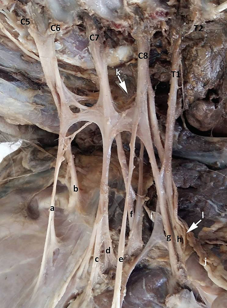
Right plexus brachialis. Dorsal aspect and separate of right neck. a, Nn. Subscapulares; b, N. suprascapularis; c, N. musculocutaneus; d, N. axillaris; e, Nn. pectorales craniales; f, N. radialis; g, N. medianus; h, N. ulnaris; i, N. thoracicus lateralis; j, N. thoracodorsalis; k, Nn. pectorales caudales; l, N. thoracicus longus; p, N. phrenicus; C5, Rami ventralis of cervical 5; C6, Rami ventralis of cervical 6; C7, Rami ventralis of cervical 7; C8, Rami ventralis of cervical 8; T1, Rami ventralis of thoracal 1; T2, Rami ventralis of thoracal 2.
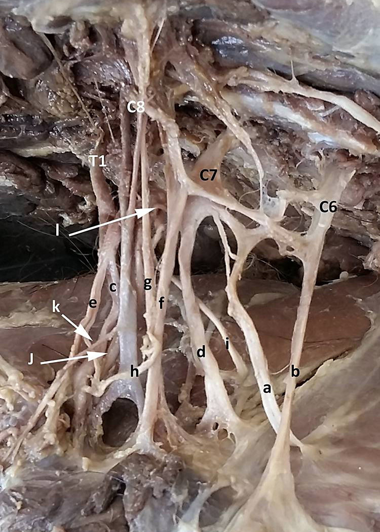
Left plexus brachialis. Dorsal aspect and separate of left neck. a, Nn. Scapulares; b, N. suprascapularis; c, N. musculocutaneus; d, N. axillaris; e, Nn. pectorales craniales; f, N. radialis; g, N. medianus; h, N. ulnaris; i, N. thoracicus lateralis; j, N. thoracodorsalis; k, Nn. pectorales caudales; l, N. thoracicus longus; p, N. phrenicus; C6, Rami ventralis of cervi cal 6; C7, Rami ventralis of cervical 7; C8, Rami ventralis of cervical 7; T1, Rami ventralis of thoracal 1.
References
- Angelica-Almeida, M., Casal, D., Mafra, M., Mascarenhas-Lemos, L., Martins-Ferreira, J., Ferraz-Oliveira, M., Amarante, J. and Goyri-O’neill, J., 2013. Brachial plexus morphology and vascular supply in the wistar rat. Acta Med. Port., 26: 243-250.
- Anonymous, 2017. Nomina anatomica veterinaria. Prepared by the International Committes on Veterinary Gross Anatomical Nomenclature and Authorized by the General Assambly of the World Association of Veterinary Anatomists, The Editorial Committee Hannover, Japan.
- Aydin, A., 2011. The spinal nerves that constitute the brachial plexus in the red squirrel (Sciurus vulgaris). Vet. Med. Czech., 56: 405-408. https://doi.org/10.17221/1556-VETMED[CrossRef]
- Aydin, A. and Karan, M., 2012. The spinal nerves forming the brachial plexus in mole-rats (Spalax leucodon). Vet. Med. Czech., 57: 430-433. https://doi.org/10.17221/6311-VETMED[CrossRef]
- Bakaloudis, D.E., Bontzorlos, V.A., Vlachos, C.G., Papakosta, M.A., Chatzinikos, E.N., Braziotis, S.G. and Kontsiotis, V.J., 2015. Factors affecting the diet of the red fox (Vulpes vulpes) in a heterogeneous Mediterranean landscape. Turk. J. Zool., 39: 1151-1159. https://doi.org/10.3906/zoo-1411-22[CrossRef]
- Cevik-Demirkan, A., Ozdemir, V., Demirkan, I. and Turkmenoglu, I., 2007. Gross morphological features of plexus brachialis in the chinchilla (Chinchilla lanigera). J. S. Afr. Vet. Ass., 78: 21-24. https://doi.org/10.4102/jsava.v78i1.281[CrossRef]
- Demiraslan, Y., Aykut, M. and Ozgel, O., 2015. Macroanatomical characteristics of plexus brachialis and its branches in martens (Martes foina). Turk. J. Vet. Anim. Sci., 39: 693-698. https://doi.org/10.3906/vet-1506-48[CrossRef]
- Demirsoy, A., 2003. Basic rules of life: Vertebrates / Amniota (reptiles, birds and mammals). Meteksan.Ankara. pp. 749-750.
- Dursun, N., Tipirdamaz, S. and Gezici, M., l994. Macroanatomical investigation of the plexus brachialis in the Kangal dog. Vet. Bil. Derg., 1: 78-80.
- Evans, H.E. and de Lahunta, A., 2013. Millers anatomy of the dog, 4th ed. Elsevier, London.
- Kikuchi, Y., Oishi, M. and Shimizu, D., 2011. Morphology of brachial plexus and axillary artery in bonobo (Pan paniscus). Anat. Histol. Embryol., 40: 68-72. https://doi.org/10.1111/j.1439-0264.2010.01040.x[CrossRef]
- König, H.E. and Liebich, H.G., 2015. Veterinary anatomy of domestic mammals, 6th ed. Stuttgart, Germany, pp. 542-543.
- Mencalha, R., Fernandes, N., Sousa, C.A.S. and Abidu-Figueiredo, M., 2014. A cadaveric study to determine the minimum volume of methylene blue to completely color the nerves of brachial plexus in cats. An update in forelimb and shoulder surgeries. Acta Bras. Cir., 29: 877-882. https://doi.org/10.1590/S0102-86502014000600006[CrossRef]
- Mohiuddin, M., Rahman, M.L., Alim, M.A., Kabir, M.B.H. and Kashem, M.A., 2011. Macro anatomical investigation of brachial plexus of the white New Zealand rabbit (Orycotolagus cuniculus). Int. J. nat. Sci., 1: 74-76.
- Ozbag, D., Gumusalan, Y., Bakaris, S., Ciralik, H., Senoglu, M. and Kalender, A.M., 2009. Morphologic and morphometric investigation of plexus brachialis in rat. Anatomy, 3: 21-28. https://doi.org/10.2399/ana.09.003[CrossRef]
- Pop, C. and Penteathe, M., 2007. Roots of the brachial and lumbosacral plexus in Myocastor coypus. Lucrari Stiintifice: Med. Vet. Timisoara, 41: 569-571. https://usab-tm.ro/vol7MV/101_vol7.pdf
- Santos-Sousa, C.A., Gomes, M.S., Da Cruz De Carvalho, N., Souza, Jr. P., Machado Dos Santos, C. and Abidu-Figueiredo, M., 2016. Origin and antimeric distribution of brachial plexus nerves in Macaca mulatta (Zimmermann, 1780) (Primates: Cercopithecidae). Ital. J. Zool., 83: 469-481.
- Sidorovich, V.E., Sidorovich, A.A. and Izotova, I., 2006. Variations in the diet and population density of the red fox Vulpes vulpes in the mixed woodlands of northern Belarus. Mammal. Biol., 71: 74-89. https://doi.org/10.1016/j.mambio.2005.12.001[CrossRef]
- Sisson, S., Grossman, J.D. and Getty, R., 1975. The anatomy of the domestic animals, vol. I, II, 5th edn. W.B. Saunders Co., Philadelphia, pp. 668-669, 1127, 1385, 1700.
- Souza, Jr. P., Carvalho, N.C., Mattos, K., Figuiredo, M.A. and Santos, A.L.Q., 2016. Brachial plexus in the pampas fox (Lycalopex gymnocercus): A descriptive and comparative analysis. Anat. Rec., 300: 537-548.
- Souza, Jr. P., Cardoso, J.R., Araujo, L.B.M., Moreira, P.C., Cruz, V.S. and Araujo, E.G., 2014a. Gross anatomy of the brachial plexus in the giant anteater (Myrmecophaga tridactyla). Anat. Histol. Embryol., 43: 341-345. https://doi.org/10.1111/ahe.12080[CrossRef]
- Souza, Jr. P., Carvalho, N.C., Mattos, K. and Santos A.L.Q., 2014b. Origins and ramifications of brachial plexus in the crab-eating-fox Cerdocyon thous (Linnaeus, 1766). Pesq. Vet. Bras., 34: 1011-1023. https://doi.org/10.1590/S0100-736X2014001000015[CrossRef]
- Yoshitomi, S., Kawashima, T., Murakami, K., Takayanagi, M., Inoue, Y., Aoyagi R. and Sato, F., 2012. Anatomical architecture of the brachial plexus in the common hippopotamus (Hippopotamus amphibius) with special reference to the derivation and course of its unique branches. Anat. Histol. Embryol., 41: 280-285. https://doi.org/10.1111/j.1439-0264.2012.01135.x[CrossRef]
Services
Article usage statistics
Loading…