
Lathyrol, a natural diterpenoid molecule is one of the major components of Semen Euphorbiae, a famous traditional Chinese medicine with a long history of clinical use in China. Very recently, lathyrol has been reported to inhibit the growth and induce apoptosis in lung cancer cells. However, the anticancer activity of lathyrol remains largely unknown in various human cancers. The present study was designed to evaluate lathyrol for its broad-spectrum anticancer activity and binding affinity with STAT3 DNA binding domain. Using CCK-8 assay kit, we showed that lathyrol reduced the cell viability of Hep-3B, MHCC97-L, A2780 and taxol resistant Hey-T30 cells in a dose-dependent fashion. Using Molecular docking study, we found that lathyrol exhibits strong binding interactions with STAT3 DNA binding domain through hydrogen bonding and hydrophobic interactions with various amino acid residues of STAT3. Using immunoblotting, we found that lathyrol did not inhibit STAT3 phosphorylation and dimerization. Moreover, we showed that lathyrol induces apoptosis as evident from a remarkably increased expression of cleaved caspase-3 in all four cancer cell lines. Taken together, our findings suggest that lathyrol is a potent STAT3 DNA binding domain inhibitor and exhibits a broadspectrum anticancer activity.
Research Article · Volume 56, Issue 6, 7 November 2024 · Open Access
Lathyrol Binds with STAT3 DNA Binding Domain and Induces Apoptosis in Multiple Human Cancer Cells
· · · · · · · ·
Affiliations & Notes ⌄
- 1Cancer research Lab, Institute of Zoology, University of the Punjab, Quaid-e-Azam Campus, Lahore, Pakistan 2School of Medicine and Holistic Integrative Medicine, Nanjing University of Chinese Medicine, Nanjing 210023, China. 3School of Biological Sciences, University of the Punjab, Quaid-e-Azam Campus, Lahore, Pakistan 4Department of Biotechnology, University of Sargodha, Sargodha, Pakistan. Abrar ul Haq and Shusheng Yin contributed equally in this paper. * Corresponding author: khan_zoologist@y
Article Info ⌄
- Journal:
- Pakistan Journal of Zoology
- Published:
- 7 November 2024

From the article
- In this study, we have identified lathyrol a natural bioactive molecule as potential STAT3 DNA binding domain inhibitor using molecular docking study.
- The data clearly showed that lathyrol binds only with DBD of STAT3.
- In conclusion, we have shown that lathyrol is a potent STAT3 DBD inhibitor and exhibits good anticancer activity against multiple human cancer cell lines.
- Finally, further in vitro study is required to validate the potential of lathyrol as STAT3 DBD inhibitor which is one of the major limitations of this study.
Selected from the article's own text.
Abstract
Introduction
Signal transducer and activator of transcription 3 (STAT3) is a key transcriptional factor of the STAT protein family, together with STAT1, 2, 4, 5a, 5b, and 6 ; . The structure of STATs is divided into different domains like a coiled-coil domain (CCD), an amino-terminal domain (ATD), a DNA-binding domain (DBD), Src homology 2 domain (SH2D), linker domain and a transactivation domain ; . Among all the members, STAT3 is the most investigated member of the STATs family. STAT3 can be triggered by different factors like growth factors, cytokines, and oncogenic proteins . Stimulation of STAT3 by growth factors and cytokines triggers the phosphorylation of STAT3 at tyrosine residue 705 (Tyr705), that results in STAT3 dimerization, and translocation into nucleus where it binds with DNA and induces transcription of certain genes ; . STAT3 potentially regulates the expressions of different genes involved in controlling the vital functions of the cell such as, cell proliferation, cell survival, metastasis and invasion, apoptosis, and development of immune suppressive tumor microenvironment. Under normal circumstances, the expression of STAT3 is highly regulated but if this balance gets disturbed then overexpression of STAT3 can lead to multiple types of cancers such as multiple myeloma, hepatocellular carcinoma, epidermoid carcinoma, prostate cancer, breast cancer, lung cancer, and pancreatic cancer etc. . It is strongly recommended that higher expression of phosphorylated STAT3 increases the rate of malignancy of the neoplasm . Therefore, inhibition of STAT3 is characterized as a striking target for the development of new antineoplastic drugs.
Indeed, several inhibitors of STAT3 have been identified over the last 15 years ; ; . Some of them even potentially inhibit the growth in xenograft tumors. However, very limited number of these inhibitors have reached the clinical trials but none of them is approved for clinical practices. The causes of such failures are still unknown. It is important to note that all these inhibitors were designed to bind with SH2D of STAT3 and thereby inhibiting STAT3 tyr705 phosphorylation and dimerization of STAT3. Such approach of drug designing to find the inhibitor of STAT3 is a little bit problematic because it is reported that unphosphorylated STAT3 even has the potential to bind to DNA and may still be efficient to perform its functions ; ; . Thus, disrupting the activation of STAT3 by inhibiting the interactions between Tyr705 phosphorylation and SH2 domain alone may not completely lower the level of STAT3. Although targeting the DBD of STAT3 can be more efficient to diminish the expressions of STAT3, it has yet not been in the main domain of research because it is expected that targeting DBD may has limited selectivity ; ; . But due to a continuous disappointment in finding the inhibitor of STAT3 by using the SH2D, now researchers started to find the inhibitors for DBD of STAT3 to develop new therapeutics for cancer , 2016; .
Plants have a great role in human community, they may be used for nutritional additives, beverages production, flavoring agents, shelter, supplier of oxygen, forage for animals and to lower the burden of various disorders such as cancer ; . Many clinically practiced anticancer therapeutics such as camptothecin, taxol, podophyllotoxins and vinca alkaloids are obtained from biological sources ; ; . Eighty-five out of 175 drugs that had been practiced as an anticancer drug between 1940-2012 were derived from mother nature . Naturally derived drugs are multitargeted, less toxic, cost-effective, and easily available in every region across the globe that’s why now a days naturally derived molecules are preferably used for the treatment of various disorders including cancer .
Euphorbia semen (ES) is a traditional Chinese medicine made by the ripe and dried seed of Euphorbia lathyris. In China it is used to increase blood circulation, eliminate blood stasis and to treat scabies and tinea, snakebites, anuria, amenorrhea, terminal schistosomiasis, and constipation . The major bioactive molecules of SE include diterpenoids, coumarins, flavonoids, amino acids, steroids, volatile oil, and fatty oil . The diterpenoids, coumarins, and flavonoids in ES show multiple pharmacological activities, such as diuresis, antibacterial, anti-inflammatory, hepatoprotective, neuroprotective, antiviral, and anti-tumor both in in vivo and in clinical practice ; .
Lathyrol, a diterpenoid component of SE ; has been suggested to exhibit different pharmacological activities including anticancer effects . Here in this study, using molecular docking study, we have first time discovered that lathyrol could exclusively binds with DBD of STAT3 and induces anticancer activity in multiple drug-sensitive and drug-resistant cancer cell lines.
Materials and Methods
Reagents and antibodies
Lathyrol (Fig. 1) was purchased from TargetMol. (Catalog No. T3S2019). The purity of lathyrol was >99%. The primary antibodies against cleaved caspase-3 (9664), Phospho-STAT3 (9145) were purchased from Cell Signaling technology (Baverly MA). The primary antibodies against STAT3 (10253-2-AP) and GAPDH (10494-1-AP) and HRP-conjugated secondary antibody (goat anti-rabbit (SA00001-2) were purchased from Proteintech (Wuhan, China).
Cell lines and cell culture
Human ovarian cancer cell line A2780 was obtained from the European Collection of Cell Culture. Human liver cancer cell lines Hep-3B, and MHCC97-L and human Hey-T30 taxol resistant cell line were purchased from ATCC (USA). A2780, Hep-3B and MHCC97-L cells were cultured in DMEM medium supplemented with 10% FBS while Hey-T30 cells were cultured in RPMI-1640 medium supplemented with 10%FBS. The culture media were supplemented with 100units/mL penicillin and 100µg/mL streptomycin.
Determination of cell viability
Cells were cultured in recommended media supplemented with 10%FBS and treated with lathyrol in a concentration-dependent manner overnight. Following drug treatment for indicated time periods, CCK-8 assay was employed to evaluate cell viability as described by us previously .
Immunoblotting
Cells were cultured in 6 well plates and treated with indicated concentration of lathyrol overnight. Total cell lysate was prepared and subjected to immunoblotting for the expression of p-STAT3, STAT3, Cleaved caspase-3 and GAPDH as described by us previously .
Molecular docking
Retrieval of ligand (Lathyrol) and target protein (STAT3)
The 3D structure of STAT3 (PDB ID: 1BG1) with resolution 2.25 Å was retrieved from the official website, Protein Data Bank (PDB) (www.rcsb.pdb.com) in the form of PDB format. The ligand molecule, lathyrol was downloaded from PubChem (PubChem CID: 15479845) (www.pubchem.com) and saved in the form of structure data file (SDF).
Preparation of target (STAT3) protein
The retrieved 3D structure of protein from PDB may contain the water molecules and nonstandard ligands that may interfere with the interaction of our desired ligand with the protein molecule during molecular docking (MD) that’s why all these molecules must be removed before running the MD. To prepare the protein we just opened the previously retrieved PDB format of STAT3 in the Biovia discovery visualizer (BDV) then, water molecules, heteroatoms, and nonstandard ligands were selected and deleted. Then we saved the crystal structure of STAT3 in PDB format.
Docking
For the MD we used the PyRx software 0.8 version. To run the molecular docking the prepared crystal structure of STAT3 (in PDB format) was imported in PyRx and selected as a macromolecule, this causes the minimization of STAT3 by using the default settings with the addition of specific charges and hydrogen bonds. Then ligand (lathyrol) was imported in PyRx in SDF format and was minimized by using default settings of PyRx and finally converted it into PDBQT, the required format of ligand to run the molecular docking.
After that the protein and ligand were selected using command Vina wizard in PyRx. The grid box (The grid box basically selects the boundary of the docking of ligand with our desired macromolecule) was prepared as follows: STAT3 [Centre: Dimension-X (22.9381:46.6372); Y (12.5582:49:4057); Z (3.7933:59.1362)] and run the docking at exhaustiveness of 8. After the completion of the docking the pose with lowest binding affinity between protein and ligand was selected.
Visualization
Ligand binding with 3D structure of STAT3 (Fig. 1) is checked by using PyMol software. Further interactions between the ligand and STAT3 residues were visualized using the BDV. To show the different interactions between the various amino acids of STAT3 and lathyrol, the 2D diagram was retrieved as in Figure 1 by using the LigPlot+ software.
ADMET studies
The drug-likeness of lathyrol was studied through the Swiss Adme (http://www.swissadme.ch/) by using canonical smiles of lathyrol obtained from pubchem to verify the Lipinski rule of five (RO5) violations. In addition, the toxicity and pharmacokinetics (distribution, absorption, etc.), of lathyrol were also explored through the pkCSM server (http://biosig.unimelb.edu.au/pkcsm/prediction) by using canonical smiles of lathyrol obtained from pubchem.
Results
Lathyrol inhibits proliferation of cancer cells
To evaluate the broad-spectrum anticancer activity of lathyrol, we used four different cancer cell lines. The cells were exposed to lathyrol overnight and cell viability was measured using CCK-8 assay. Our data demonstrated that lathyrol decreased cell viability of all four different cancer cell lines in a similar fashion. It is important to mention that lathyrol exhibited no toxicity against any cell lines upto 20µM. However, a dose-dependent anticancer activity has been noted when cells were treated with >20 µM lathyrol (Fig. 2A-D).
Molecular docking
The basic result in the studies of MD is binding energy (BE). It provides a deep insight into the affinity of the ligand-receptor (protein) interactions. The binding energy is calculated in the form of negative value. The greater the negative value of the BE, the stronger and more stable will be the interactions between the ligand and receptor . In the present study, the results of molecular docking clearly suggested a strong interaction between the atoms of lathyrol and amino acid residues of STAT3 (Fig. 3A). The best hit pose between lathryol and residues of STAT3 gives the BE -7.5 kcal/mol, which also supported to a very stable interaction between selected ligand and protein molecule. To further confirm the interactions between STAT3 residues and lathyrol, 2D diagram was derived by using LigPlot+ which also clearly supported our hypothesis of lathyrol as a potential inhibitor for STAT3 DBD. As in Figure 3B, 2D diagram shows that lathyrol strongly interacted with the various residues of STAT3 such as Ser 381, Lys 383, Glu 415 and Gln 416 by forming H-bonds as represented with green dotted lines in the diagram. The bond distances of all the H-bonds among lathyrol and various amino acids are in the range of 3.0-3.21Å, that describe the stability of the H-bonds between the ligand and receptor. Interestingly all the H-bonds between lathyrol and STAT3 are in the range of 320 to 494 amino acid residues which is exactly the region of DBD of STAT3. These results of MD potentially identified the lathyrol as an inhibitor of DBD of STAT3.
Table I. ADME properties (Lipinski’s RO5).
| Parameter | Standard | Calculated value |
| Molecular weight g/mol | ≤500 | 334.45 |
| Hydrogen bond acceptors | <10 | 4 |
| Hydrogen bond donors | <5 | 3 |
| Log p (Lipophilicity) | ≤5 | 2.30 |
| Polar surface area Å | <140 | 77.76 |
ADMET profile of lathyrol
RO5 is a rule that is designed as a standard to confirm the drug likeness of any molecule and it can easily be calculated by using online web server (swissadme). Results of RO5 for lathyrol were calculated by using swissadme and shown in Table I. These results clearly suggest that our desired compound has a strong potential to be used as a drug, as lathyrol successfully followed all the standards of RO5 with zero violation of any of the parameter, as shown in Table I. Pharmacokinetic characteristics of the lathyrol show good gastrointestinal absorption explored by the pkCSM.
Lathyrol does not affect STAT3 Tyr705 activation and dimerization
Our molecular docking study revealed that lathyrol could bind with DNA binding domain of STAT3. However, STAT3 is activated upon phosphorylation at tyr705 in SH2 domain. Upon phosphorylation, STAT3 dimerizes and translocated into nucleus where it binds with DNA using DNA binding domain. We wanted to know if lathyrol binding with DNA binding domain of STAT3 could affect phosphorylation in SH2 domain. For this we measured the activation of STAT3 using immunoblotting. That data demonstrated that lathyrol did not inhibit STAT3 tyr705 activation in any cancer cell line (Fig. 4) indicating that lathyrol mainly binds with DNA binding domain of STAT3.
Lathyrol induces apoptosis in multiple human cancer cell lines
Since lathyrol reduced the cell viability and have been shown to bind with STAT3 DNA binding domain, we were interested to know if these effects lead to apoptotic cell death. Therefore, we treated all four types of cancer cells with lathyrol in a dose-dependent manner and measured the expression of cleaved caspase-3 which is the hallmark of apoptotic cell death. The data showed that lathyrol increased the expression of cleaved caspase-3 in all four types of cancer cells dose-dependently (Fig. 4) confirming that lathyrol-induced cell death is apoptotic cell death.
Discussion
In this study, we have identified lathyrol a natural bioactive molecule as potential STAT3 DNA binding domain inhibitor using molecular docking study. Our in silico data further indicate that lathyrol exclusively binds with DBD of STAT3. The crystal structure of STAT3 suggests that STAT3 DBD is composed of amino acid resides from 321 to 494 . Our docking data obtained by PyRx demonstrates that lathyrol makes hydrogen bonds with Ser381, Lys383 and Glu415 residues of STAT3. The 2D representation plotted by LigPlot+ shows that lathyrol is predicted to form stable hydrogen bonds with Ser381, Lys383, Glu415 and Gln416. The data clearly showed that lathyrol binds only with DBD of STAT3. STAT3 inhibitors targeting SH2 domain of STAT3 have been reported to inhibit STAT3 tyr705 phosphorylation and dimerization , 2018). On the other hand, STAT3 inhibitors targeting exclusively DBD of STAT3 do not affect STAT3 phosphorylation, dimerization and nuclear translocation , 2016). In line with previously published data, lathyrol did not inhibit STAT3 activation and dimerization as evident from Western blot data.
Previous studies have shown that bioactive molecules targeting DBD of STAT3 and inhibiting STAT3 binding with DNA have the potential to suppress growth and induce apoptosis in cancer cells , 2018; . Since lathyrol is predicted to bind exclusively with DBD of STAT3, we were interested to know if lathyrol could also inhibit growth and induce cytotoxicity in cancer cells. For this we measured the cytotoxicity of lathyrol in 4 different human cancer cell lines. The data showed that lathyrol inhibited the cell proliferation in a dose-dependent manner in all four different cell lines. Anticancer drugs as well as natural bioactive molecules has been reported to inhibit growth and induce cell death through multiple mechanism in cancer cells including cell cycle arrest, induction of apoptosis, necrosis, autophagy and necroptosis and ferroptosis . Among various modes of cell death, apoptosis is considered one of the major mechanisms activated in response to chemotherapy. Apoptosis is highly sophisticated mode of cell death in which a series of cellular events come into play to set the cells on the road to death. DNA damage, plasma membrane blebbing and cleavage of caspases are characteristic features of apoptotic cell death . To further decipher the mode of lathyrol-induced cell death, we investigated the effect of lathyrol on cleavage of caspase-3, which is considered the classical marker of apoptosis . Lathyrol exhibited a suppressive effect on growth and promoted cleavage of caspase-3 in all four cancer cell clines. Our findings are further supported by a recent study by Chen et al. (2023) which has shown that lathyrol inhibits the proliferation and induces mitochondrial apoptosis by inducing caspase-3 cleavage .
conclusion
In conclusion, we have shown that lathyrol is a potent STAT3 DBD inhibitor and exhibits good anticancer activity against multiple human cancer cell lines. The anticancer activity is mainly attributed by its potential to cleave caspase-3, a classical markers of apoptotic cell death. The natural source and anticancer mechanism of lathyrol has been shown in Figure 5. Finally, further in vitro study is required to validate the potential of lathyrol as STAT3 DBD inhibitor which is one of the major limitations of this study.
Funding
This study is supported by a research grant from Higher Education Commission (HEC) of Pakistan to Muhammad Khan (Project No.: 20-15729/NRPU/RandD/HEC/2021 2021).
Statement of conflict of interest
The authors have declared no conflict of interest.
Figures
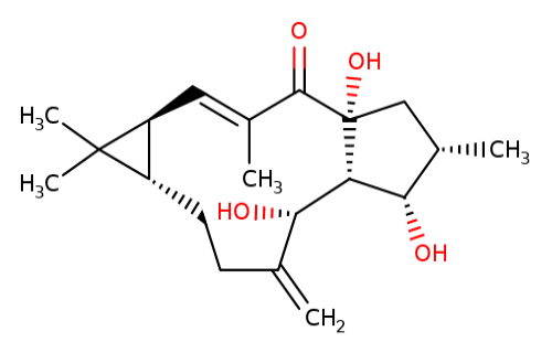
Chemical structure of lathyrol.
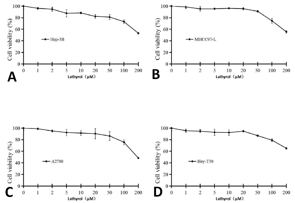
Lathyrol decreases cell viability of multiple human cancer cell lines. Cells were cultured in 96-well plates in triplicates and treated with given concentrations of lathyrol overnight. The cell viability was determined using CCK-8 assay kit.
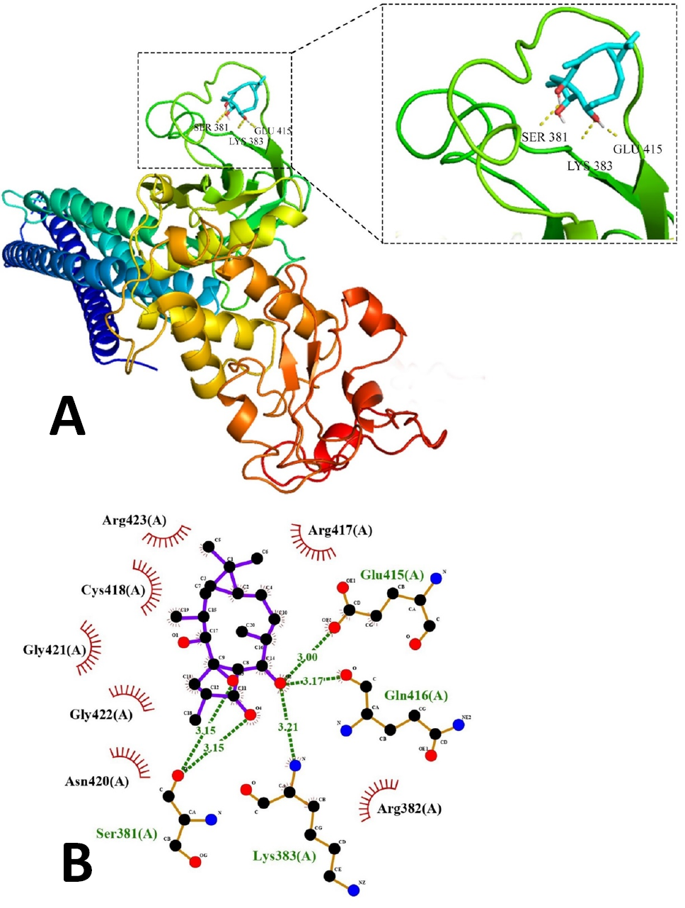
Lathyrol is predicted to bind with DNA binding domain of STAT3. (A) 3D Interaction of lathyrol with crystal structure of STAT3 obtained by using PyMol. (B) 2D diagram obtained by LigPlot+ (version 2.2), showing the hydrogen bond in green dotted lines between lathyrol and different amino acid residues of STAT3.
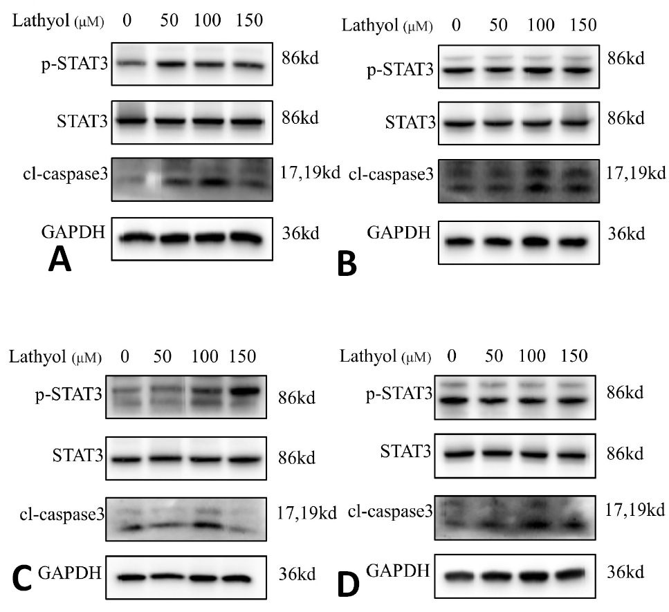
Effect of lathyrol on activation and expression of STAT3 and cleavage of caspase-3. (A) Hep-3B, (B) MHCC97-L, (C) A2780 and (D) taxol-resistant Hey-T30 cells were treated with lathyrol overnight and cell lysates were prepared using RIPA lysis buffer supplemented with 10%PMSF. The cell lysates were subjected to immunoblotting for the expression of p-STAT3, STAT3, and cleaved caspase-3. GAPDH was used a loading control.
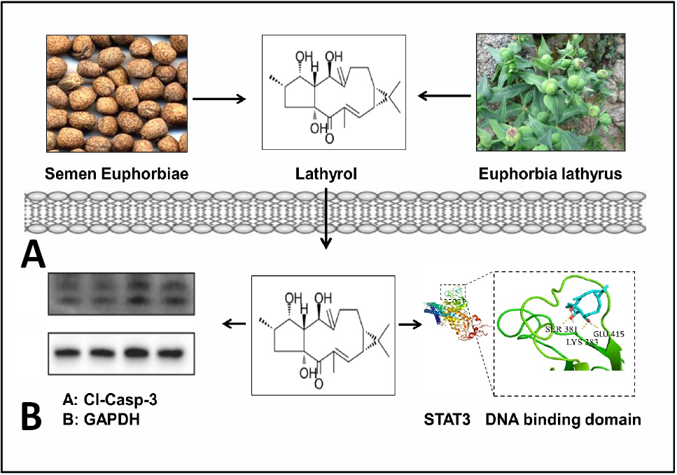
A schematic diagram representing natural sources and cellular targets of lathyrol in multiple human cancer cell lines. Semen Euphorbiae is a traditional Chinese medicine prepared from ripen fruits of Euphorbi lathyrus. Lathyrol is the major components of Semen Euphorbiae and Euphorbi lathyrus. Lathyrol binds with DNA binding domain of STAT3 and induces cleavage of caspase-3 in cancer cells to induce apoptosis.
References
- Aja, P.M., Agu, P.C., Ezeh, E.M., Awoke, J.N., Ogwoni, H.A., Deusdedit, T., Ekpono, E.U., Igwenyi, I.O., Alum, E.U., Ugwuja, E.I., Ibiam, A.U., Afiukwa, C.A. and Adegboyega, A.E., 2021. Prospect into therapeutic potentials of Moringa oleifera phytocompounds against cancer upsurge: De novo synthesis of test compounds, molecular docking, and ADMET studies. Bull. natl. Res. Cent., 45: 99. https://doi.org/10.1186/s42269-021-00554-6[CrossRef]
- Arora, L., Kumar, A. P., Arfuso, F., Chng, W.J. and Sethi, G., 2018. The role of signal transducer and activator of transcription 3 (STAT3) and its targeted inhibition in hematological malignancies. Cancers, 10: 327. https://doi.org/10.3390/cancers10090327[CrossRef]
- Ashaq, A., Maqbool, M., Maryam, A., Khan, M.S., Shakir, H.A., Irfan, M., Qazi, J.I., Li, Y. and Ma, T., 2021. Hispidulin: A novel natural compound with therapeutic potential against human cancers. Phytother. Res., 35: 771–789. https://doi.org/10.1002/ptr.6862[CrossRef]
- Bao, M., Cao, Z., Yu, D., Fu, S., Zhang, G., Yang, P., Pan, Y., Yang, B., Han, H. and Zhou, Q., 2012. Columbamine suppresses the proliferation and neovascularization of metastatic osteosarcoma U2OS cells with low cytotoxicity. Toxicol. Lett., 215: 174–180. https://doi.org/10.1016/j.toxlet.2012.10.015[CrossRef]
- Caboni, L. and Lloyd, D., 2013. Beyond the ligand-binding pocket: Targeting alternate sites in nuclear receptors. Med. Res. Rev., 33: 1081–1118. https://doi.org/10.1002/med.21275[CrossRef]
- Chan, K.S., Li, K., Liu, S.J., Chu, K.H., Toh, M. and Xie, W., 2010. Cucurbitacin B inhibits STAT3 and the Raf/MEK/ERK pathway in leukemia cell line K562. Cancer Lett., 289: 46–52. https://doi.org/10.1016/j.canlet.2009.07.015[CrossRef]
- Chen, P., Li, Y., Zhou, Z., Pan, C. and Zeng, L., 2023. Lathyrol promotes ER stress-induced apoptosis and proliferation inhibition in lung cancer cells by targeting SERCA2. Biomed. Pharmacother., 158: 114123. https://doi.org/10.1016/j.biopha.2022.114123[CrossRef]
- Chen, X., Li, H., Tian, L., Xiao, X., Luo, J., and Zhang, Y., 2020. Analysis of the physicochemical properties of acaricides based on Lipinski’s rule of five. Comput. Biol., 27: 1397–1406. https://doi.org/10.1089/cmb.2019.0323[CrossRef]
- D’Arcy, M.S., 2019. Cell death: A review of the major forms of apoptosis, necrosis and autophagy. Cell Biol. Int., 43: 582-592. https://doi.org/10.1002/cbin.11137[CrossRef]
- Debnath, B., Xu, S. and Neamati, N., 2012. Small molecule inhibitors of signal transducer and activator of transcription 3 (STAT3) protein. J. med. Chem., 55: 6645–6668. https://doi.org/10.1021/jm300207s[CrossRef]
- Funakoshi-Tago, M., Tago, K., Nishizawa, C., Takahashi, K., Mashino, T., Iwata, S., Inoue, H., Sonoda, Y. and Kasahara, T., 2008. Licochalcone A is a potent inhibitor of TEL-Jak2-mediated transformation through the specific inhibition of Stat3 activation. Biochem. Pharmacol., 76: 1681–1693. https://doi.org/10.1016/j.bcp.2008.09.012[CrossRef]
- Gul, S., Maqbool, M., Zheng, D., Li, Y., Khan, M.S. and Ma, T., 2022. Alpinetin: A dietary flavonoid with diverse anticancer effects. Appl. Biochem. Biotechnol., 194: 4220–4243. https://doi.org/10.1007/s12010-022-03960-2[CrossRef]
- Huang, W., Dong, Z., Wang, F., Peng, H., Liu, J.Y. and Zhang, J.T., 2014. A small molecule compound targeting STAT3 DNA-binding domain inhibits cancer cell proliferation, migration, and invasion. ACS Chem. Biol., 9: 1188-1196. https://doi.org/10.1021/cb500071v[CrossRef]
- Huang, W., Liu, Y., Wang, J., Yuan, X., Jin, H., Zhang, L., Zhang, J., Liu, Z. and Cui, J., 2018. Small-molecule compounds targeting the STAT3 DNA-binding domain suppress survival of cisplatin-resistant human ovarian cancer cells by inducing apoptosis. Eur. J. med. Chem., 157: 887–897. https://doi.org/10.1016/j.ejmech.2018.08.037[CrossRef]
- Huang, W., Zhang, R., Chen, Y., Wang, F., Wang, C. H., Peng, H., He, Y., Hangoc, G., Pollok, K., Sandusky, G. E., Fu, X., Broxmeyer, H.E., Zhang, Z.P., Liu, J.B. and Zhang, J., 2016. Small-molecule inhibitors targeting the DNA-binding domain of STAT3 suppress tumor growth, metastasis and STAT3 target gene expression in vivo. Oncogene, 35: 783–792. https://doi.org/10.1038/onc.2015.215[CrossRef]
- Jiao, W., Wan, Z., Chen, S., Lu, R., Chen, X., Fang, D., Wang, J., Pu, S., Huang, X., Gao, H. and Shao, H., 2015. Lathyrol diterpenes as modulators of P-glycoprotein dependent multidrug resistance: Structure-activity relationship studies on Euphorbia factor L3 derivatives. J. med. Chem., 58: 3720–3738. https://doi.org/10.1021/acs.jmedchem.5b00058[CrossRef]
- Kashyap, D., Tuli, H.S., Yerer, M.B., Sharma, A.K., Sak, K., Srivastava, S., Pandey, A., Garg, V., Sethi, G. and Bishayee, A., 2021. Natural product-based nano formulations for cancer therapy: Opportunities and challenges. Semin. Cancer Biol., 69: 5–23. https://doi.org/10.1016/j.semcancer.2019.08.014[CrossRef]
- Khan, M., Maryam, A., Qazi, J.I. and Ma, T., 2015. Targeting apoptosis and multiple signaling pathways with Icariside II in cancer cells. Int. J. biol. Sci., 11: 1100–1112. https://doi.org/10.7150/ijbs.11595[CrossRef]
- Khan, M., Maryam, A., Saleem, M.Z., Shakir, H.A., Qazi, J.I., Li, Y. and Ma, T., 2020. Brevilin A induces ROS-dependent apoptosis and suppresses STAT3 activation by direct binding in human lung cancer cells. J. Cancer, 11: 3725-3735. https://doi.org/10.7150/jca.40983[CrossRef]
- Kim, J.H., Lee, Y.S., Shin, D., Jeon, S.K., Son, K., Han, D.S., Jung, S., Oh, T.K. and Kwon, B., 2011. Cosmomycin C inhibits signal transducer and activator of transcription 3 (STAT3) pathways in MDA-MB-468 breast cancer cell. Bioorg. med. Chem., 19: 7582–7589. https://doi.org/10.1016/j.bmc.2011.10.025[CrossRef]
- Leung, C., Chan, D.W., Pui-Yan, V.M.A. and Ma, D., 2013. DNA-binding small molecules as inhibitors of transcription factors. Med. Res. Rev., 33: 823–846. https://doi.org/10.1002/med.21266[CrossRef]
- Lin, L., Hutzen, B., Zuo, M., Ball, S., Deangelis, S., Foust, E., Pandit, B., Ihnat, M.A., Shenoy, S.S., Kulp, S.K., Li, P., Li, C., Fuchs, J.R. and Lin, J., 2010. Novel STAT3 phosphorylation inhibitors exhibit potent growth-suppressive activity in pancreatic and breast cancer cells. Cancer Res., 70: 2445–2454. https://doi.org/10.1158/0008-5472.CAN-09-2468[CrossRef]
- Liu, C., Wang, T., Khan, M., Cui, X. and Li, Y., 2023. Alantolactone inhibits A2780 cell growth through glycolysis inhibition and ros-induced apoptosis and overcomes cisplatin resistance. Pakistan J. Zool., 55: 1199-1206.
- Maryam, A., Mehmood, T., Yan, Q., Li, Y., Khan, M.S. and Ma, T., 2018. Proscillaridin A promotes oxidative stress and ER stress, inhibits STAT3 activation, and induces apoptosis in A549 lung adenocarcinoma cells. Oxid. Med. Cell, 2018: 1–17. https://doi.org/10.1155/2018/3853409[CrossRef]
- Maryam, A., Mehmood, T., Zhang, H., Li, Y., Khan, M. and Ma, T., 2017. Alantolactone induces apoptosis, promotes STAT3 glutathionylation and enhances chemosensitivity of A549 lung adenocarcinoma cells to doxorubicin via oxidative stress. Sci. Rep., 7: 6242. https://doi.org/10.1038/s41598-017-06535-y[CrossRef]
- Mohan, C.D., Rangappa, K.S., Preetham, H.D., Nayaka, S.C., Gupta, V.K., Basappa, B., Sethi, G. and Rangappa, K.S., 2020. Targeting STAT3 signaling pathway in cancer by agents derived from mother nature. Semin. Cancer Biol., 80: 157–182. https://doi.org/10.1016/j.semcancer.2020.03.016[CrossRef]
- Newman, D. and Cragg, G.M., 2012. Natural products as sources of new drugs over the 30 years from 1981 to 2010. J. Nat. Prod., 75: 311–335. https://doi.org/10.1021/np200906s[CrossRef]
- Son, D.H., Zheng, J., Jung, Y.J., Hwang, C.J., Lee, H.C., Woo, J.Y., Baek, S.E., Ham, Y.R., Kang, M., Shong, M., Kweon, G.R., Song, M., Jung, J., Han, S., Kim, B.Y., Yoon, Y.S., Choi, B.Y. and Hong, J.T., 2017. MMPP attenuates non-small cell lung cancer growth by inhibiting the STAT3 DNA-binding activity via direct binding to the STAT3 DNA-binding domain. Theranostics, 7: 4632–4642. https://doi.org/10.7150/thno.18630[CrossRef]
- Timofeeva, O.A., Chasovskikh, S., Lonskaya, I., Tarasova, N.I., Khavrutskii, L., Tarasov, S.G., Zhang, X., Korostyshevskiy, V.R., Cheema, A.K., Zhang, L., Dakshanamurthy, S., Brown, M.L. and Dritschilo, A., 2012. Mechanisms of unphosphorylated STAT3 transcription factor binding to DNA. J. biol. Chem., 287: 14192–14200. https://doi.org/10.1074/jbc.M111.323899[CrossRef]
- Wong, J., De Rond, T., D’Espaux, L., Van Der Horst, C., Dev, I., Rios-Solis, L., Kirby, J.T., Scheller, H.V. and Keasling, J.D., 2018. High-titer production of lathyrane diterpenoids from sugar by engineered Saccharomyces cerevisiae. Metab. Eng., 45: 142–148. https://doi.org/10.1016/j.ymben.2017.12.007[CrossRef]
- Yang, J., Liao, X., Agarwal, M.M., Barnes, L.E., Auron, P.E. and Stark, G.R., 2007. Unphosphorylated STAT3 accumulates in response to IL-6 and activates transcription by binding to NFκB. Genes Dev., 21: 1396–1408. https://doi.org/10.1101/gad.1553707[CrossRef]
- Yang, J., Wang, L., Guan, X. and Qin, J., 2022. Inhibiting STAT3 signaling pathway by natural products for cancer prevention and therapy: In vitro and in vivo activity and mechanisms of action. Pharmacol. Res., 182: 106357. https://doi.org/10.1016/j.phrs.2022.106357[CrossRef]
- Yarla, N.S., Bishayee, A., Sethi, G., Reddanna, P., Kalle, A.M., Dhananjaya, B.L., Dowluru, K.S.V.G.K., Chintala, R. and Duddukuri, G.R., 2016. Targeting arachidonic acid pathway by natural products for cancer prevention and therapy. Semin. Cancer Biol., 40–41: 48–81. https://doi.org/10.1016/j.semcancer.2016.02.001[CrossRef]
- Zhang, J. and Liu, J., 2016. Drugging the undruggable DNA-binding domain of STAT3. Oncotarget, 7: 66324–66325. https://doi.org/10.18632/oncotarget.12181[CrossRef]
- Zhu, A. and Zhang, T., 2018. The phytochemistry, pharmacokinetics, pharmacology and toxicity of Euphorbia semen. J. Ethnopharmacol., 227: 41–55. https://doi.org/10.1016/j.jep.2018.08.024[CrossRef]
Cited by 1 article
In journals published by ResearchersLinks.
Services
Article usage statistics
Loading…