Peripheral blood of animals taken at a volume of 2 mL from the lateral vein of the forelimb into vacuum tubes containing EDTA potassium salt as anticoagulant.
Haematological panel: was performed using the Cell Dyn 3700 automatic haematology analyser (Abbott, USA). Of the general spectrum of haematological data, the following parameters were used: 1) WBC absolute content in G/L, 2) Neutrophils stab (Neu st), Neutrophils segmented (Neu se), Lymphocytes (Lymph), and Monocytes (Mon) relative content (in %), 3) erythrocyte sedimentation rate (ESR) in mm/hour.
The determination of the WBC blast forms in blood was carried out through analysis of a microscopic blood smear.
According to the haematological panel results, all animals were divided into two groups: (1) cats in remission of chronic leukaemia and (2) cats with initial signs of leukaemic process activation (increased WBC count).
Biochemical blood tests: were performed using the URIT-800 VET (URIT Medical Electronic Co., Ltd., China) biochemical analyser. The following parameters were tested: creatinine (in μmol/L), urea (in mmol/L), residual nitrogen (in mmol/L), glucose (in mmol/L), total cholesterol (mmol/L), activity of amylase and AST and ALT transaminases (in U/ml), and total blood protein (in g/L).
FeLV p27 virus-specific antigen testing: in blood plasma was conducted with the ELISA method, using the FeLV p27 antigen ELISA diagnostic kit (Demeditec, Germany) under the manufacturer’s recommendations.
Molecular genetic examination of FeLV provirus in the cat WBC: The extraction of total nuclear DNA from WBC was carried out using a set of «DNA sorb B» («Amplisens», Russia) following the manufacturer’s recommendations. Analysis of the concentration and quality of isolated DNA was carried out using the spectrophotometer NanoDrop 2000C («Thermo Scientific», USA). The FeLV provirus was detected with an application of the FRT PCR with the Thermocycler Real-Time CFX96 Touch (BioRAD, USA) and the LEIKIS diagnostic kit (Amplisens, Russia), following the manufacturer’s recommendations.
Along with a qualitative assessment of the presence of provirus FelV in WBC, it was quantified. To this end, a positive reference sample with a known viral DNA concentration was titrated (dilution coefficient = log2), and a calibration curve was generated based on Ct indicators. The data were approximated for an R2 of 0.95. The calculated values obtained were expressed in relative units of the provirus concentration of FeLV per 1 mL DNA sample.
Analysis of target gene expression: Extraction of the total RNA from the cat WBC was performed with an application of the ExtractRNA reagent (Eurogen, Russia), according to the manufacturer’s recommendations. The concentration and quality of the isolated RNA were analysed with a NanoDrop 2000C spectrophotometer (Thermo Scientific, USA). Reverse transcription needed to obtain a single-stranded cDNA on the mRNA matrix was performed with an application of the MMLV RT reagent kit (Eurogen, Russia) with the Oligo(dT) primer. All procedures were performed according to the manufacturer’s recommendations.
The expression of the following feline genes was analysed: (a) type I IFN (ifnα6, ifnα7 and ifnβ1), (b) type II IFN (ifnγ), (c) type III IFN (ifnλ1), (d) p53 and gadd45g genes of the cell cycle control, (e) gapdh (glyceraldehyde 3-phosphate dehydrogenase).
Primer-BLAST NCBI and SnapGene 4.1.8 were used to design the primers. The following pairs of forward (F) and reverse (R) gene-specific primers were used (Table 1).
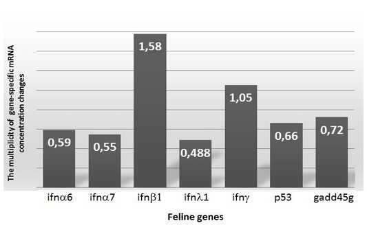
Gene expression analysis was carried out using the qPCR mix HS SYBR («Eurogen», Russia) by qPCR, applying the thermocycler CFX96 («BIORAD», USA). The gapdh gene was used as a normaliser gene. Samples of cDNA derived from mRNAs of practically healthy cats were used as control. The relative differences in the expression of each gene in Group 2 compared to Group 1 were obtained by calculating the multiplicity of expression variation (fold) according to a previously described method (Livak and Schmittgen, 2001).
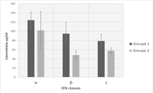
Quantitative analysis of IFN classes α, β and γ in blood plasma of cats was performed by ELISA, using Cat IFNα ELISA KIT, Cat IFNβ ELISA KIT and Cat IFNγ ELISA KIT (Bioassay Technology Lab., China) according to the manufacturer’s recommendations. Analysis results were detected on the microplate photometer ELx808iu (BioTek, USA) at a wavelength of 450 nm; IFN concentrations were expressed in µg/mL of blood plasma.
Statistical processing of the data was carried out using the SPSS 11.0 statistical analysis software with the determination of the Fisher-Student confidence criterion. Differences at p < 0.05 were considered significant.
The results were expressed as arithmetic mean and standard deviation (M±SEM). The Fisher-Student confidence criterion was used to evaluate the significance of results. Differences were considered statistically significant at p<0.05.