
Transforming growth factor-alpha (TGF-α) is a mitogen polypeptide that regulates the pre and post-natal development, structure and function of salivary glands. The localization of TGF-α in post-natal submandibular gland (SMG) of rats and humans is well-studied.However, less is known regarding post-natal sublingual salivary gland (SLG) development and expression of TGF-α. The comparison of TGF-α expression in SLG with that of its expression in SMG during post-natal rat salivary gland development has not been documented in the literature so far. Following ethical approval, a total of twelve rats were divided into two groups. Group A1 consisted of six animals taken at the age of four weeks and sacrificed at age of five weeks. Group A2 contained six animals taken at age of eight weeks and sacrificed at age of nine weeks. The post-natal submandibular and sublingual salivary glands of both the groups were processed for comparison of immunohistochemistry for TGF-α expression at week five and week nine. Expression of TGF-α was noted inthe ductal epithelial cells of SLG that was found to be comparatively increased at the age of five weeks and reduced at age of nine weeks. The expression of TGF-α between both glands at age of nine weeks showed a decreased expression in SLG as comparedto that of SMG.A decrease in the expression of TGF-α in SLG occurs with progression of post-natal salivary gland development. This decrease in TGF-α expression in SLG occurs earlier and is moreas compared to that of the SMG.
Research Article · Volume 51, Issue 3, 21 April 2019 · Open Access
Immunohistochemical Expression of Transforming Growth Factor-Alpha between Rat Submandibular and Sublingual Salivary Glands during Post-Natal Development
· ·
Affiliations & Notes ⌄
- 1Department of Oral Biology, University of Health Sciences, Lahore 2Department of Morbid Anatomy and Histopathology, University of Health Sciences, Lahore , * Corresponding author: sarahghafoor@uhs.edu.pk
Article Info ⌄
- Journal:
- Pakistan Journal of Zoology
- Published:
- 21 April 2019

From the article
- To our knowledge, it is the first time that expression of TGF-α in SLG is being compared with the SMG at different time-in-point during post-natal development of the salivary glands.
- According to the current study, a significant difference in expression of TGF-α between submandibular and sublingual salivary glands of rats of group A2 which were sacrificed at the age of 9 weeks was found.
- It was also observed that the expression of TGF-α in ductal epithelial cells of SLG decreased with age much earlier as compared to SMG.
- The expression of TGF-α is stronger in both salivary glands during the early post-natal growth period but its expression decreases with the advancing age.
Selected from the article's own text.
Abstract
Introduction
Exocrine glands such as salivary glands, lacrimal glands and mammary glands secrete a variety of peptide growth factors which are crucial for their development, cellular proliferation, differentiation and healing . In rats, major salivary gland is the submandibular gland (SMG), and it is considered as an excellent model to study pre- and post-natal development of salivary glands . SMG is involved in secretion of many peptide growth factors such as epidermal growth factor (EGF), transforming growth factor-alpha (TGF-α), nerve growth factor (NGF), insulin-like growth factor (IGF), hepatocyte growth factor (HGF), platelet-derived growth factor (PDGF) . The development of salivary glands continues post-nataly to reach their final functional form and all these growth factors play essential roles in regulating their pre-natal and post-natal development .
TGF-α is a 50 amino acid mitogenic polypeptide that has been detected from a variety of exocrine glands of body including lacrimal glands, pancreas and in the striated ducts of both human submandibular and sublingual salivary glands ; ; Humprey-Beher et al., 1994; ; ; Dernyk, 1988).
TGF-α regulates epithelial proliferation and branching morphogenesis during prenatal and post-natal growth of salivary glands ; ; ; . TGF-α is also important for maintaining taste bud morphology and also found in ductal cells of Von Ebner glands . In rats, TGF-α expression has been reported in all three major salivary glands and is found throughout the entire ductal system excluding the acinar cells . In the SMG, the levels of TGF-α are maximum immediately after birth (during early post-natal development) and decline post-natally by 10th week .
As the development of rodent salivary glands does not complete prenatally therefore it must undergo post-natal development to function as a fully mature adult gland. It is reported that the salivary glands of rats complete their morphological development by 7-10 weeks . During this post-natal growth period, a temporal variation in expression of TGF-α has been reported such as the content of TGF-α is found to be high during early post-natal period of SMG and decreases around 5th week becoming very low at 10th week of post-natal development .
Much work has been done on submandibular glands of rats to elucidate various aspects of pre and post-natal growth and development. However, little is known regarding post-natal development of sublingual salivary gland. Therefore, the current study was designed to investigate the temporal expression of TGF-α in ductal epithelial cells of sublingual gland during early and late post-natal developmental period and also to compare temporal differences in expression of TGF-α between SMG and SLG.
Materials and methods
In order to study the variation in TGF-α expression during post-natal SLG development, and to compare its expression between SMG and SLG, we sacrificed male Wistar rats at week 5 and week 9. The ethical approval of this experimental study was taken from the institutional review board of University of Health Sciences, Lahore. A total of twelve healthy male Wistar rats (Rattus norvegicus) were divided into two groups (group A1 and group A2). The animals of group A1 were taken at the age of four weeks and sacrificed at the age of five weeks. While the animals of group A2 were taken at the age of eight weeks and sacrificed at the age of nine weeks. The care and housing of all the animals was done as described previously , 2018). The salivary gland tissues were processed for immunohistochemistry. The immunohistochemistry was performed as described previously using anti-TGF-α (Abcam, ab16768) as primary antibody and biotinylated horseradish conjugate (Abcam, ab16768) as secondary antibody with DAB chromogen used for detection of color .
The grading for immuno-histochemical intensity score of TGF-α at week five and week nine was carried out by keeping the following scale; 4+ (highly positive), 3+ (positive), 2+ (weakly positive), l+ (minimally positive) and - (undetectable reaction) . Normal skin tissue was taken as a positive control. The acinar cells showing absence of immuno-positive reaction to TGF-α antibody were taken as negative control ; .
Statistical analysis
The analysis and interpretation of the data was done using Statistical Package for Social Sciences version 20 (SPSS-20). Fisher’s exact test was used to determine the comparison of TGF-α expression between SMG and SLG.
Results
Temporal expression of TGF-α in sublingual gland
A strong expression of TGF-α in ductal epithelial cells of SLG was found at week five with the post-natal growth of SLG. However, with the advancing age, the expression of TGF-α was reduced and found to be very low at week 9 (Fig. 1).
Comparison of TGF-α expression between SMG and SLG at week 5
The ductal epithelial cells of SMG and SLG of group A1 at week five were compared with each other for analysis of TGF-α expression. It was noted that Fisher’s exact test showed a non-significant association between the two salivary gland cells. The expression of TGF-α in SMG was found to be similar to that of the SLG at week five and the p-value was found to be 0.06 (Fig. 2).
Comparison of TGF-α expression between SMG and SLG at week 9
Contrary to the results seen at week 5, when the expression of TGF-α was compared between SMG and SLG of group A2 at week 9, Fisher’s exact test showed a significant association between the two salivary glands with regards to TGF-α expression. The expression of TGF-α was much reduced in ductal epithelial cells of SLG as compared to the SMG (Fig. 3). The percentage is given in graphs shown in Figure 4.
Discussion
In order to study the variation in TGF-α expression during post-natal SLG development, we sacrificed healthy male rats at week 5 and week 9. To our knowledge, it is the first time that expression of TGF-α in SLG is being compared with the SMG at different time-in-point during post-natal development of the salivary glands. According to the current study, a significant difference in expression of TGF-α between submandibular and sublingual salivary glands of rats of group A2 which were sacrificed at the age of 9 weeks was found. It is already known that SMG develops one day ahead of SLG . It is also known that rat SMG is a pure serous gland and secrets watery saliva while SLG is predominantly a mucous gland and secrets thick saliva . Although not much is known regarding differences in development and molecular regulation of SLG and is believed to be similar, however keeping in view the developmental, structural and functional differences between the two glands, we speculate that all these differences may account for difference in expression of TGF-α in both salivary glands.
It was also observed that the expression of TGF-α in ductal epithelial cells of SLG decreased with age much earlier as compared to SMG. As, Mogi et al (1995) showed that the content of TGF-α in ductal epithelial cells of SMG is very high during early post-natal development. It peaks till week 3 and starts to decrease from week 5 and is low at week 10 . SLG although starts to develop later than SMG but due to its smaller size may complete its development earlier and the expression of TGF-α reduces earlier. Other compensatory mechanisms like other members of its family EGF may come into play and take over its functions. We found changes in expression of TGF-α in the SLG but it should be explored that which factors regulate TGF-α expression in SLG to better understand the mechanism governing its postnatal development.
Conclusions
The expression of TGF-α is stronger in both salivary glands during the early post-natal growth period but its expression decreases with the advancing age. This decrease in expression is more evident and earlier in the SLG as compared to the SMG.
Acknowledgements
The authors would like to thank all the staff at the animal-care facility of the university, laboratory assistants and bio-statistician of the University of Health Sciences, Lahore, for their help and guidance in the research project. The authors are also thankful to the assistant librarian of the university. The M. Phil research of SB was funded by the University of Health Sciences, Lahore.
Statement of conflict of interest
The authors declare no conflict of interest
Figures
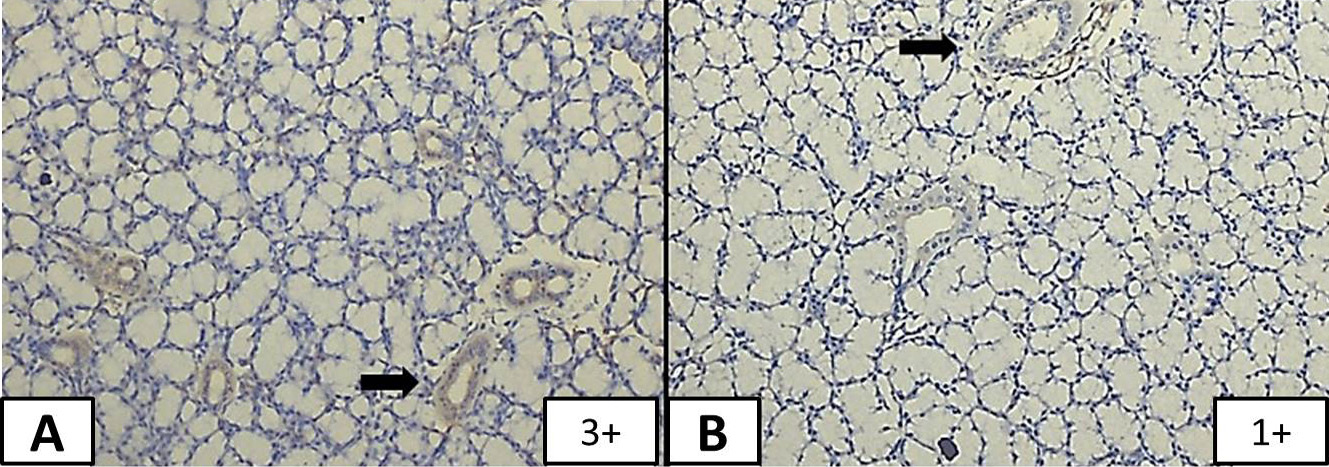
TGF-α expression in SLG: A, longitudinal section of ductal epithelial cells of SLG of group A1 showing TGF-α intensity score of positive (3+) under 10 x magnifications; B, Ductal epithelial cells of SLG of group A2 with TGF-α intensity score of minimally positive (1+) under 10x magnification. Black arrows marks striated ducts.
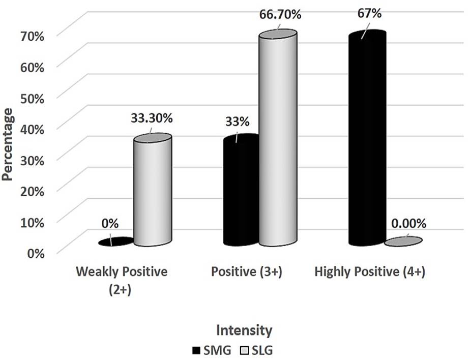
Comparison between SMG and SLG of group A1 (week 5): 67% (n=4/6) of SMGs showed intensity score of 4+ (highly positive) and 33% (n=2/6) showed 3+ (positive). While 67% (n=4/6) SLGs showed a score of 3+ (positive) and 33% (n=2/6) showed 2+ (weakly positive) showing that TGF-α expression in ductal epithelial cells of SMG is almost similar to that of SLG. Fisher’s exact test showed that p-value was found to be 0.06.
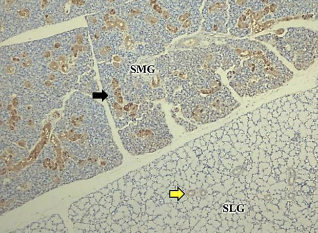
Comparison of expression of TGF-α between SMG and SLG of group A2: Immunohistochemically stained section of SMG and SLG showing a highly positive staining (4+) of TGF-α in the ducts of SMG (black arrow) while the ducts of SLG (yellow arrow) are stained minimally (1+) with TGF-α suggesting that TGF-α is highly expressed in SMG as compared to SLG at week 9.
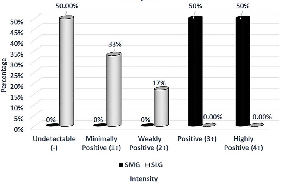
Comparison between SMG and SLG of control Group A2 (week 9): Among SMGs 50% (n=3/6) showed intensity score of 4+ (highly positive) and 50% (n=3/6) showed a score of 3+ (positive). While among the SLGs 17% (n=1/6) showed a score of 2+ (weakly positive), 33% (n=2/6) showed a score of 1+ (minimally positive) and 50% (n=3/6) showed an undetectable score showing that TGF-α expression is more in ductal epithelial cells of SMG as compared to SMG. When TGF-α expression of SMG and SLG of control group (A2) at week 9 was compared with each other, Fisher’s exact test showed a statistically significant association between them. P-value was found to be p=0.006.
References
- Amano, O., Mizobe, K., Bando, Y. and Sakiyama, K., 2012. Anatomy and histology of rodent and human major salivary glands-Overview of the Japan Salivary Gland Society-Sponsored Workshop. Acta Histochem. Cytochem., 45: 241-250. https://doi.org/10.1267/ahc.12013[CrossRef]
- Bancroft, J.D. and Gamble, M., 2008. Bankroft’s theory and practice of histological techniques. Elsevier Health Sciences, pp. 674.
- Bano, S., Naseem, N. and Ghafoor, S., 2018. Effect of thyroid hormone on the histology of rat submandibular salivary gland during postnatal development. J. Pakistan Dental Assoc., 27: 37-42. https://doi.org/10.25301/JPDA.271.37[CrossRef]
- Bano, S., Ghafoor, S. and Naseem, N., 2017. Cytological changes in rat’s sublingual salivary gland development produced by thyroid hormone administration during postnatal period. Biomedica, 33: 206-210.
- Chiang, C., Chen, C., Liu, B., Sun, A., Leu, J. and Wang, J., 2001. Expression of transforming growth factor-α in adenoid cystic carcinoma of the salivary gland. J. Formos med. Assoc., 100: 471-477.
- Derynck, R., 1988. Transforming growth factor α. Cell, 54: 593-595. https://doi.org/10.1016/S0092-8674(88)80001-1[CrossRef]
- Humphreys-Beher, M.G., Macauley, S.P., Chegini, N.A.S.S.E.R., van Setten, G., Purushotham, K., Stewart, C., Wheeler, T.T. and Schultz, G.S., 1994. Characterization of the synthesis and secretion of transforming growth factor-alpha from salivary glands and saliva. Endocrinology, 134: 963-970. https://doi.org/10.1210/endo.134.2.8299590[CrossRef]
- Jaskoll, T. and Melnick, M., 1999. Submandibular gland morphogenesis: Stage-specific expression of TGF-α/EGF, IGF, TGF-β, TNF, and IL-6 signal transduction in normal embryonic mice and the phenotypic effects of TGF-β2, TGF-β3, and EGF-r null mutations. Anat. Rec., 256: 252-268. https://doi.org/10.1002/(SICI)1097-0185(19991101)256:3<252::AID-AR5>3.0.CO;2-6[CrossRef]
- Kouidhi, W., Desmetz, C., Nahdi, A., Bergès, R., Cravedi, J.P., Auger, J., El-May, M. and Canivenc-Lavier, M.C., 2012. In utero and lactational exposure to low-dose genistein-vinclozolin mixture affects the development and growth factor mRNA expression of the submandibular salivary gland in immature female rats. Toxicol. Pathol., 40: 593-604. https://doi.org/10.1177/0192623311436183[CrossRef]
- Mogi, M., Matsuura, S., Suzuki, K., Inagaki, H., Minami, M., Kojima, K. and Harada, M., 1995. Differential expression of transforming growth factor-α and epidermal growth factor during postnatal development of rat submandibular gland. Biochem. biophys. Res. Commun., 217: 271-277. https://doi.org/10.1006/bbrc.1995.2774[CrossRef]
- Morris-Wiman, J., Sego, R., Brinkley, L. and Dolce, C., 2000. The effects of sialoadenectomy and exogenous EGF on taste bud morphology and maintenance. Chem. Senses, 25: 9-19. https://doi.org/10.1093/chemse/25.1.9[CrossRef]
- Ogbureke, K., Macdaiel, R., Jacob, R. and Durban, E., 1995. Distribution of immunoreactive transforming growth factor-alpha in non-neoplastic human salivary glands. Histol. Histopathol., 10: 691-696.
- Patel, V.N., Rebustini, I.T. and Hoffman, M.P., 2006. Salivary gland branching morphogenesis. Differentiation, 74: 349-364. https://doi.org/10.1111/j.1432-0436.2006.00088.x[CrossRef]
- Rougeot, C., Rosinski-Chupin, I., Mathison, R. and Rougeon, F., 2000. Rodent submandibular gland peptide hormones and other biologically active peptides. Peptides, 21: 443-455. https://doi.org/10.1016/S0196-9781(00)00158-3[CrossRef]
- Sabbadini, E. and Berczi, I., 1995. The submandibular gland: A key organ in the neuro-immuno-regulatory network? Neuroimmunomodulation, 2: 184-202. https://doi.org/10.1159/000097197[CrossRef]
- Tucker, A., 2007. Salivary gland development. Seminars in Cell and Developmental Biology, Elsevier, pp. 237-244.
- Tucker, A.S. and Miletich, I., 2010. Salivary glands: Development, adaptation and disease. Front. Oral Biol., 14: 1-20. https://doi.org/10.1159/isbn.978-3-8055-9407-3[CrossRef]
- Wang, X., Qi, S., Wang, J., Xia, D., Qin, L., Zheng, Z. and Fan, Z., 2014. Spatial and temporal expression of c-Kit in the development of the murine submandibular gland. J. mol. Histol., 45: 381-389. https://doi.org/10.1007/s10735-014-9570-7[CrossRef]
- Wu, H.H., Kawamata, H., Wang, D.D. and Oyasu, R., 1993. Immunohistochemical localization of transforming growth factor α in the major salivary glands of male and female rats. Histochem. J., 25: 613-618. https://doi.org/10.1007/BF00157875[CrossRef]
- Yasui, W., Ji, Z.Q., Kuniyasu, H., Ayhan, A., Yokozaki, H., Ito, H. and Tahara, E., 1992. Expression of transforming growth factor alpha in human tissues: Immunohistochemical study and Northern blot analysis. Virchows Arch. A, 421: 513-519. https://doi.org/10.1007/BF01606881[CrossRef]
Cited by 1 article
In journals published by ResearchersLinks.
Services
Article usage statistics
Loading…