Research Article · Volume 13, Issue 4, 4 April 2025 · Open Access
Immunohistochemical Estimation of Apoptotic Activity and Testosterone Level in the Testes of Rats after Chronic Exposure to Uranium Acetate
· ·
Affiliations & Notes ⌄
- 1Department of Pathology and Poultry Diseases, Collage of Veterinary Medicine, Al-Qasim Green University, Babylon, Iraq. *Correspondence | Heba Saleh Shaheed, Department of Pathology and Poultry Diseases, Collage of Veterinary Medicine, Al-Qasim Green University, Babylon, Iraq
- 2Email: Hebasaleh@vet.uoqasim.edu.iq
Article Info ⌄
- Journal:
- Advances in Animal and Veterinary Sciences
- Published:
- 4 April 2025

Abstract
INTRODUCTION
One of the intensive nature occurring elements and valued for its radioactive properties is Uranium ; , because of the radioactive and chemical properties, uranium is distinguished as an element with a toxic effect . One of the targeted organs of chronic DU exposure is reproductive organs . Uranyl acetate were administrate to female mice by using gavage through gestation, parturition, and nursing had an enhance number of dead young per-litter Rodent reproduction and development can be adversely affect by natural uranium when It is probably exposure to the high doses of Uranium so in these research led to reproductive toxicity provide as an additional evidence of effect of Uranium approved . Uranium has indicated to cause many effects like testicular toxicity, maternal toxicity, fetotoxic elevated developmental difference, and growth impediment independent of maternal toxic response when dosing to rodents ; . Uranium, was given as uranyl nitrate, postponement development of mouse embryos in culture and was correlated with lower embryo cell numbers through the hatched blastocyst stage, suggested there are severe alterations in DNA synthesis due to exposure to uranium ; . In the chronic exposure, there were some reports on rodents clarify a negative impact of uranium on male reproductive function, including a reduction in male fertility and in the spermatic number per-testis with a few histopathological effects on the seminiferous tubules and interstitium . As well as the damage of DNA can be as consequence due to apoptosis cell death, which can be caused by metals either indirectly through the generation of free radicals, or by direct interaction between the metal and DNA (Ce´line, et al., 2007). Rodents chronically exposed to 10–80 mg/kg/ day of uranium approaching few testicular histopathological abnormalities and minimized in spermatic number and pregnancy rate .
Experimental Design
Experimental animals: Twenty albino male rats aged 10-15 weeks and weighted 150 -200 gm were gained from (Animal house colony of National Center for Drug Control and Research) Ministry of health/ Baghdad, housed in plastic cages 50×30×10 cm and placed in the room for 10 days for adaptation. The temperature of the room was preserved at 21 ± 3 ˚C, by using exhausted fan the air of the room was changed continuously. The bedding of the cages was replaced every 7 days. The animals accommodate in animal house of Veterinary Medicine College at Al-qasim Green University and were fed on pellet ad libitum.
Chemicals: Uranyl acetate (UA) ([UO₂ (CH₃CO₂) ₂H₂O)] H₂O is yellow to green crystals) was obtained from SPI-Chem (U.S.A).
Dose Calculation for Uranyl Acetate
The dose was calculated depending on the value (LD50 1/5) 40 mg/kg.bw . Melt in normal saline and quaff to the rats by oral intubations using a stomach tube for 90 days.
Histopathological Study
Testes removed from the animals after sacrificing by using chloroform and fixed immediately in 10% buffered formalin solution before being histologically examined and embedded in paraffin. The slides were then routinely stained using Hematoxylin and Eosin stain after the histological sections were cut using a rotary microtome .
Estimation of Apoptotic Activity
The apoptosis activity in paraffin embedded tissue section was determined by using a tunnel assay kit from novous biotechne brand.
Measurement of Testosterone Level
- The MILE card of the kit is placed in its place in the Mini-Vida’s system, the machine automatically recognizes the test so that the system can show the result and then print it.
- A single STR and SPR strip was used for each sample of serum, control and standard solution and placed in a designated place in the system.
- 200 microns are pulled from the serum sample and placed in their own well On the STR strip as well as for control and standard solution.
- Follow the steps for the machine to start the calibration process automatically, which takes about 40 minutes.
- After the calibration, results were printed.
Statistical Analysis
SAS was used to examine the data (Statistical Analysis System - version 9.1). P 0.001 was regarded as statistically significant and Least Significant Differences (LSD) were performed to increased significant differences among means .
RESULTS
Histopathological Study
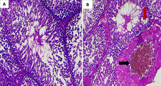
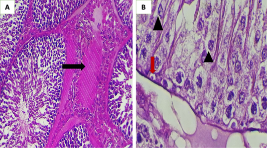
Histopathological examination of testes sections was showed normal histological structures in GI group (Figure 1A). Whereas the GII group showed severe thickening in blood vessel wall and congestion in their lumen. Also, vacuolation in the germinal cells (Figure 1B), also interstitial thickening with hyalinization (Figure 2A) and severe necrosis (nuclear changes) in the germinal cells and apoptosis also seen (Figure 2B).
Immunohistochemical Study
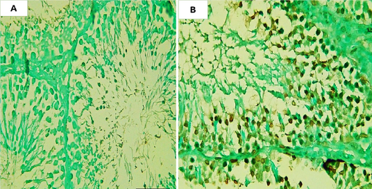
The result of estimation of apoptotic activity in the testes showed increased in the numbers of apoptosis in the spermatogonia that appeared as brown in color indicate positive reaction as in (Figure 3).
Estimation of Testosterone Level
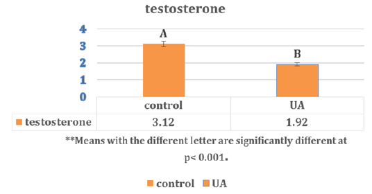
The results for estimation of testosterone level in the serum of rat showed in Figure 4. The level of testosterone was significantly (p< 0.001) decline to (1.92 ± 0.03) compared with control group (3.12 ± 0.016).
DISCUSSION
Recent decades have seen an increase in the amount of evidence supporting a possible link between exposure to toxicants of the environmental and unfavorable consequences for reproductive health . Our histopathological results showed many of degenerative changes such as vacuolation of spermatogonia, and severe necrosis and apoptosis in seminiferous tubules. This result might be attributed to uranium’s capacity to damage reproduction because of its chemical and radioactive characteristics . It has also been shown in previous investigations that uranium accumulates in the reproductive organs. Moreover, our findings imply that DU may originate from testicular fluid, which would allow it to avoid the blood-epididymal barrier and/or the BTB. Male reproductive organs are well-protected by both barriers . According to the Sertoli cells’ ability to sustain testicular homeostasis, germ cell death through apoptosis plays a role in maintaining it. This process may also be connected to vacuolization . The two distinct pathways of testicular apoptosis—the mitochondrial-mediated pathway and the receptor-mediated Fas/FasL pathway—play a crucial role in preserving the germ cell population. The immunohistochemical and histopathological sections of the current study demonstrated that exposure to low doses of UA resulted in increased apoptotic activity. The mitochondrial apoptosis route and the Fas/FasL-mediated apoptotic signal transduction pathway can both be induced by reactive oxygen species . According to a research by Monleau et al. (2006), these findings reveal that the generation of ROS and inflammatory processes contributed to some of the DNA damage, and that recurrent exposure to insoluble DU particles may have a potentiation impact. Gonadotropins and steroid hormones precisely control the hormonal balance essential for male reproductive function, including spermatogenesis and BTB integrity. Nonetheless, our data showed a decrease in testosterone levels following a 90-day exposure to UA; this finding was consistent with earlier research ; , which also noted changes in sexual hormones following DU exposure. While both DU groups had lower intratesticular levels of estradiol, there was no significant change in intratesticular or plasma testosterone levels. Furthermore, a toxin may potentially impact rodent reproduction and fetal development through a number of different routes. Mutagenicity and genotoxicity, disruptions in cell division, modifications or inhibition of protein or steroid synthesis, disruption or inhibition of enzyme systems, and distortion of behavior patterns associated in normal reproduction are among the harmful effects of DU alloy. These mechanisms can result in altered morphogenetic pattern formation, decreased or increased cell death, disrupted cell-to-cell contact, decreased biosynthesis, or tissue disruption that can cause aberrant pathogenesis in the developing fetus or reproductive system ; .
CONCLUSIONS AND RECOMMENDATIONS
In Conclusion of this study showed that the chronic exposure to UN can induce reproductive toxicity in male rats, mainly in the spermatogonia by increased the apoptotic activity, also decreased the testosterone level.
ACKNOWLEDGMENTS
The authors are thankful the staff of College Collage of Veterinary Medicine, Al-Qasim Green University for their cooperation and the facilities they provided during parameters measurement.
NOVELTY STATEMENT
This study investigation the exposure to uranium acetate and Immunohistochemical Estimation of Apoptotic Activity and Testosterone Level In The Testes of Rats.
AUTHOR’S CONTRIBUTIONS
Ghusoon Abdul Kareem Neamah, created and designed the study. Muna Abdul Sahib Alkhfaji, sorted and compiled the references examined and evaluated the information. Heba Saleh Shaheed prepared the first draft of the text, and gave insightful feedback that helped to mold the work into its present state. The final manuscript was critically examined and approved by all writers. All authors have critically reviewed and approved the final draft and are responsible for the content and similarity index of the manuscript.
Conflict of Interest
The authors have declared no conflict of interest.
Figures

Histopathological section of rat testis; A: for GI group showing normal histological structures, B: for GII group showing severe thickening in blood vessels wall and congestion in their lumen (black arrow). Also vacuolation in the germinal cells (red arrow) (H and E, stains, 200x).

Histopathological section of rat testis for GII group; A: showing interstitial thickening with hyalinization (black arrow) (H and E, stains, 200x); B: severe necrosis (nuclear changes) in the germinal cells (head arrow) and apoptosis also seen (red arrow) (H and E, stains, 400x).

Immunohistochamical section of rat testes; A: for GI group; B: for GII group showing positive rection for apoptotic activity appear as dark brown color of spermatogonia (DAB chromogen stain, 200x).

levels (mean±SE) of testosterone in serum (nmol/ml) of rats treated with Uranyl acetate for 90 day.
References
- Angenard G, Muczynski V, Coffigny H, Duquenne C, Frydman R, Habert R, Livera G, Rouiller-Fabre V (2011). In vitro effects of Uranium on human fetal germ cells. Reprod. Toxicol., 31: 470–476. https://doi.org/10.1016/j.reprotox.2010.12.058[CrossRef]
- Bancroft JD, Gamble M (2008). Theory and practice of histology techniques. Churchill Livingstone Elsevier, London. 83-134.
- Brugge D, de Lemos JL, Bui C (2007). The Sequoyah Corporation fuels release and the Church Rock spill: unpublicized nuclear releases in American Indian communities. Am. J. Public Health, 97:1595–1600. https://doi.org/10.2105/AJPH.2006.103044[CrossRef]
- Brugge D, deLemos JL, Oldmixon B (2005). Exposure pathways and health effects associated with chemical and radiological toxicity of natural uranium: a review. Rev. Environ. Health, 20:177–193. https://doi.org/10.1515/REVEH.2005.20.3.177[CrossRef]
- Campion S, Catlin N, Heger N, McDonnell EV, Pacheco SE, Saffarini C, Sandrof MA, Boekelheide K (2012). Male reprotoxicity and endocrine disruption. (101): 315–360. https://doi.org/10.1007/978-3-7643-8340-4_11[CrossRef]
- Ce´line T, Marie C, Sarah M, Ange l, Laure A, Barbara G (2007). Uranium Induces Apoptosis and Is Genotoxic to Normal Rat Kidney (NRK-52E) Proximal Cells. Toxicol. Sci., 98(2): 479–487. https://doi.org/10.1093/toxsci/kfm130[CrossRef]
- Chandra J, Samali A, Orrenius S (2000). Triggering and modulation of apoptosis by oxidative stress. Free Radic. Biol. Med., 29: 323–333. https://doi.org/10.1016/S0891-5849(00)00302-6[CrossRef]
- Domingo JL (2001). Reproductive and developmental toxicity of natural and depleted uranium: a review. Reprod. Toxicol., 15:603–609. https://doi.org/10.1016/S0890-6238(01)00181-2[CrossRef]
- Domingo JL, Llobet JM, Tomas JM, Corbella J (1987). Acute toxicity of uranium in rats and mice. Bull. Environ. Contam. Toxicol. 39: 168–174. https://doi.org/10.1007/BF01691806[CrossRef]
- Duncan DB (1955). Multiple Range and Multiple F-Tests. Biometrics, 11: 1-42. https://doi.org/10.2307/3001478[CrossRef]
- Grignard E, Gueguen Y, Grison S, Lobaccaro JM, Gourmelon P, Souidi M (2008). Contamination with depleted or enriched uranium differently affects steroidogenesis metabolism in rat. Int. J. Toxicol., 27: 323–328. https://doi.org/10.1080/10915810802367057[CrossRef]
- Keith LS, Faroon OM (2022). Handbook on the toxicology of metals. Volume II: Specific Metals, Fifth Edition.
- Lu B, Ran Y, Wang S, Li J, Zhao Y, Ran X, Li R, Hao Y (2021). Chronic oral depleted uranium leads to reproductive damage in male rats through the ROS-hnRNP A2/B1-COX-2 signaling pathway. Toxicology, 449: 152666. https://doi.org/10.1016/j.tox.2020.152666[CrossRef]
- Grison S, Legendre A, Svilar L, Elie C, Kereselidze D, Gloaguen C (2022). Multigenerational exposure to uranium changes sperm metabolome in rats. Int. J. Mol. Sci., 23:8349. https://doi.org/10.3390/ijms23158349[CrossRef]
- Hindin R, Brugge D, Panikkar B (2005). Teratogenicity of depleted uranium aerosols: a review from an epidemiological perspective. Environ. Health, 4:17. https://doi.org/10.1186/1476-069X-4-17[CrossRef]
- Kundt M, Ubios AM, Cabrini RL (2000). Effects of uranium poisoning on cultured preimplantation embryos. Biol. Trace Elem. Res., 75: 235-44. https://doi.org/10.1385/BTER:75:1-3:235[CrossRef]
- Lagos-Cabre R, Moreno RD (2012). Contribution of environmental pollutants to male infertilty: a working model of germ cell apoptosis induced by plasticizers. Biol. Res., 45: 5–14. https://doi.org/10.4067/S0716-97602012000100001[CrossRef]
- Linares V, Albina ML, Bellés M, Mayayo E, Sánchez DJ, Domingo JL (2005). Combined action of uranium and stress in the rat: II. Effects on male reproduction. Toxicol. Lett., 158: 186–195. https://doi.org/10.1016/j.toxlet.2005.03.015[CrossRef]
- Linares V, Albina ML, Belles M, Mayayo E, Sánchez DJ, Domingo JL (2005). Combined action of uranium and stress in the rat. II. Effects on male reproduction. Toxicol. Lett., 158(3): 186-195. https://doi.org/10.1016/j.toxlet.2005.03.015[CrossRef]
- Ma M, Wang R, Xu L, Xu M, Liu S (2020). Emerging health risks and underlying Emerging health risks and underlying toxicological mechanisms of uranium contamination: Lessons from the past two decades. Environ. Int., 145:106107. https://doi.org/10.1016/j.envint.2020.106107[CrossRef]
- Mital P, Hinton BT, Dufour JM (2011). The blood-testis and blood-epididymis barriers are more than just their tight junctions. Biol. Reprod., 84: 851–858. https://doi.org/10.1095/biolreprod.110.087452[CrossRef]
- Monleau M, De Méo M, Paquet F, Chazel V, Duménil G, Donnadieu-Claraz M (2006). Genotoxic and inflammatory effects of depleted uranium particles inhaled by rats. Toxicol Sci., 89(1):287-95. https://doi.org/10.1093/toxsci/kfj010[CrossRef]
- Peters PW, Garbls-Berkvens JM (1996). Study Unit 31: general reproductive toxicology. In Niesink, R.J.M., de Vries, J. and Hollinger, M.A., editors, Toxicology: principles and applications. Boca Raton: CRC Press, 937.
- Schardein JL (2000). Chemically induced birth defects, third edition, Revised and Expanded. New York: Marcel Decker, 1109. https://doi.org/10.3109/9780203909904[CrossRef]
- Yellowhair M, Romanotto M.R, Stearns DM, Clark Lantz R (2018). Uranyl acetate induced DNA single strand breaks and AP sites in Chinese hamster ovary cells. Toxicol. Appl. Pharmacol., 349:29-38. https://doi.org/10.1016/j.taap.2018.04.022[CrossRef]
Services
Article usage statistics
Loading…