Special Issue: Emerging and Re-Emerging Animal Health Challenges in Low and Middle-Income Countries · Volume 12, Issue 1, 11 December 2024 · Open Access
Histological and Histochemical Comparative Study of the Tongue in White-Eared Bulbul (Pycnonotus leucotis) and Bronze Fallow Cockatiel (Nymphicus hollandicus)
·
Affiliations & Notes ⌄
- 1Department of Anatomy and Histology, College of Veterinary Medicine, Al-Qasim Green University, 51013, Babylon, Iraq. *Correspondence | Siraj Moner Al-Kafagy, Department of Anatomy and Histology, College of Veterinary Medicine, Al-Qasim Green University, 51013, Babylon, Iraq
- 2Email: drsirajmoner@vet.uoqasim.edu.iq
Article Info ⌄
- Journal:
- Journal of Animal Health and Production
- Published:
- 11 December 2024

Abstract
Introduction
Birds are classified according to the relationship of diet to the bird’s tongue, as they found five functions provided by the bird’s tongue (collecting food, eating, swallowing, tasting and touching, and building the nest). They found that the tongue has great adaptations to the diet, as it can eat a variety of foods, including insects, small seeds, herbs, grains, etc. this varies depending on the species and the shape of the tongue . The white-eared bulbul (Pycnonotus leucotis) it is found in South-western Asia from India to the Arabian Peninsula and bronze fallow cockatiel (Nymphicus hollandicus) is endemic to north-central Tanzania, the tongue is localized at the entrance of the gastrointestinal system at the base of the beak and plays an important role in the intake of food and intake of diet affects the shape of tongue epithelia (Al-Jebori, ٢٠٢٠; . The mucosa of tongue is formed of the stratified squamous epithelia keratinize which lined lateral border of the dorsal surface, while the medial border lines by nonkeratinize stratified squamous epithelia ; .
Glandular secretions include various types of large molecules, such as neutral mucins, carboxylated proteins, sulfated, sialomucins, sulfomucins, and glycogen, which have significant effects on the oral cavities and upper digestive tract, including protecting the tissue and facilitating the absorption of food ; Saggsou et al., 2013).
The present study aims to elucidate the differences in histological structure and histochemical characters between the tongue of two birds based on the different food habits. The finding of the comprehensive analysis identified that there are fundamental differences in histological structure and histochemical characteristics between the tongue of two birds.
Materials and Methods
The tongue was removed from the mouth, and samples from various sections (tip, middle, base) were preserved in Bouin’s solution for 48 hours. Afterwards, the samples were dehydrated in increasing concentrations of ethanol, clarified in xylene, and encased in paraffin wax . The sections, which were 5μm thick, were cut, placed on glass slides, and stained with Haematoxylin and Eosin (H and E) for light microscopy as usual . Alcian blue stain and periodic acid shiff to determine the secretion type of lingual gland. Photographs were taken with a Moticam Images Plus 2.0 Digital Camera (Motic China Group Limited).
Statistical analysis
The data were represented by Mean±SE and statistical analysis was performed using SPSS, version 24.0.
Result and Discussion
The apex of the tongue
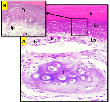
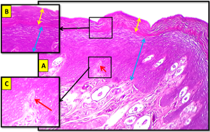
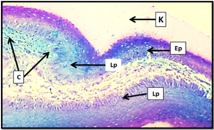
Histological result show that the apex of the tongue of two birds (bulbul and cockatiel)was composed of three histological layer (tunica mucosa, tunica submucosa, tunica muscularis), the cockatiel was have the same histological structure. In bulbul the apex of tongue was lined with stratified squamous epithelium keratinized, the keratinized layer was lined all three surface of apex (dorsal, ventral, lateral) keratinized layer in dorsal surface was more thickness than in lateral and ventral surface. While in cockatiel the apex was lined with keratinized layer only at dorsal and ventral surface (Figures ١, ٢, 9 and 10).
Tunica mucosa in bulbul was thin layer at dorsal surface and increased in thickness gradually to the lateral surface, in ventral surface the tunica mucosa was more in thickness than that in dorsal and lateral surface. While in cockatiel there is no different in thickness between the surface of the apex and the keratinized layer was thick layer at dorsal and ventral surface. The papillary projection was absent in tunica mucosa of apex in bulbul. while in cockatiel there is papillary projection found at epithelial layer. Lamina properia-sub mucosa in bulbul was thick layer at dorsal surface from the ventral surface. While in cockatiel the lamina properia-submucosa was more in thickness at ventral surface from the dorsal surface (Figures ٣, ٤ and Table 1).
The finding of Mohammad and Goodarzi (2020) in common pheasant, ; ; was agree results of bulbul and cockatiel, who mention the dorsum linguae were covered with a thick keratinized stratified squamous epithelium which decreased in thickness towards the radix linguae.
The finding of in chucker partridge, in gallinula cholorpus, in little tern and in common buzzard. was disagree results of bulbul and cockatiel. who mention the dorsal epithelium of the tongue is covered with thick keratinized stratified squamous epithelium.
The body of the tongue
The dorsal surface of the body of bulbul tongue was lining with stratified squamous epithelium keratinized, thickness of epithelial layer was decreased gradually from the cranial end of the body to the caudal end, and it was less in thickness from that in dorsal surface of the apex. In cockatiel lining epithelium of the dorsal surface have the same histological structure in bulbul with differences in thickness of lining epithelia and present of keratinized layer. The ventral surface was lined with keratinized epithelium and less in thickness from the dorsal surface (Figures 5 and 6).
Table 1: The thickness of (tunica mucosa, keratinized layer, lining epithelium, lamina properia) in dorsal and ventral surface of the apex in bulbul and cockatiel tongue.
| Birds | Surface | Thickness of Tunica mucosa (mean ± SE) µm | Thickness of Keratinized layer (mean ± SE) µm | Thickness of Epithelium (mean ± SE) µm | Thickness of Lamina properia (mean ± SE) µm |
| Bulbul | Dorsal | 311.265±12.975* | 90.138 ± 6.979 | 155.684 ± 15.093* | 37.182±5.909* |
| Ventral | 237.518±19.839* | 29.190±2.365 | 173.362±7.968* | 27.906±3.319* | |
| Cockatiel | Dorsal | 521.821±21.523* | 112.623 ± 3.821 | 399.616 ± 33.656* | 110.842±9.342* |
| Ventral | 579.212±21.496* | 98.861 ± 3.491 | 381.796±14.640* | 180.822±14.322* |
Table 2: The thickness of (tunica mucosa, keratinized layer, lining epithelium, lamina properia) in dorsal and ventral surface of the body in bulbul and cockatiel tongue.
| Birds | Surface | Thickness of Tunica mucosa (mean ± SE) µm | Thickness of Keratinized layer (mean ± SE)µm | Thickness of Epithelium (mean±SE) µm | Thickness of Lamina properia (mean ± SE) µm |
| Bulbul | Dorsal | 301.142±13.663* | 83.321 ± 5.732 | 155.684± 15.093* | 36.812±7.611* |
| Ventral | 197.126±8.988* | 161.386 ± 1.786 | 163.188±9.479* | 29.210±2.342* | |
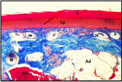
| Cockatiel | Dorsal | 449.409±16.223* | 152.44 ± 2.963 | 399.616± 33.656* | 54.022±4.395* |
| Ventral | 522.674±11.995* | 85.268±5.725 | 284.552±6.705* | 160.560±4.810* |
The finding of in coturnix bird, was disagree results of bulbul and cockatiel. who mention the histological showed that dorsal surface of lingual body covered with non-keratinized stratified squamous epithelium. Such observations resemble those of white-tailed eagle and domestic pigeon .
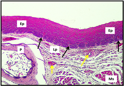
The epithelial layer of the body of bulbul tongue at dorsal surface have papillary projection into the layer. The papillary projection was absent at the ventral surface. While in cockatiel there was absent of the papillary projection at the tunica mucosa of the dorsal surface, while the papillary projection in large number of the tunica mucosa of the ventral surface. the thickness of tunica mucosa in cockatiel dorsal surface was greater than that in bulbul (Figures 5 and 6).
The finding of Igwebuike and Anagor (2013) in tongue of the muscovy duck, was disagree results of bulbul and agree with that finding in cockatiel. who mention beneath the epithelium on both the dorsal and ventral surfaces of the tongue body there is dense irregular fibrous connective tissue, which penetrates the epithelial layers in the form of connective tissue papillae. lingual papillae extend from the lateral margins of the tongue body as epithelial evaginations, with connective tissue cores.
The tunica muscularis in the body of bulbul tongue was fill the space between the dorsal and ventral surface with present of adipose tissue and collagen bundle so that the paraglossum, it was highly vascularized with present of numerous nerve fibers. the tunica muscularis of the body of cockatiel was have the same histological structure of the bulbul (Table 2).
The finding of in the domestic duck, was agree results of bulbul and cockatiel. who mention a characteristic feature of the tongue in the domestic duck was the presence of the yellow adipose tissue under the lamina propria of the mucosa on the body, the adipose tissue covered the internal skeleton of the tongue formed by the elongated entoglossum cartilage of the hyoid apparatus.
The root of the tongue
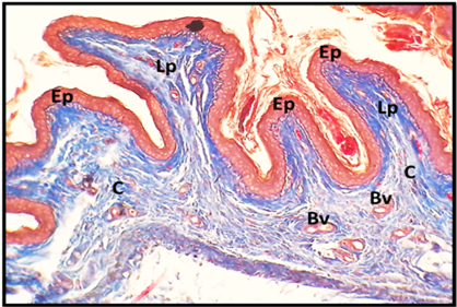
The histological study shows that the tunica mucosa at the dorsal surface of the root of bulbul lined with stratified squamous epithelium non-keratinized with present of large number of conical papillae and it less in thickness from the ventral surface the papillary projection was absent at two surface. in cockatiel the tunica mucosa has the same histological structure and the number of conical papillae was less than that in bulbul. The tunica mucosa at the ventral surface was more in thickness from that in dorsal (Figures 7).
Table 3: The thickness of (tunica mucosa, keratinized layer, lining epithelium, lamina properia) in dorsal and ventral surface of the root in bulbul and cockatiel tongue.
| Birds | Surface | Thickness of Tunica mucosa (mean±SE) µm | Thickness of Keratinized layer (mean±SE) µm | Thickness of Epithelium (mean±SE) µm | Thickness of Lamina properia (mean±SE) µm |
| Bulbul | Dorsal | 78.561±4.112* | -- | 49.182±3.571* | 24.064±3.873* |
| Ventral | 138.877±8.978* | -- | 96.888±13.445* | 36.488±6.276* | |
| Cockatiel | Dorsal | 193.281±6.634* | -- | 138.390±7.643* | 49.568±10.415* |
| Ventral | 197.722±9.314* | -- | 138.390±7.643* | 61.636±3.634* |
The finding of in white headed duck, was agree results of bulbul and cockatiel. Who mention the non-keratinized epithelium lacking the keratinized layer covered the surface area of the root and some zones on the lingual prominence. This type of epithelium was not found on the ventral lingual surface.
The tunica muscularis was thicker layer from that in apex and body in bulbul and cockatiel in both birds present the central paraglossum in the tunica muscularis there is highly vascularized and present large number of nerve bundles.
The finding of ; in in poultry species, was agree results of bulbul and cockatiel. who mention a muscular layer (Tunica muscularis) was reported under the lamina propria in poultry species. in the guinea fowl, it was determined that the muscles of the tongue, which are skeleton-specific, were found just below the lamina propria in the corpus and radix sections.
In bulbul at the dorsal surface present of the taste bud within the tunica mucosa and increased in number under the conical papillae. While in cockatiel the number of taste bud was larger than that in bulbul, the taste bud in the two birds was distributed at the tunica mucosa of the dorsal surface of the root and absent at the ventral surface (Figures 7, 8 and Table 3).
The results of our study also agree with the results of the researchers study, which was mentioned in its results that intra-epithelial taste buds are present in chickens, while reported that intra-epithelial taste buds are present in the pied crow (Erdoğan et al., 2012) reported that intra-epithelial taste buds are also present in the partridge.
The mechanoreceptors
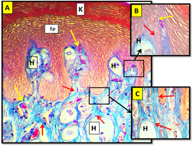
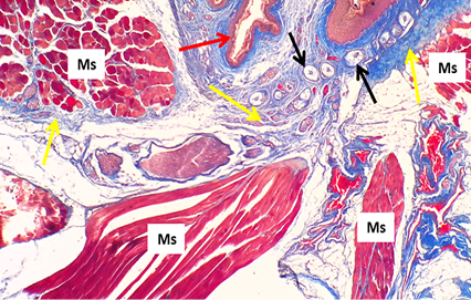
The histological result of the tongue of cockatiel showed that the lamina properia under the lining epithelium contains circular to oval-shaped particles with central axon surrounded by several sheets by supported epithelial collagen fibers, this sheets are composed of collagen fiber called herbiest corpuscle which are particles contain nerve ending to supply the tongue with sensory receptors and contain nerve cell the herbiest corpuscles located at dorsal and ventral surface of the apex and root. In body of the tongue the herbiest corpuscle was absent. In bulbul the herbiest corpuscle was absent in all part of the tongue (apex, body, root) (Figures 9).
The results disagree with the finding of a previous study who mention the lamina properia of the lingual comb, the edges of the body, and the rostral edges of the lingual prominence have two types of specifically arranged mechanoreceptors the grandry and herbst corpuscles. An interesting feature was the mutual arrangement of these corpuscles.
Histochemical finding
The histochemical result show differences in carbohydrate distribution and it is reaction with special stain, the differences were dependent on the amount of the neutral and acidic mucopolysaccharides.
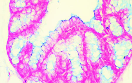
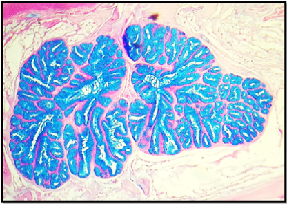
In the bulbul’s tongue, the lingual gland exhibited a weak to moderate reaction to the PAS stain (while in the cockatiel’s gland, produce a negative response to the PAS stain, this means that the gland will produce a small number of neutral polysaccharides in the bulbul, which is absent in the cocktail. The lingual gland at both birds (bulbul and cocktail) exhibits a positive response to the AB stain that is indicative of the secretion of a large number of acidic polysaccharides (Figures 10 and 11).
The earlier revelation that the tongue is grainivorous and insectivorous, was a disagreement with the results of bulbul and cockatiel. who discuss, in the red jungle fowl, the zebra finch, the black francolin, and the common kestrel, their salivary glands are said to have a strong positive response to the PAS reaction, this indicates that they have a content of neutral mucin ; .
Acknowledgement
We thankful to all staff of the anatomy and histology department, especially to the head of the department.
Novelty Statement
Development of histological and anatomical of Tongue In White-Eared Bulbul (Pycnonotus leucotis) and Bronze Fallow Cockatiel (Nymphicus hollandicus) is studied for the first time.
Author’s Contribution
Both authors contributed equally for completing of this article.
Ethical approval
Ethical approval was sought and accepted from the medical research ethical committee of Al-Qasim Green University, Babylon, Iraq.
Conflict of interest
The authors have declared no conflict of interest.
Figures

(A) Cross histological section of the apex of tongue in white eared bulbul show: Stratified squamous epithelium (Ep), keratinized layer (K), lamina properea (Lp), blood vessels (B), Hyaline cartilage (H). H&E stain. 20X. (B). Magnified section. 100x.

Longitudinal histological section of the apex of tongue in cockatiel show: Keratinized layer (yellow double head arrow), stratified squamous epithelium (blue double head arrow), papillary projection (red arrow). H&E stain. 20x. (B) and (C) magnified section. H&E stain. 40x.

Cross histological section of the apex of tongue in white eared bulbul show: Keratinized layer (K), stratified squamous epithelium (Ep), lamina properia (Lp), collagen fibers (C). Masson trichrom stain. 20x.

(A) Cross histological section of the apex of tongue in cockatiel show: Keratinized layer (K), stratified squamous epithelium (Ep), collagen fibers (red arrow), blood supply (yellow arrow), herbiest corpuscle (H). Masson trichrom stain. 20x. (B) and (C): Magnified section 40x.

Cross histological section of the body of tongue in bulbul dorsal surface show: Stratified squamous epithelium non keratinized (Ep), lamina properea (Lp), papillary projection (black arrow), blood vessels (yellow arrow), skeletal muscle (Ms), paraglossum (P). H&E stain. 10x.

Cross histological section of the body of tongue in cockatiel show: stratified squamous epithelium (Ep), keratinized layer (K), connective tissue (C), skeletal muscle (Ms), herbiest corpuscles (H). Masson trichrom stain. 10x.

Cross histological section of the root of tongue in bulbul show: stratified squamous epithelium non keratinized (Ep), lamina properia (Lp), blood vessels (B), collagen bundles (C). H&E stain. 4x.

Cross histological section of the body of tongue (ventral surface) in cockatiel show: Skeletal muscle (Ms), stratified squamous epithelium (red arrow), herbiest corpuscle (black arrow), collagen bundles (yellow arrow).Masson trichrom stain. 10x.

Cross histological section of the tongue in cockatiel show: The distribution of the herbiest corpuscle H&E stain. 10x. A: Apex of the cockatiel tongue B: Root of the cockatiel tongue.

Histological section for lingual gland of bulbul show: The ligual gland was give positive reaction for Alcian blue stain that men the gland was have acidic mucopolysaccharid secretion. AB stain. 40x.

Histological section for lingual gland of cockatiel show: the ligual gland was give positive reaction for Alcian blue stain that men the gland was have acidic mucopolysaccharid secretion. AB stain. 10x.
References
- Akbari G, Hassanzadeh B, Madadi MS, Babaei M (2018). Morphological reflections of evolutionary adaptations in the tongue of the white-headed duck. Anatom. Sci. Int., 93: 469-477. https://doi.org/10.1007/s12565-018-0438-x[CrossRef]
- Al-Jebori AKH, Al-Kafagy SM, Ghazi J (2020). Comparative histological and histochemical study of small intestine in European roller (Coracias garrulus) and Fischer’s lovebird (Agapornis fischeri). 20(2): 4539-4542.
- Al-Jumaily IS, Mnati EM, Mutlak BH, Dauod HA (2013). Morphological and histological study of the tongue in rock pigeon Columba livia gaddi Gemlin, 1789. J. Madenat Alelem Univ. College, 5(2): 94-104.
- Al-Kafagy SM, Al-Jebori AK, Alseady YY (2022). Histochemical study of proventriculus in pre-hatch and post-hatch days in northern bobwhite quail (Colinus virginianus). https://doi.org/10.33899/ijvs.2022.135832.2529[CrossRef]
- Alkafagy SM, Ghazi J, Alhaaik AG (2019). Histomorphological study of kidney in adult kestrel, Falco tinnunculus. 19(2): 3927-3933.
- Aytekin Ö (2016). Systema digestorium. Vet. Special Histol., 4th ed., İstanbul. Nobel Publication, pp. 151-156.
- Crole MR, Soley JT (2014). Comparative distribution and arrangement of Herbst corpuscles in the oropharynx of the ostrich (Struthio camelus) and emu (Dromaius novaehollandiae). Anat. Rec., 297: 1338–1348. https://doi.org/10.1002/ar.22933[CrossRef]
- El-Badry DA (2022). Comparative histological and ultra structural studies on the tongue of Gallinula cholorpus and Coturnix coturnix. J. Environ. Sci. Mansoura Univ., 51(1): 1-9. https://doi.org/10.21608/joese.2022.123051.1012[CrossRef]
- El-Beltagy AM (2013). Comparative studies on the tongue of white throated king fisher and common Buzzard. Egypt. Acad. J. Biol. Sci., 4(1): 1-14. https://doi.org/10.21608/eajbsd.2013.14137[CrossRef]
- Elyasi SMP, Goodarzi N (2020). Morphology of the tongue and laryngeal entrance in the Common pheasant (Phasianus colchicus); a light and scanning electron microscopic study. Kermanshah, Iran. https://doi.org/10.1111/ahe.12595[CrossRef]
- Erdoğan S, Iwasaki SI (2014). Function-related morphological characteristics and specialized structures of the avian tongue. Annals Anat. Anat. Anzeiger, 196(2-3): 75-87. https://doi.org/10.1016/j.aanat.2013.09.005[CrossRef]
- Erdogan S, Sagsz H, Akbalık M (2012). Anatomical and histological structure of the tongue and histochemical characteristics of the lingual salivary glands in the Chukarpartridge (Alectoris chukar, Gray 1830). Br. Poult. Sci., 53(3): 307–315. https://doi.org/10.1080/00071668.2012.700507[CrossRef]
- Igwebuike UM, Anagor TA (2013). The morphology of the oropharynx and tongue of the muscovy duck (Cairina moschata). Vet. Arh., 83(6): 685-693.
- Igwebuike UM, Eze UU (2010). Anatomy of the oropharynx and tongue of the African pied crow (Corvus albus). Vet. Arh., 80(4): 523-531. In the peregrine falcon and common kestrel. Okajimas Folia Anat Jpn. 85: 11–15.
- Iwasaki S (1992). Fine structure of the dorsal lingual epithelium of the little tern. Sterna Albifrons Pallas (Aves, Lari). J. Morphol., 212: 13–26. https://doi.org/10.1002/jmor.1052120103[CrossRef]
- Jackowiak H, Andrzejewski W, Godynicki S (2006). Light and scanning electron microscopic study of the tongue in the cormorant Phalacrocorax carbo (Phalacrocoracidae, Aves). Zool. Sci., 23(2): 161-167. https://doi.org/10.2108/zsj.23.161[CrossRef]
- Jackowiak H, Godynicki S (2005). Light and scanning electron microscopic study of the tongue in the white tailed eagle (Haliaeetus albicilla, Accipitridae, Aves). Anat. Anzeiger, 187: 251-259. https://doi.org/10.1016/j.aanat.2004.11.003[CrossRef]
- Kadhim KK, Zuki ABZ, Babjee SMA, Noordin MM, Zamri-Saad M (2011). Morphological and histochemical observations of the red jungle fowl tongue Gallus gallus. Afr. J. Biotechnol., 10(48): 9969-9977. https://doi.org/10.5897/AJB11.955[CrossRef]
- Kudo KI, Nishimura S, Tabata S (2008). Distribution of taste buds in layer-type chickens: Scanning electron microscopic observations. Anim. Sci. J., 79(6): 680-685. https://doi.org/10.1111/j.1740-0929.2008.00580.x[CrossRef]
- Luna LG (1968). Manual of histologic staining methods of armed forces institule of pathology 3rd. New York, U. S.A. pp. 123.
- Mohammad PES, Goodarzi, N. (2020). Morphology of the tongue and laryngeal entrance in the Common pheasant (Phasianus colchicus); a light and scanning electron microscopic study. Anatomia, histologia, embryologia, 50(1): 23-31.
- Nazema SA-M, Safwat A, Mohamed A, Samy F, Mahmoud A, Megeid NS, Ali S, Abdo M, Mahmoud SF (2021). Histo-morphological comparison of the tongue between grainivorous and insectivorous birds. Int. J. Morphol., 39(2): 592-600. https://doi.org/10.4067/S0717-95022021000200592[CrossRef]
- Parchami A, Dehkordi RF (2011). Lingual structure in the domestic pigeon (Columba livia Domestica): A light and scanning electron microscopic study. World Appl. Sci. J., 12: 1517–1522.
- Sagsoz H, Erdogan S, and Akbalik M (2013). Histomorphological structure of the palate and histochemical profiles of the salivary palatine glands in the Chukar partridge (Alectoris chukar, Gray 1830). Acta Zool., (Stockholm), 94(4): 382–391. https://doi.org/10.1111/j.1463-6395.2012.00566.x[CrossRef]
- Samar ME, Ávila RE, Esteban FJ, Olmedo L, Dettin L, Massone A, Peinado MA (2002). Histochemical and ultrastructural study of the chicken salivary palatine glands. Acta Histochem., 104(2): 199-207. https://doi.org/10.1078/0065-1281-00627[CrossRef]
- Schindala MK (1999). Anesthetic effect of ketamine, ketamine with diazepam in chicken, Iraqi. J. Vet. Sci., 12: 261-265.
- Skieresz-Szewczyk K, Jackowiak H (2016). Morphofunctional study of the tongue in the domestic duck (Anas platyrhynchos f. domestica, Anatidae): LM and SEM study. Zoomorphology, 135: 255-268. https://doi.org/10.1007/s00435-016-0302-2[CrossRef]
- Suhaib AHA-T, Azhar SK (2022). (Histomorphological study of the tongue in adult starling birds (Sturnus valguris). SI(1): 116-122.
Cited by 3 articles
In journals published by ResearchersLinks.
- Comparative Histochemical Study of the proventriculus Between Bronze Fallow Cockatiel and White Eared BulbulJournal of Animal Health and Production · 2025
- Morphological and Histochemical Study of the Duodenum in Post- Hatching and Adult (Columba livia)Journal of Animal Health and Production · 2025
- Comparative Histochemical Study of Duodenum Between Bronze Fallow Cockatiel and White Eared BulbulJournal of Animal Health and Production · 2025
Services
Article usage statistics
Loading…