
Feline panleukopenia (FP) is a highly contagious viral disease of cats caused by the feline panleukopenia virus (FPV) (also called Feline Parvovirus (FPV)) that affects gastrointestinal, immune and nervous system. The present study was conducted to know the prevalence of FP in cats of Barishal region and also to know the frequency of different factors associated with FP. The study was performed at Upazila Livestock Office and Veterinary Hospital, Barishal Sadar and Pet Clinic, Barishal between November 01, 2023 to 10 April, 2024. A total of 131 case data were investigated that were extracted from the paper-based recording system in both place of which 53 cases were diagnosed as FP. Next, we calculated the prevalence, mortality rate, and case fatality of FPV infection in the domestic cats of different age groups and sexes. In this study, 40.45% cats were found to be infected with FPV, where mortality rate and case fatality rate were 23.66% and 58.49%, respectively. FPV cases were most frequently recorded in non-vaccinated young local male domestic cats. Anorectic condition, frequent vomiting, diarrhea, and different level of dehydration were recorded as the prominent clinical signs in FPV infection. Fluid therapy (5% dextrose saline and 0.9% saline), antiemetic (ondansetron), proton pump inhibitor (pantoprazole/esomeprazole), antibiotics (ceftriaxone or metronidazole and ceftriaxone) and multivitamins were suggested as the supportive treatment of the disease. Overall, it is understood that a timely vaccination is imperative to prevent the disease.
Research Article · Volume 11, 24 August 2024 · Open Access
Epidemiology of Feline Panleukopenia Virus Infection in Domestic Cats of Barishal District, Bangladesh
· ·
Affiliations & Notes ⌄
- 1Faculty of Animal Science and Veterinary Medicine, Patuakhali Science and Technology University, Bangladesh
- 2Department of Microbiology and Public Health, Faculty of Animal Science and Veterinary Medicine, Patuakhali Science and Technology University, Barishal-8210, Bangladesh. *Correspondence | Mohammad Enamul Hoque Kayesh, Department of Microbiology and Public Health, Faculty of Animal Science and Veterinary Medicine, Patuakhali Science and Technology University, Barishal-8210, Bangladesh
- 3Email: mehkayesh@pstu.ac.bd
Article Info ⌄
- Journal:
- Hosts and Viruses
- Published:
- 24 August 2024
From the article
- There were also gender and breed variations in FPV prevalence in cats, a higher prevalence was recorded in male (42.02%) than those of female (38.70%) cats, and a higher prevalence of FPV was recorded in indigenous (43.20%) and cross-breed (40.00%) cats, compared to Persian cats (30.00%).
- The prevalence of FPV infection was higher in pet cats (41.07%) than that of stray cats (36.74%) (Table 1).
- Timely vaccination is critical for preventing FPV infection in cats.
- As supportive treatment of FPV infection in cats, fluid therapy (5% dextrose saline and 0.9% saline), antiemetic (ondansetron), proton pump inhibitor (pantoprazole or esomeprazole), antibiotics (metronidazole and ceftriaxone or only ceftriaxone) and multivitamins preparation can be suggested.
Selected from the article's own text.
Abstract
Introduction
Panleukopenia in cats is a highly contagious disease. The feline panleukopenia virus (FPV), belonging to the Parvoviridae family, is the causal agent of feline panleukopenia (FP) . FP is also known as feline distemper in cats. FPV is a non-enveloped, single-stranded DNA virus. Cats infected with FPV exhibit a range of clinical signs, including diarrhea, depression, vomiting, and dehydration . It is characterized by enteritis, leukopenia, and aberrant development . The FPV, which is related to canine parvovirus type 2 (CPV-2) and other parvoviruses of carnivores, is the cause of feline panleukopenia, a serious disease that affects cats . A feline ataxia syndrome has been identified, which is caused by lytic viral replication impairing cerebellar development in the infected kitten’s Purkinje cells .
Due to the highly contagious nature of the disease, it spreads by direct contact between sick and healthy cats. The FPV can infect animals for months or even years. It can be found in cages, food bowls, litter boxes, and other public areas. The virus can be spread by ingestion of contaminated feeds, as well as by indirect contact of fomites, and through intrauterine route . Viruses can spread in the environment through excrement, vomit fluid, urine, and saliva . When exposed to contaminated environments, FPV can survive for weeks or even months due to its great resistance to both physical and chemical agents . The most frequent way that FPV spreads among susceptible animals is through direct contact with infected cats or their secretions. When the disease is active, it is excreted from all bodily secretions; however, FPV is most frequently found in the colon and excrement . The infected young cats (three to five months old) show different clinical symptoms, including hemorrhagic diarrhea, anorexia, vomiting, and a decrease in white blood cells (neutropenia and lymphopenia), which may become fatal in unvaccinated cats, and kittens under 12 months show the highest morbidity and mortality rates, and based on the severity of clinical signs, mortality ranges from 25 to 100% ; . In addition, fetal infection may cause a type of immunological tolerance that allows kittens shedding the virus long after their birth .
FP is most commonly found in domestic, young, immunocompromised, and multi-cat families aggressive and male cats . The majority of FPV cases are reported in cats under a year old. However, cats of all ages that are unvaccinated or incorrectly/improperly vaccinated remain susceptible to FPV infection. It was also reported that the disease only struck vaccinated cats who had not gotten a booster shot after 12 weeks of age, with a median age of 4 months for the affected cats . Seasonal increases in vulnerable newborn kitten populations were correlated with outbreaks of FPV in cats. The majority of FPV cases are found in households with many cats, particularly in enclosed shelter settings. However, cats living outside, such as barn, feral, and stray cats may experience it .
An early diagnosis of FPV infection is critical for isolating the affected cats and shielding susceptible animals from contracting secondary infections. A conclusive diagnosis cannot be made only on the basis of signs and clinical history. The test protocol is straightforward and may be carried out by both owners and veterinarians. However, the immunochromatography assay is the most rapid field diagnostic kit (FPV Ag) used in the clinical practice .
A few supportive and symptomatic treatments for FPV-affected cats, such as nutritional supplements, antiemetic medications, removing dehydration, correcting electrolyte imbalances, and antibiotics to control secondary bacterial infections, have also been suggested. . Vaccination is recommended to prevent the disease, and the commercially available vaccines for preventing the FPV infection include Purevax feline 4, Biofel, Nobivac feline 4, the modified live virus, or an inactivated virus vaccine. Both an inactivated and a modified live virus vaccination are available for preventing the FPV infection . It is administered in two shots at 8–9 weeks of age and 3–4 weeks later, followed by a first booster shot one year later . Due to high contagious nature and fatality of FPV infection in cats, a proper understanding of the prevalence and management practices is critical for its prevention and control. However, the prevalence, treatment practices and vaccination status of cats against FP in Barishal district, Bangladesh are lacking. Therefore, in this study we investigated the prevalence, mortality rate, and case fatality of FP in domestic cats in Barishal district. In addition, we also investigated the frequency of different factors like age, breed, sex, as well as the frequently occurring clinical signs and common therapeutic practices in case of FP.
Materials and Methods
Study area
The study was conducted to observe the epidemiology and diagnosis of FPV at Upazila Livestock Office, Veterinary Hospital Barishal Sadar, and Pet Clinic, Barishal, where patients were brought from different areas of the Barishal district (Figure 1).
Study period and animals
The study was conducted based on collecting samples from admitted patients (cats) to the hospital from November 1, 2023, to April 10, 2024. During the study, a total of 131 samples were collected from sick cats and patients’ information were recorded. In this study, different cat breeds were recorded, including indigenous, Persian, and cross-breed cats. The pet cats were kept indoors and looked after by the owners, not going outside, whereas stray cats were wondering in the forest, bush, and roadside, as well as in the in the homestead area, for food. The body weight of the cats ranged between 0.3 kg and 3.5 kg. Some of the cats had a history of vaccination against FPV infection.
Data collection
After initial registration and history-taking, each case was clinically examined. The clinico-epidemiological findings of each case were recorded in the structured recordkeeping sheet. Data includes address, date, total population, housing system, species, breed, age, sex, body weight, body condition score, vaccination, de-worming, previous disease history with treatment, duration of illness, defecation, micturition, and vomiting, along with client demographic information (age, sex, education, and job). Pulse, respiration, and rectal temperature were taken; a skin fold test, examination of the mucous membrane, and different organs of the body were performed by using the methods of palpation, percussion, and auscultation. Diagnosis and drug prescription data were also recorded in the structured recordkeeping sheet. Drug data is made up of the trade names of the drugs, main and supportive drugs, dose, route, and duration.
Diagnostic approach
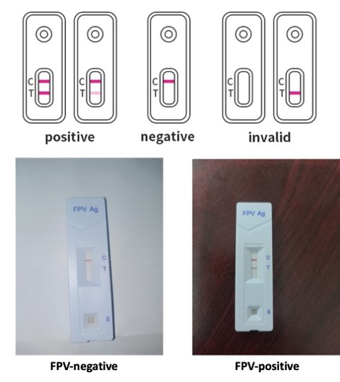
The diagnosis of each case was performed based on the findings of the clinical-epidemiological history, examination, and rapid FPV diagnostic test kit result. The cats that manifested frequent vomiting, anorexia, foul-smelling diarrhoea, depression, and severe weakness were initially suspected of FP. Finally, it was confirmed by using a rapid diagnostic Feline Parvovirus Ag (FPV Ag) test kit following the manufacturer’s instructions (Figure 2). This kit is a chromatographic immunoassay for the qualitative detection of the FPV Ag in feline feces. In brief, the cat’s feces or vomit sample was collected by a sterile swab. After that, the swab sample was placed into an assay buffer tube, stirred thoroughly, and allowed to sit for 2 minutes. Then the cassette from the foil pouch was opened and placed on a flat, clean surface. Later, samples were collected in a dropper from the buffer-specimen mixture,
Table 1: Results of rapid test kit for the detection of FPV infection in relation to age, sex, breed, type of cats.
| Variables | Category level | No. of sample tested | Test Results Positive No | No. of dead | Prevalence | Mortality rate | Case fatality |
| Age | Less than 3 months | 62 | 29 | 19 | 46.77% | 30.64% | 65.51% |
| 3 months to 8 months | 25 | 11 | 6 | 44.00% | 24.00% | 54.54% | |
| >8 months to 1.3 years | 24 | 8 | 4 | 33.33% | 16.66% | 50.00% | |
| > 1.3 years | 20 | 5 | 2 | 25.00% | 10.00% | 40.00% | |
| Sub total | 131 | 53 | 31 | 40.45% | 23.66% | 58.49% | |
| Sex | Male | 69 | 29 | 17 | 42.02% | 24.63% | 58.62% |
| Female | 62 | 24 | 14 | 38.70% | 22.58% | 58.33% | |
| Sub total | 131 | 53 | 31 | 40.45% | 23.66% | 58.49% | |
| Breed | Indigenous | 81 | 35 | 24 | 43.20% | 29.62% | 68.57% |
| Persian | 20 | 6 | 2 | 30.00% | 10.00% | 33.33% | |
| Cross | 30 | 12 | 5 | 40.00% | 16.66% | 41.66% | |
| Sub total | 131 | 53 | 31 | 40.45% | 23.66% | 58.49% | |
| Type | Pet (owned) cats | 112 | 46 | 28 | 41.07% | 25.00% | 60.86% |
| Stray (free roaming) cats | 19 | 7 | 3 | 36.84% | 15.78% | 42.85% | |
| Sub total | 131 | 53 | 31 | 40.45% | 23.66% | 58.49% |
and 3 drops of the specimen were added to the hole of the cassette. After 5 minutes, the result was read. If both the control and test lines were produced, it was considered positive.
Treatment for FPV-positive case
All of the FPV Ag test-positive cats (n=53) were given treatment that included the below regimens:
Fluid therapy: Each individual diseased cat was checked clinically and received the estimated amount of fluid therapy according to the degree of dehydration.
- Antiemetic: Ondansetron in the form of injection (Emistat) (8 mg/4 ml vial intravenously (IV) or subcutaneously (SC) by a rate of 15 mg/kg body weight.
- Antibacterial drug: ceftriaxone in the form of injection (Trizon vet, Ceftron vet) at 25–50 mg/kg body weight; metronidazole in the form of injection at 15 mg/kg body weight; or combination (ceftriaxone + metronidazole).
Prevalence, mortality and case fatality rate calculation
The proportionate prevalence of the disease was calculated using the number of identified cases divided by the total number of cases. The proportionate mortality rate of the disease was calculated using the number of deaths divided by the total number of cases. The proportionate case fatality of the disease was calculated using the number of deaths divided by the total number of identified cases.
Statistical analysis
All the data obtained were entered into Microsoft Excel 2007 (USA). Data were cleaned, sorted, and coded in MS Excel 2007 before being exported to STATA-14 (Stata Corp., 4905 Lakeway Drive, College Station, Texas 77845, USA) for descriptive analysis.
Results and Discussion
The study was conducted to observe the epidemiology and diagnosis of FPV infection in cats at Upazila Livestock Office, Veterinary Hospital Barishal Sadar, and Pet Clinic, Barishal, where patients were brought from different areas of the Barishal district. During November 1, 2023 to April 10, 2024. Out of 131 cases, 53 cases were diagnosed as positive for FP.
Results of rapid test kit for detection of FPV in relation to age, sex, breed, type, of cats
In this study, the overall prevalence of FPV infection was 40.45% (Table 1). The mortality rate and case fatality rate were 23.66% and 58.49%, respectively (Table 1). A total of 131 cases were categorized into ages (Table 1). There were varied prevalence of FPV infection based on the different age groups, where highest prevalence (46.77%) was found in cats of less than 3 months, followed by cats of 3 months to 8 months (44.00%), over 8 months to 1.3 years (33.33%), and over 1.3 year (25.00%) (Table 1). There were also gender and breed variations in FPV prevalence in cats, a higher prevalence was recorded in male (42.02%) than those of female (38.70%) cats, and a higher prevalence of FPV was recorded in indigenous (43.20%) and cross-breed (40.00%) cats, compared to Persian cats (30.00%). The prevalence of FPV infection was higher in pet cats (41.07%) than that of stray cats (36.74%) (Table 1).
The mortality rate in different groups of cats was 30.64%, 24.00%, 16.66%, and 10.00% in less than 3 months, 3 months to 8 months, > 8 months to 1.3 years, and > 1.3 years, respectively (Table 1). A comparatively higher mortality rate of FPV was recorded in male (24.63%) than female (22.58%) cats, and a higher mortality rate of FPV was recorded in indigenous cats (29.62%) compared with cross-breed cats (16.66%) and Persian cats (10.00%). The mortality rate of pet cats (25.00%) was higher than that of stray cats (15.78%) (Table 1).
Case fatalities of different groups of cats were also recorded. The case fatality rate in different groups of cats was 65.51%, 54.54%, 50.00%, and 40.00% in less than 3 months, 3 months to 8 months, >8 months to 1.3 years, and > 1.3 years, respectively (Table 1). Comparatively higher case fatalities of FPV infection were recorded in male (58.62%) than female (58.33%) cats, and a higher case fatality of FPV infection was recorded in indigenous cats (68.57%) compared with cross-breed cats (41.66%) and Persian cats (33.33%). The case fatality of the pet cat (60.86%) was higher than that of the stray cat (42.85%) (Table 1).
A higher prevalence of FPV infection was recorded in unvaccinated cats (42.27%) than in vaccinated cats (12.50%). A higher mortality rate of FPV infection was recorded in unvaccinated cats (25.20%) than in vaccinated cats (00.00%) (Table 2). A higher case fatality of FPV infection was recorded in unvaccinated cats (59.61%) than in vaccinated cats (0.00%) (Table 2).
Results of a rapid test kit for the detection of FPV in relation to the health status of cats
Vomiting (88.67%) and diarrhea (92.45%) were the most frequent clinical signs in FP (Table 3). Temperature varied in affected cats, where the majority of the cats (71.69%) had a subnormal temperature, followed by fever (20.75%) and normal (7.54%), respectively (Table 3). Anorexia was found in 86.79% cases (Table 3). Dehydration was categorized as normal, mild, and moderate (Table 3). The majority of the cats showed moderate dehydration (66.03%), followed by mild (28.30%) and normal hydration status (5.66%), respectively (Table 3).
Frequency distribution of the given treatment for feline panleukopenia in cats
Antibiotics, fluid therapy, proton pump inhibitors, antiemetics, and multivitamins were mainly prescribed for feline panleukopenia in cats. Combined antimicrobials (ceftriaxone and metronidazole) (62.26%) were highly used antimicrobials, followed by Ceftriaxone (28.30%) and only metronidazole (9.43%), respectively, and the recovery rate of combined antimicrobials (ceftriaxone and metronidazole) (48.48%) was higher than Ceftriaxone (30.00%) (Table 4). The most frequently used fluid therapy was 5% dextrose (50.94%) saline, followed by normal saline (30.18%) and Hartmann’s solutions (18.86%) to maintain fluid and electrolyte imbalance in the patients, but the recovery rate of Hartmann’s solutions (50.00%) was higher than others (Table 4). Pantoprazole (56.60%) was chosen mostly over esomeprazole (37.73%) as a proton pump inhibitor, and the recovery rate of Pantoprazole (43.33%) was higher than that of esomeprazole (40.00%) (Table 4). Antiemetics (ondansetron) and multivitamins were used in 88.67% and 77.35%, respectively, with recovery rates of 44.68% and 48.78% in FPV cases, respectively (Table 4).
Feline panleukopenia is one of the highly infectious and contagious diseases and often lethal for cats. This study examined the frequency of various FPV variables, the prevalence of FPV infection in cats, commonly
Table 2: Results of rapid test kit for detection of FPV in relation to vaccination status of cats.
| Variables | Category level | No. of sample tested | Test Results Positive No | No. of dead | Prevalence | Mortality rate | Case fatality |
| Vaccination | Yes | 8 | 1 | 0 | 12.50% | 00.00% | 00.00% |
| No | 123 | 52 | 31 | 42.27% | 25.20% | 59.61% | |
| Sub total | 131 | 53 | 31 | 40.45% | 23.66% | 58.49% |
Table 3: Results of rapid test kit for detection of FPV in relation to health status of cats.
| Variables | Category level | No. of patient into Test Results Positive. | No. of dead | FPV | Mortality rate |
| Dehydration status | Normal | 3 | 0 | 5.66% | 00.00% |
| Moderate | 35 | 27 | 66.03% | 50.94% | |
| Mild | 15 | 3 | 28.30% | 05.66% | |
| Diarrhoea | Yes | 49 | 30 | 92.45% | 56.60% |
| No | 4 | 1 | 7.54% | 01.88% | |
| Temperature | Subnormal | 38 | 28 | 71.69% | 52.83% |
| Normal | 4 | 0 | 07.54% | 00.00% | |
| Fever | 11 | 3 | 20.75% | 05.66% | |
| Feeding history | Normal | 7 | 2 | 13.20% | 03.77% |
| Anorexia | 46 | 29 | 86.79% | 54.71% | |
| Vomiting | Yes | 47 | 29 | 88.67% | 54.71% |
| No | 6 | 2 | 11.32% | 25.2% |
Table 4: Frequency distribution of given treatment for feline panleukopenia in cats.
| Treatment options | Name of drugs | No. of treated patient (n=53) | Recovery rate |
| Antibiotics/antimicrobials | Ceftriaxone(C) | 15(28.30%) | 5 (30.00%) |
| Metronidazole(M) | 5(9.43%) | 1(20.00%) | |
| Combined(C+M) | 33(62.26%) | 16 (48.48%) | |
| Fluid therapy | DNS 5% | 27(50.94%) | 12 (44.44%) |
| NS | 16(30.18%) | 5 (31.25%) | |
| HS | 10(18.86%) | 5 (50.00%) | |
| Proton pump inhibitor | Pantoprazole | 30(56.60%) | 13 (43.33%) |
| Esomeprazole | 20(37.73%) | 8 (40.00%) | |
| None | 3(5.66%) | 1 (33.33%) | |
| Antiemetic | Ondansetron | 47(88.67%) | 21(44.68%) |
| None | 6(11.32%) | 1 (16.66%) | |
| Multivitamin | Aminovit plus vet | 41(77.35%) | 20 (48.78%) |
| None | 12(22.64%) | 2 (16.66%) |
Note: DNS- Dextrose normal saline; NS- Normal saline; HS- Hartmann’s Solution
observed clinical symptoms, and the prescriptions given in FPV infection. FP is endemic in Bangladesh, and an epidemic outbreak of FPV infection may occur ; . A previous study reported an overall 22.4% prevalence of FPV infection in cats of Bangladesh , however in this study we observed a much higher prevalence of FPV infection in cats of Barishal district, Bangladesh (40.45%). This variation could be attributed to seasonal variations, where Islam et al. (2010) conducted their study in the spring season, and ours was performed in winter.
In our study we found that FPV infection is most prevalent (46.77%) in young cats of aged less than 3 months. Previous studies also reported a higher prevalence (over 25%) of FPV infection in young cats ; . Young age groups remain more prone to infection due to less immunity and also for unvaccinated status of the cats. On the other hand, vaccination, recovered from infection and some other environmental factors might influence older cats to acquire immunity against viruses that reduce the chance of infection .
We found that male cats acquired more FPV infection (42.02%) than female cats (38.70%%), which are consistent with the previous report . In our study 41.07% domesticated cats and 36.84% stray or rescued cats were infected with FPV, however a previous study reported a higher FPV infection (62.1%) in domesticated cats but FPV infection in rescued cats (37.9%) was almost similar . In inconsistent with the findings of the previous study , we also found a higher FPV infection in indigenous cats (43.20%).
Vaccinated cats (12.50%) had a lower prevalence of FPV than non-vaccinated (42.27%) ; .
In this study we found majority of the cats (71.69%) had a subnormal temperature, whereas a previous study reported 38.5% had a subnormal temperature . Similar to other studies, we also found anorexia, frequent vomiting, diarrhea, weakness, and different level of dehydration as the prominent clinical signs in FPV infection . Similar to other previous studies ; ; ; , the diagnosis of FPV infection in our study was also done by observing clinical signs and using the rapid FPV kit test result.
In the current study antibiotics (ceftriaxone and metronidazole), fluid therapy (5% dextrose saline and 0.9% saline), antiemetic (ondansetron), proton pump inhibitor (pantoprazole, esomeprazole), antibiotics (metronidazole and ceftriaxone) and multivitamins were used as the supportive treatment. Treatment with ceftriaxone and metronidazole (48.48%) was higher than ceftriaxone (30.00%) which was very similar to previous report with few exceptions. Besides, whole blood transfusion and antiviral therapy were suggested in hypo-proteinaemic cats. Additionally, proton pump inhibitor was used in this study to reduce gastritis .
Conclusions and Recommendations
The study was conducted to know the epidemiology of FPV infection in cats at Upazila Livestock Office and Veterinary Hospital Barishal Sadar and at Pet clinic, Barishal. FPV infection was most commonly found in young non-vaccinated local male domestic cats. Timely vaccination is critical for preventing FPV infection in cats. As supportive treatment of FPV infection in cats, fluid therapy (5% dextrose saline and 0.9% saline), antiemetic (ondansetron), proton pump inhibitor (pantoprazole or esomeprazole), antibiotics (metronidazole and ceftriaxone or only ceftriaxone) and multivitamins preparation can be suggested.
Acknowledgements
The author is grateful to Dr. Pradip Kumar Biswas, Upazila Livestock Officer, Upazila Livestock Office and Veterinary Hospital Barishal Sadar and Hospital attendants of Upazila Veterinary Hospital, Babuganj for their cooperation.
Author’s Contributions
MIH and SRAB collected and analyzed the data. SRAB collected the sample and tested it with test kits. MIH designed the research and wrote the manuscript. MEHK edited the manuscript and supervised activities. All authors read and approved the final version of the manuscript.
Conflict of Interest
The authors have declared no Conflict of Interest.
Figures

Study area.
References
- Awad, R.A., Hassan, S.A., and Martens, B. (2019). Treatment of Feline panleukopenia virus infection in naturally infected cats and its assessment. J. Biol. Sci., 19(2), 155-160. https://doi.org/10.3923/jbs.2019.155.160[CrossRef]
- Chandler, E.A., Gaskell, C.J., R. M. and Gaskell, R.M. (2008). “Feline Medicine and Therapeutics: Third Edition.” Feline Medicine and Therapeutics: Third Edition 1–724.
- Chisty, N.N., Belgrad, J.P., Al Sattar, A., Akter, S., and Hoque, M.A. (2020). Clinico-epidemiological investigation of feline panleukopenia and parvoviral enteritis in the two largest pet hospitals in Bangladesh. J. Adv. Vet. Anim. Res., 7(4), 726. https://doi.org/10.5455/javar.2020.g474[CrossRef]
- Clemens, D.L., and Carlson, J.O. (1989). Regulated expression of the feline panleukopenia virus P38 promoter on extrachromosomal FPV/EBV chimeric plasmids. J. Virol., 63(6), 2737-2745. https://doi.org/10.1128/jvi.63.6.2737-2745.1989[CrossRef]
- Daure, E., L. Ross, and C. R. L. Webster. (2017). “Gastroduodenal Ulceration in Small Animals: Part 2. Proton Pump Inhibitors and Histamine-2 Receptor Antagonists.” Journal of the American Animal Hospital Association 53 (1): 11–23.
- Gaskell, R.M., Dawson, S., and Radford, A.D. (2006). Duration of immunity (DOI)—The regulatory issues. Vet. Microbiol., 117(1), 80-85. https://doi.org/10.1016/j.vetmic.2006.04.014[CrossRef]
- Gore, T.C., Lakshmanan, N., Williams, J.R., Jirjis, F.F., Chester, S.T., Duncan, K.L., and Sterner, F.J. (2006). Three-year duration of immunity in cats following vaccination against feline rhinotracheitis virus, feline calicivirus, and feline panleukopenia virus. Vet. Therapeutics, 7(3), 213.
- Greene C.E. and Addie D.D. (2005). Feline panleukopenia. In: Greene C.E., editor. Infectious diseases of the dog and cat. WB Saunders Company; Philadelphia, pp. 78-88.
- Islam, M.A., M. Rahman, S. Rony, M.J. Uddin, and A.K.M.A. Rahman. (2010). Antigenic detection of feline panleukopenia virus in local breed cats at Tangail district in Bangladesh. International Journal of Biological Research 2 (11), 25-28.
- Kilham, L., Margolis, G., and Colby, E.D. (1971). Cerebellar ataxia and its congenital transmission in cats by feline panleukopenia virus.
- Kruse, B.D., Unterer, S., Horlacher, K., Sauter-Louis, C., and Hartmann, K. (2010). Prognostic factors in cats with feline panleukopenia. J. Vet. Int. Med., 24(6), 1271-1276. https://doi.org/10.1111/j.1939-1676.2010.0604.x[CrossRef]
- Mahendra, Y.N., Yuliani, M. G. A., Widodo, A., Diyantoro, D., and Sofyan, M. S. (2020). A case study of feline panleukopenia in cats at the educational animal hospital of Universitas Airlangga. J. Appl. Vet. Sci. Technol., 1(1), 6-10. https://doi.org/10.20473/javest.V1.I1.2020.6-10[CrossRef]
- Mosallanejad, B., Avizeh, R., and Ghorbanpoor Najafabadi, M. (2009). Antigenic detection of Feline Panleukopenia virus (FPV) in diarrhoeic companion cats in Ahvaz area. Iran. J. Vet. Res., 10(3), 289-293.
- Pandey, S. (2022). Feline panleukopenia infections: Treatment and control in Nepal. Eu. J. Vet. Med., 2(1), 10-14. https://doi.org/10.24018/ejvetmed.2022.2.1.19[CrossRef]
- Pedersen, N. C. 1987: Feline panleukopenia virus. In: Appel MJ (ed.). Virus Infections of Carnivores. pp. 247-254, Elsevier Science, Amsterdam.
- Richards, J. R., Elston, T. H., Ford, R. B., Gaskell, R. M., Hartmann, K., Hurley, K. F., and Sparkes, A. H. (2006). The 2006 American association of feline practitioners feline vaccine advisory panel report. J. Am. Vet. Med. Assoc., 229(9), 1405-1441. https://doi.org/10.2460/javma.229.9.1405[CrossRef]
- Scott, F.W., and Geissinger, C.M. (1999). Long-term immunity in cats vaccinated with an inactivated trivalent vaccine. Am. J. Vet. Res., 60(5), 652-658. https://doi.org/10.2460/ajvr.1999.60.05.652[CrossRef]
- Stuetzer, B., and Hartmann, K. (2014). Feline parvovirus infection and associated diseases. Vet. J., 201(2), 150-155. https://doi.org/10.1016/j.tvjl.2014.05.027[CrossRef]
- Sultana, R.N., Uddin, A. S., Asmaul, H., Yesmin, R.N., Sabina, Y., ATM, B., and Jahengir, A. K. (2016). Prevalence of diseases in pet animals at Dhaka city of Bangladesh. Ann. Vet. Anim. Sci., 3, 1-5.
- Truyen, U., Addie, D., Belák, S., Boucraut-Baralon, C., Egberink, H., Frymus, T., and Horzinek, M. C. (2009). Feline panleukopenia. ABCD guidelines on prevention and management. J. Feline Med. Surg., 11(7), 538-546. https://doi.org/10.1016/j.jfms.2009.05.002[CrossRef]
- Uttenthal, Å., Lund, E., and Hansen, M. (1999). Mink enteritis parvovirus: Stability of virus kept under outdoor conditions. Apmis, 107(1-6), 353-358. https://doi.org/10.1111/j.1699-0463.1999.tb01564.x[CrossRef]
Cited by 2 articles
In journals published by ResearchersLinks.
- Prevalence of Clinical Conditions of Livestock and Pets at Veterinary Teaching Hospital of Gazipur Agricultural University, GazipurVeterinary Sciences: Research and Reviews · 2025
- Investigation of Clinical Diseases and Surgical Cases in Cats of Dhaka City, BangladeshVeterinary Sciences: Research and Reviews · 2025
Services
Article usage statistics
Loading…